

Discovery of a new class of ionotropic glutamate receptor antagonists by the rational design of (2S,3R)-3-(3-carboxyphenyl)-pyrrolidine-2-carboxylic acid
- PMID: 22778860
- PMCID: PMC3369726
- DOI: 10.1021/cn100093f
Discovery of a new class of ionotropic glutamate receptor antagonists by the rational design of (2S,3R)-3-(3-carboxyphenyl)-pyrrolidine-2-carboxylic acid
Abstract
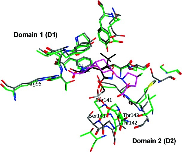
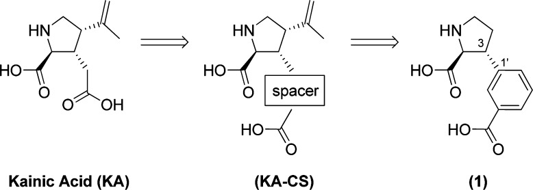
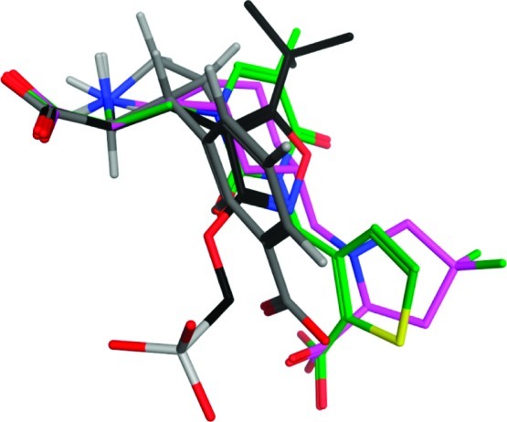
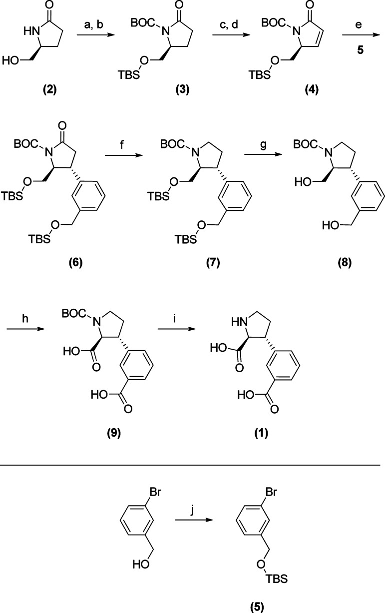
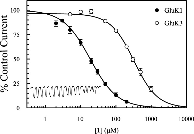
The kainic acid (KA) receptors belong to the class of glutamate (Glu) receptors in the brain and constitute a promising target for the treatment of neurological and/or psychiatric diseases such as schizophrenia, major depression, and epilepsy. Five KA subtypes have been identified and named GluK1-5. In this article, we present the discovery of (2S,3R)-3-(3-carboxyphenyl)-pyrrolidine-2-carboxylic acid (1) based on a rational design process. Target compound 1 was synthesized by a stereoselective strategy in 10 steps from commercially available starting materials. Binding affinities of 1 at native ionotropic Glu receptors were determined to be in the micromolar range (AMPA, 51 μM; KA, 22 μM; NMDA 6 μM), with the highest affinity for cloned homomeric KA receptor subtypes GluK1,3 (3.0 and 8.1 μM, respectively). Functional characterization of 1 by two electrode voltage clamp (TEVC) electrophysiology at a nondesensitizing mutant of GluK1 showed full competitive antagonistic behavior with a K(b) of 11.4 μM.
Keywords: Glutamate receptors; antagonist; kainic acid receptors; rational design.
Figures
Similar articles
-
Structure-Activity Relationship Study of Ionotropic Glutamate Receptor Antagonist (2S,3R)-3-(3-Carboxyphenyl)pyrrolidine-2-carboxylic Acid.J Med Chem. 2015 Aug 13;58(15):6131-50. doi: 10.1021/acs.jmedchem.5b00750. Epub 2015 Jul 22. J Med Chem. 2015. PMID: 26200741
-
Design and Synthesis of a Series of l-trans-4-Substituted Prolines as Selective Antagonists for the Ionotropic Glutamate Receptors Including Functional and X-ray Crystallographic Studies of New Subtype Selective Kainic Acid Receptor Subtype 1 (GluK1) Antagonist (2S,4R)-4-(2-Carboxyphenoxy)pyrrolidine-2-carboxylic Acid.J Med Chem. 2017 Jan 12;60(1):441-457. doi: 10.1021/acs.jmedchem.6b01516. Epub 2016 Dec 22. J Med Chem. 2017. PMID: 28005385
-
Rational design, synthesis and pharmacological evaluation of the (2R)- and (2S)-stereoisomers of 3-(2-carboxypyrrolidinyl)-2-methyl acetic acid as ligands for the ionotropic glutamate receptors.ChemMedChem. 2011 Mar 7;6(3):498-504. doi: 10.1002/cmdc.201000543. Epub 2011 Jan 25. ChemMedChem. 2011. PMID: 21268287
-
Ionotropic Glutamate Receptors in Epilepsy: A Review Focusing on AMPA and NMDA Receptors.Biomolecules. 2020 Mar 18;10(3):464. doi: 10.3390/biom10030464. Biomolecules. 2020. PMID: 32197322 Free PMC article. Review.
-
Glutamate receptor agonists: stereochemical aspects.Curr Top Med Chem. 2011;11(7):887-906. doi: 10.2174/156802611795164990. Curr Top Med Chem. 2011. PMID: 21291400 Review.
Cited by
-
Blood-brain barrier permeability and brain uptake mechanism of kainic acid and dihydrokainic acid.Neurochem Res. 2015 Mar;40(3):542-9. doi: 10.1007/s11064-014-1499-4. Epub 2014 Dec 9. Neurochem Res. 2015. PMID: 25488153
-
Redox-neutral α-functionalization of pyrrolidines: facile access to α-aryl-substituted pyrrolidines.RSC Adv. 2024 Apr 15;14(17):11986-11991. doi: 10.1039/d4ra00983e. eCollection 2024 Apr 10. RSC Adv. 2024. PMID: 38623291 Free PMC article.
-
Augmentation of Anticancer Drug Efficacy in Murine Hepatocellular Carcinoma Cells by a Peripherally Acting Competitive N-Methyl-d-aspartate (NMDA) Receptor Antagonist.J Med Chem. 2017 Dec 14;60(23):9885-9904. doi: 10.1021/acs.jmedchem.7b01624. Epub 2017 Dec 5. J Med Chem. 2017. PMID: 29205034 Free PMC article.
-
Structure, Function, and Pharmacology of Glutamate Receptor Ion Channels.Pharmacol Rev. 2021 Oct;73(4):298-487. doi: 10.1124/pharmrev.120.000131. Pharmacol Rev. 2021. PMID: 34753794 Free PMC article. Review.
-
Kainate Receptor Antagonists: Recent Advances and Therapeutic Perspective.Int J Mol Sci. 2023 Jan 18;24(3):1908. doi: 10.3390/ijms24031908. Int J Mol Sci. 2023. PMID: 36768227 Free PMC article. Review.
References
-
- Kaczor A. A.; Matosiuk D. (2010) Molecular structure of ionotropic glutamate receptors. Curr. Med. Chem. 17, 2608–2635. - PubMed
-
- Ferraguti F.; Shigemoto R. (2006) Metabotropic glutamate receptors. Cell Tissue Res. 326, 483–504. - PubMed
-
- Bartolotto; Clarke Z. A. (1999) Kainate receptors are involved in synaptic plasticity. Nature 402, 297. - PubMed
-
- Mellor J. R. (2006) Synaptic plasticity of kainate receptors. Biochem. Soc. Trans. 34, 949–951. - PubMed
Publication types
MeSH terms
Substances
LinkOut - more resources
Full Text Sources
Other Literature Sources
