

CT-guided radiofrequency ablation of osteoid osteoma in the long bones of the lower extremity
- PMID: 22778881
- PMCID: PMC3391674
- DOI: 10.4329/wjr.v4.i6.278
CT-guided radiofrequency ablation of osteoid osteoma in the long bones of the lower extremity
Abstract
Aim: To present our initial experience with computed tomography guided radiofrequency ablation (RFA) of osteoid osteoma (OO) in our institution.
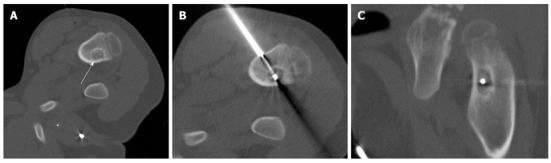
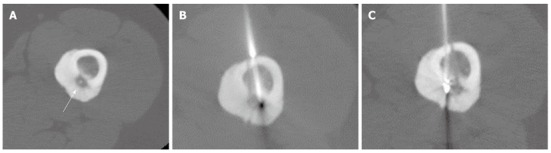
Methods: RFA was performed on eight patients (5 males and 3 females) with clinically and radiologically diagnosed OO (femoral neck, n = 4; femoral diaphysis, n = 2; tibial diaphysis, n = 1; fibular diaphysis, n = 1). Ablation was performed using an electrode with a 10-mm exposed tip for a total of 4-6 min at a targeted temperature of 90 degrees Celsius. No cooling system was used. The intervention was accepted as technically successful if the tip of the electrode could be placed within the center of the nidus. We defined clinical success as a disappearance within 2 wk after treatment of symptoms that had manifested at presentation.
Results: All procedures were technically successful. No major or immediate complications were observed. Clinical success was achieved in six of eight patients in the first procedure. A second procedure was performed for two patients who had recurrent or continued pain, and one of these cases was successfully treated. The overall rate of success was 87.5% (7/8). No complication was observed.
Conclusion: Our preliminary results indicate a favorable success rate and no complications and are compatible with the previous reports of RFA of OO.
Keywords: Ablation; Computed tomography; Osteoid osteoma.
Figures
Similar articles
-
CT-guided radiofrequency ablation of osteoid osteoma: established concepts and new ideas.Br J Radiol. 2020 Oct 1;93(1114):20200266. doi: 10.1259/bjr.20200266. Epub 2020 Jun 24. Br J Radiol. 2020. PMID: 32520586 Free PMC article. Review.
-
Percutaneous CT-guided radiofrequency ablation of osteoid osteoma: Potential Pitfalls and complications and how to avoid them.J Clin Orthop Trauma. 2022 Apr 16;28:101869. doi: 10.1016/j.jcot.2022.101869. eCollection 2022 May. J Clin Orthop Trauma. 2022. PMID: 35494487 Free PMC article. Review.
-
Role of percutaneous computed tomography-guided radiofrequency ablation in treatment of osteoid osteoma.South Asian J Cancer. 2017 Oct-Dec;6(4):139-140. doi: 10.4103/sajc.sajc_58_17. South Asian J Cancer. 2017. PMID: 29404285 Free PMC article.
-
The safety and the efficacy of computed tomography guided percutaneous radiofrequency ablation of osteoid osteoma.Acta Orthop Traumatol Turc. 2019 Sep;53(5):360-365. doi: 10.1016/j.aott.2019.06.001. Epub 2019 Jul 29. Acta Orthop Traumatol Turc. 2019. PMID: 31371131 Free PMC article.
-
Percutaneous radiofrequency ablation of osteoid osteoma using cool-tip electrodes without the cooling system.Jpn J Radiol. 2011 Feb;29(2):138-43. doi: 10.1007/s11604-010-0529-7. Epub 2011 Feb 27. Jpn J Radiol. 2011. PMID: 21359939
Cited by
-
A comparison of percutaneous trephine excision and open surgery in the treatment of osteoid osteoma.Int Orthop. 2016 Jul;40(7):1481-7. doi: 10.1007/s00264-015-3044-8. Epub 2015 Nov 16. Int Orthop. 2016. PMID: 26572883
-
CT-guided radiofrequency ablation of osteoid osteoma using a multi-tined expandable electrode system.Acta Biomed. 2017 Oct 18;88(4S):31-37. doi: 10.23750/abm.v88i4-S.6791. Acta Biomed. 2017. PMID: 29083350 Free PMC article.
-
Challenges in Diagnosing Juxt-Articular Osteoid Osteoma of the Talus: A Case Report.Cureus. 2023 Jun 28;15(6):e41068. doi: 10.7759/cureus.41068. eCollection 2023 Jun. Cureus. 2023. PMID: 37519524 Free PMC article.
-
CT-guided radiofrequency ablation of osteoid osteoma: established concepts and new ideas.Br J Radiol. 2020 Oct 1;93(1114):20200266. doi: 10.1259/bjr.20200266. Epub 2020 Jun 24. Br J Radiol. 2020. PMID: 32520586 Free PMC article. Review.
-
Percutaneous CT-guided radiofrequency ablation of osteoid osteoma: Potential Pitfalls and complications and how to avoid them.J Clin Orthop Trauma. 2022 Apr 16;28:101869. doi: 10.1016/j.jcot.2022.101869. eCollection 2022 May. J Clin Orthop Trauma. 2022. PMID: 35494487 Free PMC article. Review.
References
-
- Woertler K, Vestring T, Boettner F, Winkelmann W, Heindel W, Lindner N. Osteoid osteoma: CT-guided percutaneous radiofrequency ablation and follow-up in 47 patients. J Vasc Interv Radiol. 2001;12:717–722. - PubMed
-
- Motamedi D, Learch TJ, Ishimitsu DN, Motamedi K, Katz MD, Brien EW, Menendez L. Thermal ablation of osteoid osteoma: overview and step-by-step guide. Radiographics. 2009;29:2127–2141. - PubMed
-
- Schulman L, Dorfman HD. Nerve fibers in osteoid osteoma. J Bone Joint Surg Am. 1970;52:1351–1356. - PubMed
-
- Venbrux AC, Montague BJ, Murphy KP, Bobonis LA, Washington SB, Soltes AP, Frassica FJ. Image-guided percutaneous radiofrequency ablation for osteoid osteomas. J Vasc Interv Radiol. 2003;14:375–380. - PubMed
-
- Parlier-Cuau C, Champsaur P, Nizard R, Hamze B, Laredo JD. Percutaneous removal of osteoid osteoma. Radiol Clin North Am. 1998;36:559–566. - PubMed
LinkOut - more resources
Full Text Sources
