

Carotid velocities determine cerebral blood flow deficits in elderly men with carotid stenosis <50%
- PMID: 22778963
- PMCID: PMC3388379
- DOI: 10.1155/2012/579531
Carotid velocities determine cerebral blood flow deficits in elderly men with carotid stenosis <50%
Abstract
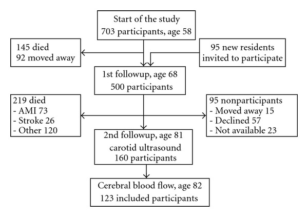
To examine if mild carotid stenosis correlates with silent vascular brain changes, we studied a prospective population-based cohort "Men born in 1914." Data from followups at ages 68 and 81, have been used. Carotid ultrasound was performed at age 81, and cerebral blood flow (CBF) was measured with SPECT at age 82. Out of 123 stroke-free patients, carotid stenosis <50% was observed in 94% in the right and 89% in the left internal carotid arteries (ICAs). In these subjects, Peak Systolic Velocities in ICA correlated negatively with CBF in a majority of several brain areas, especially in mesial temporal area. Results were limited to normotensive until their seventies, who developed late-onset hypertension with a subsequent blood pressure, pulse pressure, and ankle-brachial index growth. Elderly with asymptomatic carotid stenosis <50% and peak systolic velocities in ICA 0.7-1.3 m/s, should be offered an intensified pharmacotherapy to prevent stroke or silent cerebrovascular events.
Figures


References
-
- Hobson RW, II, Weiss DG, Fields WS, et al. Efficacy of carotid endarterectomy for asymptomatic carotid stenosis. The New England Journal of Medicine. 1993;328(4):221–227. - PubMed
-
- Endarterectomy for asymptomatic carotid artery stenosis. Executive Committee for the Asymptomatic Carotid Atherosclerosis Study. Journal of the American Medical Association. 1995;273(18):1421–1428. - PubMed
-
- Halliday A, Mansfield A, Marro J, et al. Prevention of disabling and fatal strokes by successful carotid endarterectomy in patients without recent neurological symptoms: randomised controlled trial. The Lancet. 2004;363(9420):1491–1502. - PubMed
-
- Palamuthusingam D, Quigley F, Golledge J. Implications of the finding of no significant carotid stenosis based on data from a regional Australian vascular unit. Annals of Vascular Surgery. 2011;25(8):1050–1056. - PubMed
-
- Kakkos SK, Sabetai M, Tegos T, et al. Silent embolic infarcts on computed tomography brain scans and risk of ipsilateral hemispheric events in patients with asymptomatic internal carotid artery stenosis. Journal of Vascular Surgery. 2009;49(4):902–909. - PubMed
LinkOut - more resources
Full Text Sources
Miscellaneous
