Selective gene expression analysis of muscular and vascular components in hearts using laser microdissection method
- PMID: 22778964
- PMCID: PMC3384972
- DOI: 10.1155/2012/863410
Selective gene expression analysis of muscular and vascular components in hearts using laser microdissection method
Abstract
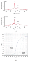
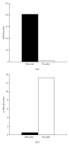
Background. The heart consists of various kinds of cell components. However, it has not been feasible to separately analyze the gene expression of individual components. The laser microdissection (LMD) method, a new technology to collect target cells from the microscopic regions, has been used for malignancies. We sought to establish a method to selectively collect the muscular and vascular regions from the heart sections and to compare the marker gene expressions with this method. Methods and Results. Frozen left ventricle sections were obtained from Wistar-Kyoto rats (WKY) and stroke-prone spontaneously hypertensive rats (SHR-SP) at 24 weeks of age. Using the LMD method, the muscular and vascular regions were selectively collected under microscopic guidance. Real-time RT-PCR analysis showed that brain-type natriuretic peptide (BNP), a marker of cardiac myocytes, was expressed in the muscular samples, but not in the vascular samples, whereas α-smooth muscle actin, a marker of smooth muscle cells, was detected only in the vascular samples. Moreover, SHR-SP had significantly greater BNP upregulation than WKY (P < 0.05) in the muscular samples. Conclusions. The LMD method enabled us to separately collect the muscular and vascular samples from myocardial sections and to selectively evaluate mRNA expressions of the individual tissue component.
Figures


References
-
- Weber KT, Brilla CG, Janicki JS. Myocardial fibrosis: functional significance and regulatory factors. Cardiovascular Research. 1993;27(3):341–348. - PubMed
-
- Nicoletti A, Michel JB. Cardiac fibrosis and inflammation: interaction with hemodynamic and hormonal factors. Cardiovascular Research. 1999;41(3):532–543. - PubMed
-
- Tachikawa T, Irie T. A new molecular biology approach in morphology: basic method and application of laser microdissection. Medical Electron Microscopy. 2004;37(2):82–88. - PubMed
-
- Kajimoto H, Kai H, Aoki H, et al. Inhibition of eNOS phosphorylation mediates endothelial dysfunction in renal failure: new effect of asymmetric dimethylarginine. Kidney International. 2012;81(8):762–768. - PubMed
-
- Kuwahara F, Kai H, Tokuda K, et al. Transforming growth factor-β function blocking prevents myocardial fibrosis and diastolic dysfunction in pressure-overloaded rats. Circulation. 2002;106(1):130–135. - PubMed
LinkOut - more resources
Full Text Sources

