. 2012 Jul;47(1):29-37.
doi: 10.1016/j.nbd.2012.03.021.

Role of the pituitary–adrenal axis in granulocyte-colony stimulating factor-induced neuroprotection against hypoxia–ischemia in neonatal rats

Affiliations

Role of the pituitary–adrenal axis in granulocyte-colony stimulating factor-induced neuroprotection against hypoxia–ischemia in neonatal rats

Mélissa S Charles et al. Neurobiol Dis. 2012 Jul.

Abstract

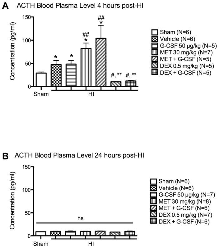
Several reports indicate that the activity of the hypothalamic–pituitary–adrenal axis (HPA) is increased after a brain insult and that its down-regulation can improve detrimental outcomes associated with ischemic brain injuries.Granulocyte-colony stimulating factor (G-CSF) is a neuroprotective drug shown in the naïve rat to regulate hormones of the HPA axis. In this study we investigate whether G-CSF confers its neuroprotective properties by influencing the HPA response after neonatal hypoxia–ischemia (HI). Following the Rice–Vannucci model, seven day old rats (P7)were subjected to unilateral carotid ligation followed by 2.5 h of hypoxia. To test our hypothesis,metyrapone was administered to inhibit the release of rodent specific glucocorticoid, corticosterone, at the adrenal level. Dexamethasone, a synthetic glucocorticoid, was administered to agonize the effects of corticosterone.Our results show that both G-CSF and metyrapone significantly reduced infarct volume while dexamethasone treatment did not reduce infarct size even when combined with G-CSF. The protective effects of G-CSF do not include blood brain barrier preservation as suggested by the brain edema results. G-CSF did not affect the pituitary released adrenocorticotropic hormone (ACTH) levels in the blood plasma at 4 h, but suppressed the increase of corticosterone in the blood. The administration of G-CSF and metyrapone increased weight gain, and significantly reduced the Bax/Bcl-2 ratio in the brain while dexamethasone reversed the effects of G-CSF. The combination of G-CSF and metyrapone significantly decreased caspase-3 protein levels in the brain, and the effect was antagonized by dexamethasone.We report that G-CSF is neuroprotective in neonatal HI by reducing infarct volume, by suppressing the HI-induced increase of the Bax/Bcl-2 ratio, and by decreasing corticosterone in the blood. Metyrapone was able to confer similar neuroprotection as G-CSF while dexamethasone reversed the effects of G-CSF. In conclusion, we show that decreasing HPA axis activity is neuroprotective after neonatal HI, which can be conferred by administering G-CSF.


Figures

Fig 1
Fig 1
Dose response of metyrapone and dexamethasone. Representative TTC stained coronal brain section of the dose response analysis for metyrapone (MET) and dexamethasone (DEX) at 24 h. (*=p<0.01 vs Vehicle, #=p<0.05 vs MET 30 mg/kg, all HI groups p<0.01 vs Sham). Each line on the left hand side of the brain images demarks 1 mm.
Fig 2
Fig 2
Infarct volume and body weight at 24 h after G-CSF, metyrapone and dexamethasone administration. A. TTC stained coronal brain section of Sham, and G-CSF, MET, DEX treated groups after HI. (*=p<0.05 vs Vehicle, #=p<0.05 vs G-CSF, all HI groups p<0.01 vs Sham). B. Body weight gain was measured on a high precision balance scale and calculated as such: (Weight 24 h post-HI)−(Weight before HI). (*=p<0.01 vs Vehicle, #=p<0.05, **=p<0.001 vs Sham, ##=p<0.01 vs MET + G-CSF). Each line on the left hand side of the brain images demarks 1 mm.
Fig 3
Fig 3
Brain water content after G-CSF, metyrapone, and dexamethasone treatment after HI. Quantification of brain water content 24 h after HI in the cerebellum, contra-lateral, and ipsilateral brain hemisphere. Brain water content was markedly increased by HI + Vehicle, DEX and DEX + G-CSF treated groups. (*=p<0.05). No significance was observed in G-CSF, MET and MET + G-CSF. DEX treated group had higher brain water content than the G-CSF treated group (##=p<0.05 vs G-CSF). No significant difference was observed amongst the groups in the cerebellum and contralateral hemisphere.
Fig 4
Fig 4
ACTH blood plasma level 4 h and 24 h after HI. A. At 4 h, ACTH levels were significantly increased by HI (*=p<0.05 vs Sham). MET and MET + G-CSF groups had markedly increased ACTH level compared to Vehicle (##=p<0.05). DEX and DEX + G-CSF had significantly decreased ACTH levels compared to MET and MET + G-CSF groups (#=p<0.01 vs MET 30 mg/kg, **=p<0.01 vs MET + G-CSF). B. ACTH blood plasma level is normalized to Sham at 24 h after HI.
Fig 5
Fig 5
Corticosterone blood plasma level 4 h and 24 h after HI. A. HI considerably increases CORT levels and G-CSF robustly decreases CORT levels at 4 h (# = p<0.05 vs Sham, * = p <0.01 vs Vehicle). MET +G-CSF decreased CORT compared to Vehicle group while the DEX + G-CSF group increased CORT levels significantly (** = p <0.05). B. CORT levels remain higher in Vehicle and DEX treated groups (* = p <0.05 vs Sham). All groups with G-CSF in their regimen have lower CORT levels than their control group (# = p<0.05 vs DEX 0.5 mg/kg).
Fig 6
Fig 6
The expression of apoptotic markers in the ipsilateral hemisphere 24 h post-HI. A. HI increased Bax protein expression. No significant difference is reported amongst all groups. B. Bcl-2 relative density normalized to actin in decreased by HI but no significant difference is observed between groups. C. Bax/Bcl-2 ratio was significantly increased by HI (# = p<0.05 vs Sham). G-CSF, MET and MET + G-CSF treated groups significantly reduced Bax/Bcl-2 ratio compared to Vehicle treated groups (* = p<0.05). D. Cleaved caspase-3 levels are markedly increased by HI (# = p<0.05 vs Sham), and relatively lowered by G-CSF and MET groups (no significance observed). MET + G-CSF significantly decreased caspase-3 levels (* = p<0.05 vs Vehicle), and DEX + G-CSF group antagonized the effects (** = p<0.05 vs MET + G-CSF).

References

    1. Allen C, Kendall JW. Maturation of the circadian rhythm of plasma corticosterone in the rat. Endocrinology. 1967 May;80(5):926–930. - PubMed
    1. Baldi E, Bucherelli C. The inverted “u-shaped” dose–effect relationships in learning and memory: modulation of arousal and consolidation. Nonlinearity Biol Toxicol Med. 2005 Jan;3(1):9–21. - PMC - PubMed
    1. Betz AL, Coester HC. Effect of steroids on edema and sodium uptake of the brain during focal ischemia in rats. Stroke. 1990 Aug;21(8):1199–1204. - PubMed
    1. Butt TK, Farooqui R, Khan MA. Risk factors for hypoxic ischemic encephalopathy in children. J Coll Physicians Surg Pak. 2008 Jul;18(7):428–432. - PubMed
    1. Chen W, Jadhav V, Tang J, Zhang JH. HIF-1alpha inhibition ameliorates neonatal brain injury in a rat pup hypoxic–ischemic model. Neurobiol Dis. 2008 Sep;31(3):433–441. - PMC - PubMed

Publication types

MeSH terms

LinkOut - more resources