. 2012 Sep;85(6):1119-32.
doi: 10.1111/j.1365-2958.2012.08163.x. Epub 2012 Jul 26.

Cationic antimicrobial peptides disrupt the Streptococcus pyogenes ExPortal

Affiliations

Cationic antimicrobial peptides disrupt the Streptococcus pyogenes ExPortal

Luis Alberto Vega et al. Mol Microbiol. 2012 Sep.

Abstract

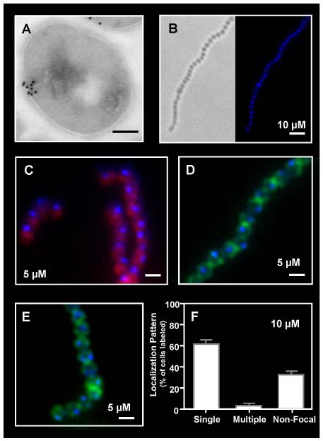
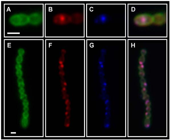
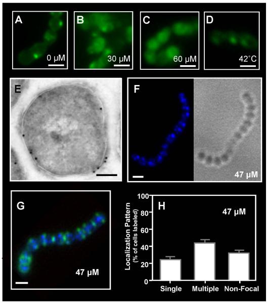
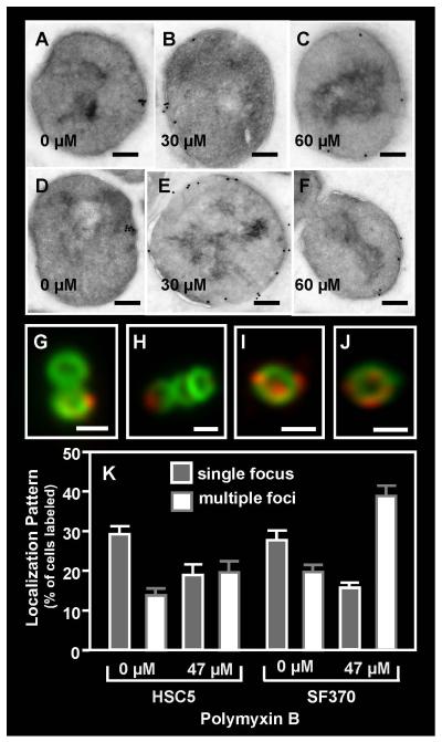
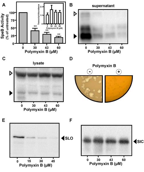
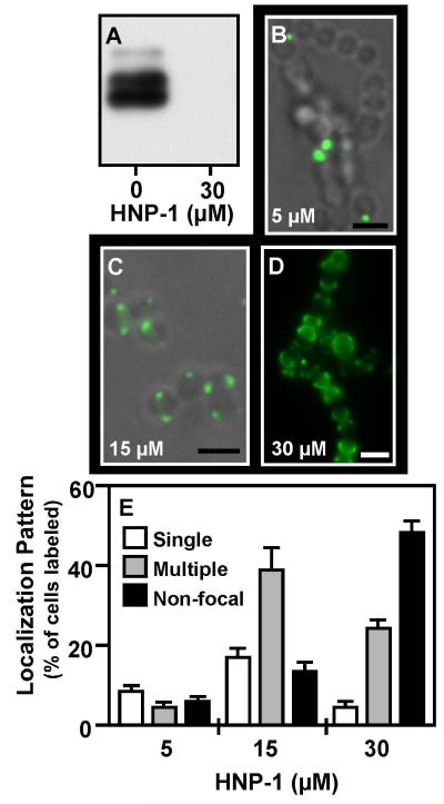
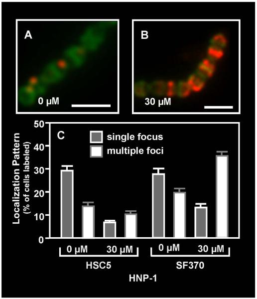
Although they possess a well-characterized ability to porate the bacterial membrane, emerging research suggests that cationic antimicrobial peptides (CAPs) can influence pathogen behaviour at levels that are sublethal. In this study, we investigated the interaction of polymyxin B and human neutrophil peptide (HNP-1) with the human pathogen Streptococcus pyogenes. At sublethal concentrations, these CAPs preferentially targeted the ExPortal, a unique microdomain of the S. pyogenes membrane, specialized for protein secretion and processing. A consequence of this interaction was the disruption of ExPortal organization and a redistribution of ExPortal components into the peripheral membrane. Redistribution was associated with inhibition of secretion of certain toxins, including the SpeB cysteine protease and the streptolysin O (SLO) cytolysin, but not SIC, a protein that protects S. pyogenes from CAPs. These data suggest a novel function for CAPs in targeting the ExPortal and interfering with secretion of factors required for infection and survival. This mechanism may prove valuable for the design of new types of antimicrobial agents to combat the emergence of antibiotic-resistant pathogens.


Figures

Figure 1
Figure 1. Focal binding of Polymyxin B to the S. pyogenes surface
The distribution of polymyxin B on the surface of S. pyogenes SF370 following sub-lethal challenge was revealed: (A) by treatment with biotinylated polymyxin B (1:10,000) and immunogold electron microscopy using staining with a streptavidin-gold conjugate (scale bar = 200nm) and (B, C, D, E) by fluorescent microscopy following challenge with dansyl-polymyxin B alone (B) at the concentration indicated in the Figure (scale bar = 1μm) or in cells counterstained with Nile Red (C), fluorescent vancomycin (D) or wheat germ agglutinin Alexa Fluor 488 conjugate (E). Staining patterns following challenge with dansyl-polymyxin B were quantitated as described in the Experimental Procedures (F). Data represents the mean and standard error of the mean (SEM) derived from at least 3 independent experiments and examination of a minimum of 1000 stained cells. The number of cells with a single focus was significantly higher than any other staining pattern (P < 0.0001).
Figure 2
Figure 2. The site of polymyxin B binding is coincident with the site of SpeB secretion
Cells of S. pyogenes SF370 were challenged with dansyl-polymyxin B (10 μM), fluorescent vancomycin stained to visualize cell wall and subjected to the red protease assay, which monitors cleavage of BODIPY TR-X-casein by the SpeB protease. Stained cells were then examined by fluorescent microscopy. Panels are as follows: (A, E) fluorescent vancomycin, (B, F) BODIPY TR-X-casein, (C, G) Dansyl-polymyxin B and (D, H) Merge of Panels A, B and C. Scale bar = 1μm.
Figure 3
Figure 3. Sub-lethal challenge with polymyxin B alters the distribution of anionic membrane lipids
Cultures of S. pyogenes SF370 were stained with NAO following challenge with 0 μM (A), 30 μM (B) or 60 μM (C) polymyxin B or heat shock at 42°C (D) and examined by fluorescent microscopy (scale bar = 1μm). The distribution of polymyxin B following higher, but still sub-lethal challenge, was revealed by treatment with biotinylated polymyxin B (1:500) and immunogold electron microscopy using staining with a streptavidin-gold conjugate (scale bar = 200nm) (E) and by fluorescent microscopy following challenge with dansyl-polymyxin B alone at the concentration indicated in the Figure (scale bar = 1μm) (F) or costained with fluorescent vancomycin (G). Staining patterns following challenge were quantitated as described previously (H). Data represents the mean and SEM derived from at least 3 independent experiments and examination of a minimum of 1000 stained cells. The number of cells with multiple foci was significantly higher than any other staining pattern (P < 0.0001) at this polymyxin B concentration.
Figure 4
Figure 4. Redistribution of ExPortal proteins following sub-lethal polymyxin B challenge
The distribution of SecA (A, B, C) and HtrA (D, E, F) on S. pyogenes SF370 was assessed by immunogold electron microscopy following challenge with polymyxin B at the concentrations indicated in the Figure (scale bar = 200nm). The distribution of HtrA was also assessed by immunofluorescent microscopy in the absence of (G, H) and following challenge with polymyxin B at 47 μM (I, J) and was quantitated as described previously (scale bar = 500nm) (K). Data represents the mean and SEM derived from at least 3 independent experiments and examination of a minimum of 1000 stained cells. In untreated cultures, the number of SF370 or HSC5 cells with a single focus was significantly higher than any other staining pattern (P < 0.05), whereas in polymyxin B-treated cultures, the number of streptococcal cells with multiple foci was significantly higher (P < 0.05).
Figure 5
Figure 5. High sub-lethal challenge with polymyxin B inhibits secretion of SpeB and SLO, but not SIC
Expression of the SpeB protease in cultures of S. pyogenes SF370 was determined following challenge with the indicated concentrations of polymyxin B by quantitation of cysteine protease activity in culture supernatant (A), by real-time RT-PCR analysis of speB transcript abundance (A, inset) by Western blot analysis of culture supernatant (B) in whole cell lysates (C) and following overnight culture on protease indicator plates containing (+) or lacking () 150 μM polymyxin B (D). Expression of SpeB results in a zone of clearing around colonies. Western blotting was also used to analyze the amount of SLO (E) and SIC (F) present in culture supernatant following challenge with the indicated concentrations of polymyxin B. All samples for SpeB analysis were harvested at 3 hrs post-challenge; samples for SLO and SIC were harvested 2hrs post-challenge. Open and filled triangles indicate the migration of the zymogen and mature form of SpeB, respectively. The migration of the SLO and SIC polypeptides are also indicated.
Figure 6
Figure 6. Focal localization and inhibition of SpeB expression by HNP-1
Cultures were challenged with HNP-1 or by 5-FAM-HNP-1 at the concentrations indicated. Expression of SpeB was evaluated by a Western blot analysis (A) and binding to the S. pyogenes surface was assessed using fluorescent microscopy (B, C, D). Presented are overlays of fluorescent and phase images (B, C) or the fluorescent image alone (D) (scale bar = 1μm). Staining patterns were quantitated as described previously (E). Data represents the mean and SEM derived from at least 3 independent experiments and examination of a minimum of 1000 stained cells. At minimal concentration the number of cells with single foci is significantly higher than all other staining patterns, whereas multiple foci and hemisphere staining predominated significantly at higher peptide concentrations (P < 0.05).
Figure 7
Figure 7. Challenge with HNP-1 results in re-distribution of HtrA
Cultures of S. pyogenes HSC5 were challenged with the indicated concentrations of HNP-1 and subjected to immunofluorescent microscopy to assess the distribution of HtrA (A, B). Cell walls were visualized by staining with fluorescent vancomycin (scale bar = 1μm). Staining patterns were quantitated as described previously (C). Data represents the mean and SEM derived from at least 3 independent experiments and examination of a minimum of 1000 stained cells. Challenge with the peptide significantly decreased the number of SF370 or HSC5 cells exhibiting HtrA at single foci (P<0.0001) and resulted in a significant increase in the number of multiple foci (P<0.0001) in the SF370 strain.

References

    1. Barak I, Muchova K, Wilkinson AJ, O’Toole PJ, Pavlendova N. Lipid spirals in Bacillus subtilis and their role in cell division. Mol Microbiol. 2008;68:1315–1327. - PMC - PubMed
    1. Boman HG. Antibacterial peptides: basic facts and emerging concepts. J Intern Med. 2003;254:197–215. - PubMed
    1. Braff MH, Zaiou M, Fierer J, Nizet V, Gallo RL. Keratinocyte production of cathelicidin provides direct activity against bacterial skin pathogens. Infect Immun. 2005;73:6771–6781. - PMC - PubMed
    1. Brenot A, King KY, Caparon MG. The PerR regulon in peroxide resistance and virulence of Streptococcus pyogenes. Mol Microbiol. 2005;55:221–234. - PubMed
    1. Brenot A, King KY, Janowiak B, Griffith O, Caparon MG. Contribution of glutathione peroxidase to the virulence of Streptococcus pyogenes. Infect Immun. 2004;72:408–413. - PMC - PubMed

Publication types

MeSH terms