. 2012 Sep;85(6):1148-65.
doi: 10.1111/j.1365-2958.2012.08165.x. Epub 2012 Jul 26.

Mycobacterium tuberculosis WhiB4 regulates oxidative stress response to modulate survival and dissemination in vivo

Affiliations
Free PMC article

Mycobacterium tuberculosis WhiB4 regulates oxidative stress response to modulate survival and dissemination in vivo

Manbeena Chawla et al. Mol Microbiol. 2012 Sep.
Free PMC article

Abstract

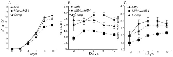
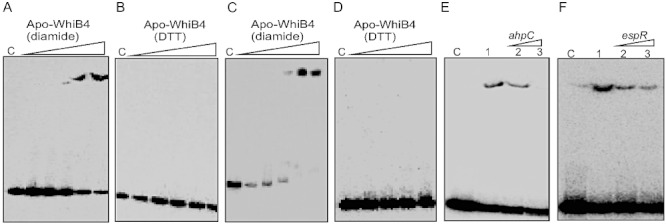
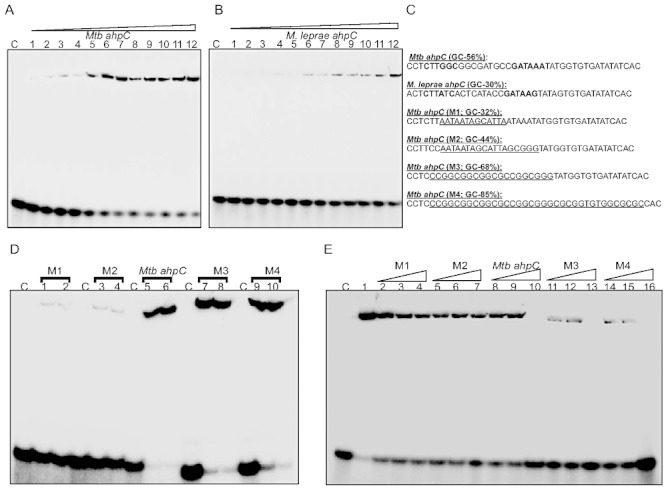
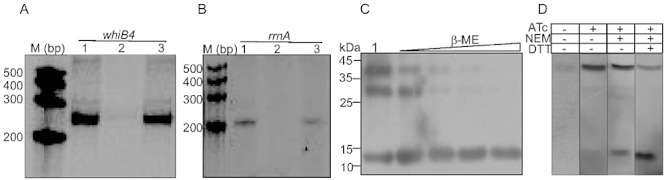
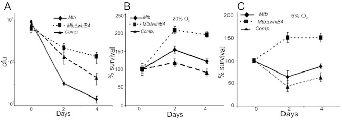
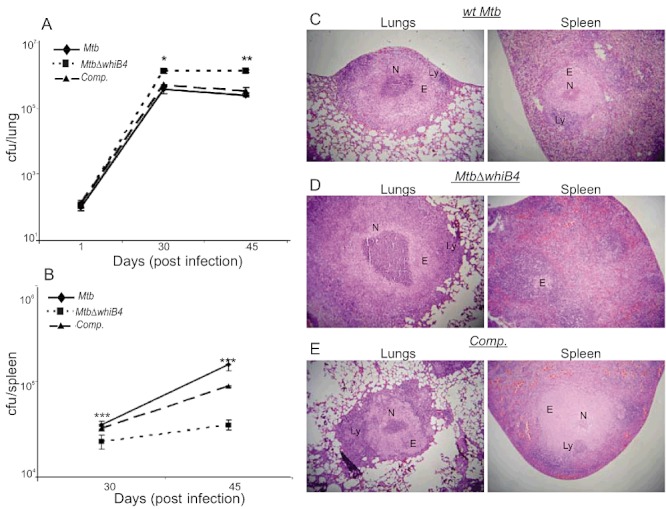
Host-generated oxidative stress is considered one of the main mechanisms constraining Mycobacterium tuberculosis (Mtb) growth. The redox-sensing mechanisms in Mtb are not completely understood. Here we show that WhiB4 responds to oxygen (O₂) and nitric oxide (NO) via its 4Fe-4S cluster and controls the oxidative stress response in Mtb. The WhiB4 mutant (MtbΔwhiB4) displayed an altered redox balance and a reduced membrane potential. Microarray analysis demonstrated that MtbΔwhiB4 overexpresses the antioxidant systems including alkyl hydroperoxidase (ahpC-ahpD) and rubredoxins (rubA-rubB). DNA binding assays showed that WhiB4 [4Fe-4S] cluster is dispensable for DNA binding. However, oxidation of the apo-WhiB4 Cys thiols induced disulphide-linked oligomerization, DNA binding and transcriptional repression, whereas reduction reversed the effect. Furthermore, WhiB4 binds DNA with a preference for GC-rich sequences. Expression analysis showed that oxidative stress repressed whiB4 and induced antioxidants in Mtb, while their hyper-induction was observed in MtbΔwhiB4. MtbΔwhiB4 showed increased resistance to oxidative stress in vitro and enhanced survival inside the macrophages. Lastly, MtbΔwhiB4 displayed hypervirulence in the lungs of guinea pigs, but showed a defect in dissemination to their spleen. These findings suggest that WhiB4 systematically calibrates the activation of oxidative stress response in Mtb to maintain redox balance, and to modulate virulence.


Figures

Fig. 1
Fig. 1
Spectroscopic characterization of WhiB4. A. Aerobically purified WhiB4 was scanned by a UV-vis spectrophotometer. Note the presence of broad peaks at ∼ 330 nm, ∼ 420 and ∼ 450 nm characteristic of a 2Fe-2S cluster. B. UV-visible spectra of anaerobically reconstituted WhiB4. Enzymatic reconstitution of WhiB4 Fe-S was carried out inside an anaerobic glove box. Note the time-dependent increase in the characteristic 4Fe-4S cluster peak at 420 nm. Reconstitution of the 4Fe-4S cluster was completed in 30 min. C. EDFS-EPR spectra of reconstituted WhiB4 after reduction with sodium dithionite (DTH). The experimental conditions were: π/2 and π pulses of 16 and 32 ns; τ = 180 ns; T = 9 K. Spectra were acquired 60 shots with a two-step cycle at a repetition rate of 1 kHz. Microwave frequency = 9.806 GHz.
Fig. 2
Fig. 2
The Mtb WhiB4 Fe-S cluster responds to O2 and NO. A. UV-visible spectra were obtained before and after exposing anaerobically reconstituted WhiB4 4Fe-4S cluster to air at various time points. Note the time-dependent decrease in the absorbance at 420 nm. B. Air-exposed samples of reconstituted WhiB4 were withdrawn immediately (5 min; green spectrum) or 30 min post exposure (blue spectrum) and analysed by EDFS-EPR. The appearance of a sharp signal at g = 2.01 indicates a [3Fe-4S]1+ cluster. Conditions for EPR spectroscopy were the same as in Fig. 1C. The influence of NO on WhiB4 was analysed by adding a 10-fold molar excess of proline NONOate (WhiB4:NO) before analysis by UV-visible and cw-EPR spectroscopy. C. UV-visible spectra were acquired before and after addition of proline NONOate. Note the increase in the characteristic monomeric DNIC peak at ∼ 350 nm in the NO-treated sample. D. cw-EPR spectra of NO-treated samples were acquired at a microwave frequency of 9.667 GHz and microwave power of 2 mW at 100, 155 and 200 K. The appearance of a sharp signal around 2.03 and the increase in the intensity of the signal at lower temperatures is consistent with the formation of monomeric DNIC.
Fig. 3
Fig. 3
WhiB4 modulates growth, redox homeostasis and membrane potential of Mtb under aerobic conditions. A. Aerobic growth phenotype of wt Mtb, MtbΔwhiB4 and complemented strains was determined by growing cells in 7H9 medium under aerobic conditions. Growth was monitored at different time intervals by cfu analysis. B. At various days post inoculation, cells were analysed for intracellular redox balance by measuring the poise of NAD+/NADH as described in Experimental procedures. C. Membrane potential of aerobically growing cells was determined by staining with DiOC2. A change in the membrane potential was detected using the average mean fluorescence intensity (Red/Green) emitted by the cells. The fluorescent intensity was normalized to the values obtained upon carbonyl cyanide m-chlorophenyl hydrazone (CCCP) treatment. All of the above experiments were carried out at least three times in triplicate and results are given as the mean values and standard deviations.
Fig. 4
Fig. 4
DNA binding activity of WhiB4. A–D. Apo-WhiB4 was prepared as described in Experimental procedures. The concentrations of apo-WhiB4 used for EMSAs were 0.1, 0.2, 0.4, 0.8 and 1 µM. EMSA reactions were performed with 0.2 nM γ-32P-labelled ahpC (A and B) and whiB4 (C and D) promoter DNA fragments. DNA binding of apo-WhiB4 in the presence of thiol-oxidant, diamide (A and C) or thiol-reductant, DTT (B and D). C: DNA binding in the absence of WhiB4 in each panel. E and F. Sequence preference of WhiB4 for DNA binding. EMSAs were performed using γ-32P-labelled ahpC promoter DNA with 800 nM of apo-WhiB4 in the presence of 50 mM diamide. The DNA binding was competed using increasing concentrations of unlabelled ahpC (specific) or espR (non-specific) promoter DNA. Lane 1 in (E) and (F): WhiB4:ahpC promoter complex. WhiB4 DNA binding was competed using 10-fold (lane 2) and 50-fold (lane 3) molar excess of either unlabelled ahpC (E) or espR (F) promoter DNA. C: DNA binding in the absence of WhiB4 in each panel.
Fig. 5
Fig. 5
WhiB4 binds to GC-rich DNA. A and B. Concentration-dependent binding of oxidized apo-WhiB4 to a 40 bp γ-32P-labelled DNA fragment derived from (A) Mtb and (B) M. leprae ahpC promoter regions containing the OxyR-binding motif. The concentrations of oxidized apo-WhiB4 were 0.25, 0.50, 0.75, 1.00, 1.25, 1.50, 1.75, 2.0, 2.25, 2.50, 2.75, 3.00 µM. The Kd for both the fragments was calculated by measuring the intensity of free and protein bound DNA using ImageJ software. Note that apo-WhiB4 binds with higher affinity to the GC-rich Mtb OxyR recognition sequence as compared with the AT-rich M. leprae OxyR recognition sequence within the ahpC promoter. C. DNA fragments containing mutations to modify the GC content of the ahpC promoter region (OxyR binding core motif is shown in bold). The mutated sequences in various DNA fragments (M1–M4) are underlined. These fragments were subjected to EMSA analysis. D. EMSAs were carried out using 0.8 and 1 µM oxidized apo-WhiB4 and 0.2 nM γ-32P-labelled DNA fragments. Note that apo-WhiB4 showed enhanced binding to M3 (68% GC) and M4 (85% GC) as compared with M1 (32% GC) and M2 (44% GC) fragments. E. Competition assay using high and low GC DNA fragments. Lane 1: oxidized apo-WhiB4:ahpC promoter complex. DNA binding was competed using 10-fold (lanes 2, 5, 8, 11 and 14), 20-fold (lanes 3, 6, 9, 12 and 15) and 50-fold (lanes 4, 7, 10, 13 and 16) molar excess of unlabelled DNA fragments as indicated in the figure. C: DNA binding in the absence of WhiB4 in each panel.
Fig. 6
Fig. 6
A and B. Effect of WhiB4 on in vitro transcription. Single round transcription assays show that RNAP-σA holoenzyme was proficient in directing transcription from whiB4 (A, lane 1) and rrnA (B, lane 1) promoters. 50 nM of whiB4 and rrnA promoter DNA fragments were pre-incubated with either 2 µM oxidized apo-WhiB4 (A, lane 2; B, lane 2) or reduced apo-WhiB4 (A, lane 3; B, lane 3) and subjected to transcription by RNAP-σA. M: RNA marker (Century™ Marker Template, Ambion). C. Disulphide bond formation induces oligomerization of apo-WhiB4 in vitro. Five micrograms of apo-WhiB4 is either oxidized by atmospheric O2 (lane 1) or reduced by 50 mM, 100 mM, 200 mM and 400 mM β-ME and resolved on a 12% non-reducing SDS-PAGE. Apo-WhiB4 bands were visualized by Western blot analysis using anti-His antibody. The ∼ 14 kDa, ∼ 28 kDa and ∼ 42 kDa bands correspond to the His-tagged apo-WhiB4-monomer, -dimer and -trimer. D. In vivo existence of disulphide-linked oligomers of apo-WhiB4. Aerobically grown Msm WhiB4 FLAG-tag strain was either uninduced or induced with ATc and 30 µg of cell-free extract was analysed by non-reducing Western blot using anti-FLAG antibody. Note that a significant portion of the FLAG-tagged apo-WhiB4 exists as a trimer in ATc induced Msm cells. To minimize the possibility of O2-induced thiol oxidation and subsequent oligomerization of apo-WhiB4 during cell-free extract preparation, Msm cells expressing FLAG-tagged WhiB4 were pretreated with the thiol-alkylating agent NEM. Note the presence of apo-WhiB4 trimer in the NEM-pretreated sample. A significant loss of apo-WhiB4 oligomerization upon DTT reduction further suggests the presence of intermolecular disulphide-linked oligomers in vivo.
Fig. 7
Fig. 7
WhiB4 regulates survival of Mtb inside macrophages. A. rIFNγ and LPS-activated Raw264.7 cells were infected with Mtb strains at a moi of 10 and growth was monitored over time by cfu. B and C. To investigate the role of O2 tension, phorbol 12-myristate 13-acetate (PMA)-stimulated THP-1 human monocytic cell lines were maintained at 20% O2 (B) and 5% O2 (C) and infected by various Mtb strains at a moi of 10. For each strain, the cfu at each time point are expressed relative to the cfu at time 0. In each case, data shown are the average of three experiments performed in triplicate; the error bars indicate standard deviation in each group.
Fig. 8
Fig. 8
WhiB4 modulates in vivo survival and pathology of Mtb in guinea pigs. Outbred Hartley guinea pigs (n = 5) given an aerosol challenge of Mtb were assessed for bacterial burden in lungs (A) and spleen (B), and for the severity of lung and spleen pathology (C–E). Statistical significance for the pulmonic and splenic bacterial load was obtained by comparing wt Mtb and MtbΔwhiB4 strains: *P < 0.05, **P < 0.01, ***P < 0.001. Haematoxylin and eosin stained lung and spleen sections (30 days post infection) from guinea pigs infected with wt Mtb (C), MtbΔwhiB4 (D) and the whiB4 complemented (Comp.) strains (E). The pathology sections show granulomas containing areas of necrosis (N), epithelioid cells (E) and lymphocytes (L). All images were taken at 4× magnification. Error bars represent the standard error of the mean.

Similar articles

Cited by

References

    1. Alam MS, Garg SK, Agrawal P. Molecular function of WhiB4/Rv3681c of Mycobacterium tuberculosis H37Rv: a [4Fe-4S] cluster co-ordinating protein disulphide reductase. Mol Microbiol. 2007;63:1414–1431. - PubMed
    1. Alam MS, Garg SK, Agrawal P. Studies on structural and functional divergence among seven WhiB proteins of Mycobacterium tuberculosis H37Rv. FEBS J. 2009;276:76–93. - PubMed
    1. Bardarov S, Bardarov S, Jr, Pavelka MS, Jr, Sambandamurthy V, Larsen M, Tufariello J, et al. Specialized transduction: an efficient method for generating marked and unmarked targeted gene disruptions in Mycobacterium tuberculosis, M. bovis BCG and M. smegmatis. Microbiology. 2002;148:3007–3017. - PubMed
    1. Betts JC, Lukey PT, Robb LC, McAdam RA, Duncan K. Evaluation of a nutrient starvation model of Mycobacterium tuberculosis persistence by gene and protein expression profiling. Mol Microbiol. 2002;43:717–731. - PubMed
    1. Betts JC, McLaren A, Lennon MG, Kelly FM, Lukey PT, Blakemore SJ, Duncan K. Signature gene expression profiles discriminate between isoniazid-, thiolactomycin-, and triclosan-treated Mycobacterium tuberculosis. Antimicrob Agents Chemother. 2003;47:2903–2913. - PMC - PubMed

Publication types

MeSH terms