Review
. 2012 Jul;6 Suppl 1(Suppl 1):S48-54.
doi: 10.1007/s12105-012-0371-6. Epub 2012 Jul 3.

The morphologic profile of HPV-related head and neck squamous carcinoma: implications for diagnosis, prognosis, and clinical management

Affiliations
Review

The morphologic profile of HPV-related head and neck squamous carcinoma: implications for diagnosis, prognosis, and clinical management

William H Westra. Head Neck Pathol. 2012 Jul.

Abstract

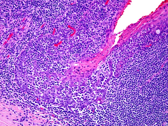
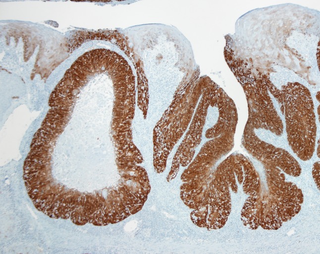
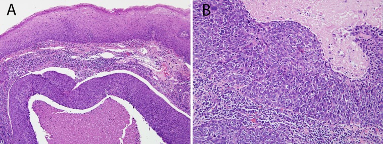
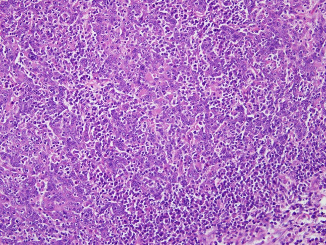
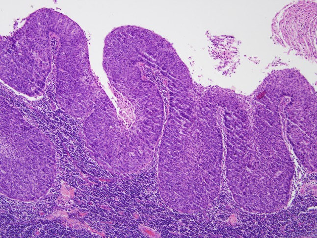
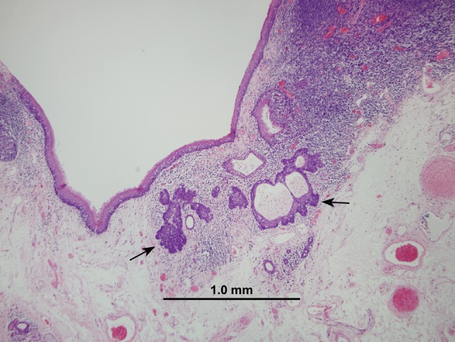
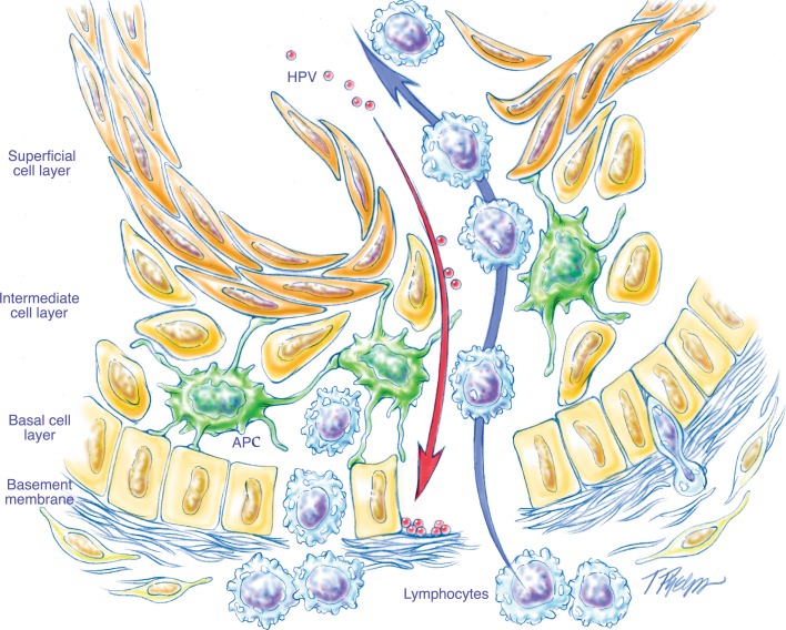
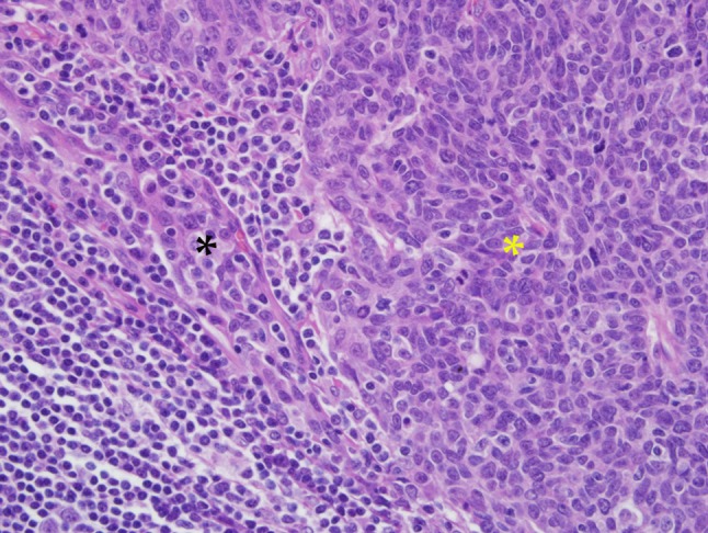
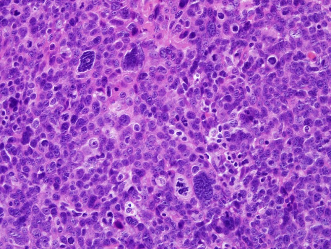
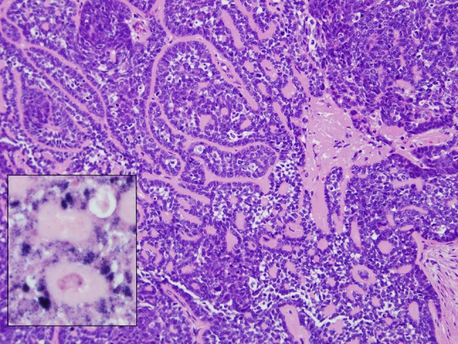
Much recent attention has highlighted a subset of head and neck squamous cell carcinomas (HNSCCs) related to the human papillomavirus (HPV) that is characterized by an epidemiologic, demographic, and clinical profile that deviates from the profile of conventional non-HPV-related HNSCC. Lost in the dash to develop and implement diagnostic assays to detect the presence of HPV in HNSCCs is the unpretentious observation that these HPV-HNSCCs are also distinctive with respect to their microscopic appearance, and that an awareness of these characteristic morphologic features can facilitate the diagnosis of HPV-related HNSCC (HPV-HNSCC). This review will delineate the microscopic appearance of HPV-HNSCC, spotlight ways in which the misinterpretation of these microscopic features can lead to diagnostic confusion, and provide recommendations for appropriate terminology when diagnosing HPV-HNSCC.


Figures

Fig. 1
Fig. 1
The crypts of the tonsils are lined by a specialized squamous epithelium known as the reticulated epithelium. The epithelium is penetrated by numerous interdigitating lymphocytes that obscure the junction between the epithelium and lymphoid stroma, and fragment the epithelium into cords of basaloid cells
Fig. 2
Fig. 2
A p16 immunohistochemical stain is used to highlight tumor distribution in the tonsil. HPV-HNSCC (as indicated by strong p16 immunohistochemical staining) tends to target the tonsillar crypts. When the surface epithelium is involved, it is usually by contiguous extension from the tonsillar crypt
Fig. 3
Fig. 3
This HPV-HNSCC infiltrates the lymphoid stroma of the tonsil as expanding tumor lobules with central necrosis. The surface epithelium lacks the dysplastic changes that typify most non-HPV-HNSCCs (a). The absence of keratinization, cell borders, and abundant cytoplasm (i.e. high nuclear-to-cytoplasmic ratio) often imparts a “basaloid” appearance (b)
Fig. 4
Fig. 4
In HPV-HNSCC, the tumor nests are often permeated by lymphocytes. In some cases, a very brisk lymphoplasmacytic infiltrate will disrupt the tumor nests into irregular cords and individual cells in a lymphoepithelial-like fashion
Fig. 5
Fig. 5
When dealing with metastatic HNSCC to a cervical lymph node, the presence of cystic change and a non-keratinizing morphology should raise the possibility of origin from the base of tongue or palatine tonsil
Fig. 6
Fig. 6
This small focus of HPV-HNSCC was detected in the tonsil of a patient who presented with bulky lymph node metastases. Some HPV-HNSCCs appear to possess the capacity to metastasize even when small in size and seemingly confined to a tonsillar crypt
Fig. 7
Fig. 7
Schematic representation of the reticulated epithelium lining the tonsillar crypts. A porous basement membrane facilitates the migration of lymphocytes and antigen presenting cells (APCs). This lack of structural integrity of the basement membrane confounds the distinction between in situ and invasive disease (figure borrowed from [8])
Fig. 8
Fig. 8
This HPV-HNSCC (yellow asterisk) maintains a close morphologic resemblance to reticulated crypt epithelium (black asterisk) from which it takes origin
Fig. 9
Fig. 9
Occasionally, large pleomorphic cells may be encountered in an HPV-HNSCC. The significance of these is not known with certainty, but they may be associated with more aggressive tumor behavior
Fig. 10
Fig. 10
HPV-HNSCC with prominent basaloid features including lobules of hyperchromatic cells separated by hyaline stromal deposits. This tumor can only be separated from the basaloid variant of HNSCC by detection of HPV. In this case, HPV in situ hybridization demonstrates the presence of abundant hybridization signals in the nuclei of the tumor cells (inset)

References

    1. Gillison ML, Koch WM, Spafford M, et al. Causal association between human papillomavirus and a subset of head and neck cancers. J Natl Cancer Inst. 2000;92:709–720. doi: 10.1093/jnci/92.9.709. - DOI - PubMed
    1. Adelstein DJ, Ridge JA, Gillison ML, et al. Head and neck squamous cell cancer and the human papillomavirus: summary of a National Cancer Institute State of the Science Meeting, November 9–10, 2008, Washington, D.C. Head Neck. 2009;31:1393–1422. doi: 10.1002/hed.21269. - DOI - PubMed
    1. Ang KK, Harris J, Wheeler R, et al. Human papillomavirus and survival of patients with oropharyngeal cancer. N Engl J Med. 2010;363:24–35. doi: 10.1056/NEJMoa0912217. - DOI - PMC - PubMed
    1. Fakhry C, Westra WH, Li S, et al. Improved survival of patients with human papillomavirus-positive head and neck squamous cell carcinoma in a prospective clinical trial. Natl Cancer Inst. 2008;100:261–269. doi: 10.1093/jnci/djn011. - DOI - PubMed
    1. Begum S, Cao D, Gillison ML, et al. Tissue distribution of HPV 16 DNA integration in patients with tonsillar carcinoma as visualized by HPV 16 in situ hybridization and p16 immunohistochemistry. Clin Caner Res. 2005;11:5694–5699. doi: 10.1158/1078-0432.CCR-05-0587. - DOI - PubMed

MeSH terms