

PET of serotonin 1A receptors and cerebral glucose metabolism for temporal lobectomy
- PMID: 22782314
- PMCID: PMC3856554
- DOI: 10.2967/jnumed.112.103093
PET of serotonin 1A receptors and cerebral glucose metabolism for temporal lobectomy
Abstract
The objective of this study was to compare 5-hydroxytryptamine receptor 1A (5-HT(1A)) PET with cerebral metabolic rate of glucose (CMRglc) PET for temporal lobectomy planning.
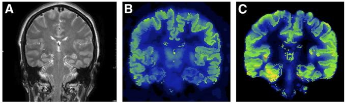
Methods: We estimated 5-HT(1A) receptor binding preoperatively with (18)F-trans-4-fluoro-N-2-[4-(2-methoxyphenyl) piperazin-1-yl]ethyl-N-(2-pyridyl) cyclohexane carboxamide ((18)F-FCWAY) PET and CMRglc measurement with (18)F-FDG in regions drawn on coregistered MRI after partial-volume correction in 41 patients who had anterior temporal lobectomy with at least a 1-y follow-up. Surgery was tailored to individual preresection evaluations and intraoperative electrocorticography. Mean regional asymmetry values and the number of regions with asymmetry exceeding 2 SDs in 16 healthy volunteers were compared between seizure-free and non-seizure-free patients. (18)F-FCWAY but not (18)F-FDG and MRI data were masked for surgical decisions and outcome assessment.
Results: Twenty-six of 41 (63%) patients seizure-free since surgery had significantly different mesial temporal asymmetries, compared with 15 non-seizure-free patients for both (18)F-FCWAY (F(1,39) = 5.87; P = 0.02) and (18)F-FDG PET (F(1,38) = 5.79; P = 0.021). The probability of being seizure-free was explained by both (18)F-FDG and (18)F-FCWAY PET, but not MRI, with a significant additional (18)F-FCWAY effect (chi(2)(2) = 9.8796; P = 0.0072) after the probability of being seizure-free was explained by (18)F-FDG. Although MRI alone was not predictive, any combination of 2 lateralizing imaging studies was highly predictive of seizure freedom.
Conclusion: Our study provides class III evidence that both 5-HT(1A) receptor PET and CMRglc PET can contribute to temporal lobectomy planning. Additional studies should explore the potential for temporal lobectomy based on interictal electroencephalography and minimally invasive imaging studies.
Figures
References
-
- Wiebe S, Blume WT, Girvin JP, Eliasziw M. Effectiveness and Efficiency of Surgery for Temporal Lobe Epilepsy Study Group: a randomized, controlled trial of surgery for temporal-lobe epilepsy. N Engl J Med. 2001;345:311–318. - PubMed
-
- Uijl SG, Leijten FS, Arends JB, Parra J, van Huffelen AC, Moons KG. Prognosis after temporal lobe epilepsy surgery: the value of combining predictors. Epilepsia. 2008;49:1317–1323. - PubMed
-
- Duncan J. The current status of neuroimaging for epilepsy. Curr Opin Neurol. 2009;22:179–184. - PubMed
-
- Richardson M. Update on neuroimaging in epilepsy. Expert Rev Neurother. 2010;10:961–973. - PubMed
-
- Toczek MT, Carson RE, Lang L, et al. PET imaging of 5-HT1A receptor binding in patients with temporal lobe epilepsy. Neurology. 2003;60:749–756. - PubMed
Publication types
MeSH terms
Substances
Grants and funding
LinkOut - more resources
Full Text Sources