. 2012 Aug;5(2):95-112.
doi: 10.1007/s12307-012-0099-6.

Clinical and biological implications of the tumor microenvironment

Affiliations

Clinical and biological implications of the tumor microenvironment

David Tarin. Cancer Microenviron. 2012 Aug.

Erratum in

  • Cancer Microenviron. 2012 Aug;5(2):113

Abstract

In normal tissues and organs, the activities of the constituent cells are strictly restricted to the tasks assigned to them during development. In addition they (with the exception of leukocytes) remain inflexibly confined to their territorial domains by regulatory interactions with their neighbors.This creates specialized local micro-environments in which structure and function are orderly, stable and tightly controlled by feed-back loops, within interacting regulatory networks.This system has considerable ability to adapt to changing conditions. In contrast, the microenvironment in regions where tumors are forming and expanding is characterized by progressive loss of specialized or differentiated cellular functions,disorderly molecular signals, and degeneration of microscopical organ structure. This, coupled with the traffic of cells into and out of the tumor, often culminates in local invasion and metastasis to other organs. The nature of these disturbed molecular and cellular interactions is, by definition,highly unstable and increasingly unpredictable as time passes.It also varies between different tumors, sometimes even leading to regression. However, systematic analysis of this dysfunction in the tumor microcosm, using multiple modern research techniques, has revealed that all actively growing primary and secondary neoplasms share an absolute dependency upon support from adjacent non-neoplastic cells of the host. This support, in turn, continuously depends upon dynamic interplay between tumor and host cell populations, via signaling molecules and surface receptors in the tumor microenvironment.Such interplay determines the fate of the growing neoplasm. Such information, described and evaluated in this article, provides important new insights into the etiology of carcinogenesis and how tumor growth, invasion and metastasis might be therapeutically arrested. The facts and concepts assembled below, regarding the cancer microenvironment, demonstrate how modern molecular findings reveal the impact of the wide range of cancer diseases upon the internal cellular, tissue and organ environments of the whole individual and how this applies to designing new work to improve human cancer diagnosis and treatment. The article discusses several specific types of experimentally-induced and clinically common cancers to derive principles useful for interpreting events in the tumor microenvironment,which apply to cancers in general and especially to human malignant disease.


Figures

Fig. 1
Fig. 1
Normal (a–d, above red line) and cancer (e–h) micro-environment. a Normal human mammary ducts (solid arrow) and glands composed of secretory acini (open arrow). Whole-mount of human mammary tissue stained with hematoxylin before fixation and viewed through a dissecting microscope.(x20). b Histological section through terminal breast ductule and secretory acini to show vimentin staining of myoepithelial layer of the epithelium (brown stain) and to show other resident cell populations composing the normal organ. The circle outlines the acini and the diverse cell types (fibroblasts, macrophages, endothelial cells, lymphocytes etc.) in the cellular community living in the collagenous stroma around the glandular acini (x 100). c View of normal human mammary tissue stained with labeled antibody to CD8+ lymphocytes (brown) to show the organ also contains cells which are transiting through its tissues (x250). d Electron micrograph of the boundary between epithelium (E) of a normal breast duct and the surrounding stroma(S). The arrow marks the basement membrane (B) which provides support and attachment for the epithelium. Note the orderly arrangement of the epithelial-stromal junction compared to Fig. 1h below. (x14,000). e Infiltrating lobular carcinoma of the breast showing cancer cells invading as strings and columns (arrow) of adherent epithelial cells through dense fibrous connective tissue containing few non-malignant stromal cells. A residual duct containing apoptotic cells is present centrally (x100). f Magnified view of invading columns of malignant cuboidal cells of the carcinoma in Fig. 2f, still displaying pathognomonic epithelial characteristics, including production of mucin droplets (white arrows), which is an exclusively epithelial property. The intervening stroma is collagenous and contains occasional non epithelial, non-malignant host mesenchymal cells (black arrowheads). No evidence of EMT (x250). g Infiltrating ductal carcinoma of the breast. Note that the tumor is invading as adherent malignant epithelial cells, still in distorted glandular formation. These are passing through sparse cellular connective tissue stroma containing non-malignant cells, lying between the malignant glands. There is no significant evidence of any inflammatory cell infiltrate, nor of EMT. (x100). h Electron micrograph of an area at the invading front of a malignant gland comparable to that within the rectangle in Fig. 1g. Invading epithelium of the gland is labeled (E). The basement membrane normally present along the whole of the epithelial stromal boundary (between the broad arrows) is completely dissolved. Also, the fibres and background matrix of the stroma (S), ahead of the invading glands, is disorganized and disintegrating, showing the disorder in the tumor microenvironment (x 2,000)
Fig. 2
Fig. 2
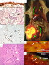
Carcinoma in situ, absence of EMT and tumor cell dissemination. a An area of severe dysplasia bordering upon carcinoma in situ in a urinary bladder biopsy. The epithelium is grossly thickened and irregularly arranged. Marked overexpression of CD44 (brown stain) is seen in its deeper layers. The stroma (S) shows increased cellularity and loosening and disorganization of collagen fibres. Stained with Hermes 3 antibody, which reacts with all isoforms of CD44 (x250). b Areas of intra-glandular carcinoma in situ (encircled) of the breast within a larger field of infiltrating lobular carcinoma. In carcinoma in situ, the neoplastic cells proliferate within the boundaries of the epithelial domain and have not yet acquired invasive properties. A gland which is partially converted to carcinoma in situ, is also present (asterisk) in this field of view and the arrow marks invading carcinoma cells in the adjacent stroma (x100). c Prostate cancer: Within a sea of unstained invading malignant glands in the stroma of this tumor there are occasional residual normal glands in which the peripheral ring of myoepithelial cells is stained with an antibody to vimentin. Some of the glands have ragged partial investment with myoepithelial cells (broad arrow) and focal invasion of epithelial cells, bursting into the stroma through the gaps, can be seen (thin arrow). No evidence of EMT. (x100). d Disseminating adherent clumps of cancer cells within veins in the liver. These show clear epithelial differentiation and some are still forming glands with central lumina (white arrows). No evidence of EMT. Occasional dissociated carcinoma cells (black arrows) are also disseminating in this patient (x250). e Survey view of an autopsy of a mouse with a red fluorescent protein-labeled human mammary carcinoma (P) xenograft in the mammary gland, generated by orthotopic inoculation of the human mammary carcinoma cell line MDA MB 435 LM3. This cancer metastasizes copiously to lymph nodes (thin green arrows) and lungs (open white arrow) but not to any other organs. The liver (L,) spleen(S) and kidneys (K) are not colonized, but do contain disseminated single living cells (Figs e and f below). A lymphatic enormously distended with tumor cells, draining the tumor is seen at bottom left (solid green arrow). f Isolated single tumor cells labeled with RFP in the liver of the mouse shown above, visible by inspection of the liver surface with a dissecting microscope under illumination with light of wavelength which excites RFP fluorescence (x 250). g Scattered labeled single tumor cells in the spleen (x250).
Fig. 3
Fig. 3
Necessity for recruitment of stromal cells for tumor formation and growth: Life history of metastatic deposits. a The arrival of the tumor cells in the lung via the blood stream. Loose epithelial tumor cells (arrow) are seen in the lumen of a pulmonary arteriole lying in thin septum between alveolar air sacs (asterisks). x250. b In a different patient, cancer cells had been disseminating for years from a carcinoma in the ovary but there were no metastases in any organ at autopsy. A small clump of malignant cells forming a rudimentary glandular structure with a lumen can be seen in a lung capillary (open arrow). Other carcinoma cells are extravasating singly from the small vessels and lying free in the extravascular tissues (solid arrows), but there is evidence of recruitment of adjacent stromal cells into an organized structure. There is also no sign of any inflammatory cell response near these tumor cells.(x250). c This picture shows the first stage in recruitment of non-malignant stromal cells into the structure of an early deposit in the lungs of the same patient as in the lesions in Fig. 3a above and 3 d below. Host mesenchymal cells and a small capillary blood vessel (open arrow) have passed through the narrow base of a papillary frond covered with malignant epithelium into its core, to form its supporting framework. Two parallel endothelial cell nuclei resembling a diagonally aligned equals (=) sign lie to the left of the open arrow. These line a vascular space which extends upwards and to the right within this space there are two nucleated blood cells. Outside the white space, stromal cell nuclei (blue/black) are visible amongst pink stromal collagen which they have recently produced and assembled. The frond projects into the lumen of a large glandular space lined by malignant epithelial cells. Asterisks mark alveolar air spaces (x250). d A later stage in the formation of a pulmonary metastasis in the same patient as the lesions in Fig. 3a and c. The tumor deposit is much larger and recruitment of host stromal cells is more advanced. This metastasis has formed many more papillary structures, containing mesenchymal cores (arrow), projecting into the central lumen of the malignant gland(x100).

References

    1. Orr J. Changes antecedent to tumour formation during the treatment of mouse skin with carcinogenic hydrocarbons. J Pathol Bacteriol. 1938;46:495–515. doi: 10.1002/path.1700460310. - DOI
    1. Tarin D. Morphological studies on the mechanism of carcinogenesis. In: Tarin D, editor. Tissue interactions in carcinogenesis. London: Academic; 1972. pp. 227–289.
    1. Tarin D. Tissue interactions in morphogenesis, morphostasis and carcinogenesis. J Theor Biol. 1972;34(1):61–72. doi: 10.1016/0022-5193(72)90054-9. - DOI - PubMed
    1. Kratochwil K. Tissue interaction during embryonic development. In: Tarin D, editor. Tissue interactions in carcinogenesis. London: Academic; 1972. pp. 1–47.
    1. Tarin D. Sequential electron microscopical study of experimental mouse skin carcinogenesis. Int J Cancer. 1967;2(3):195–211. doi: 10.1002/ijc.2910020302. - DOI - PubMed

LinkOut - more resources