. 2012;98(3):195-211.
doi: 10.1002/bip.22030. Epub 2012 Feb 3.

N-glycosylation of enhanced aromatic sequons to increase glycoprotein stability

Affiliations

N-glycosylation of enhanced aromatic sequons to increase glycoprotein stability

Joshua L Price et al. Biopolymers. 2012.

Abstract

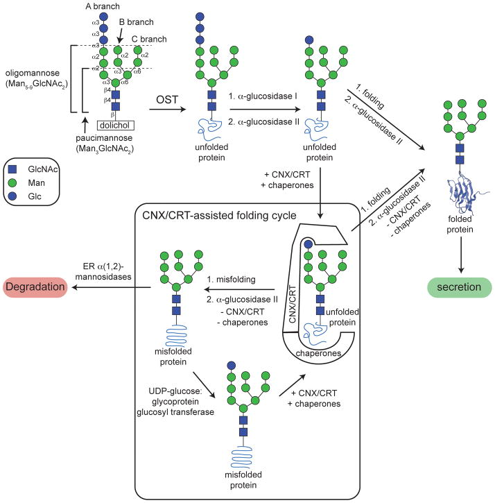
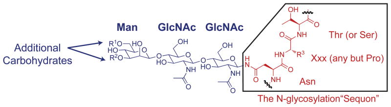
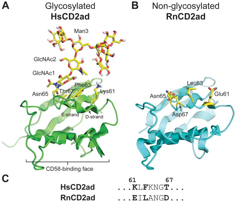
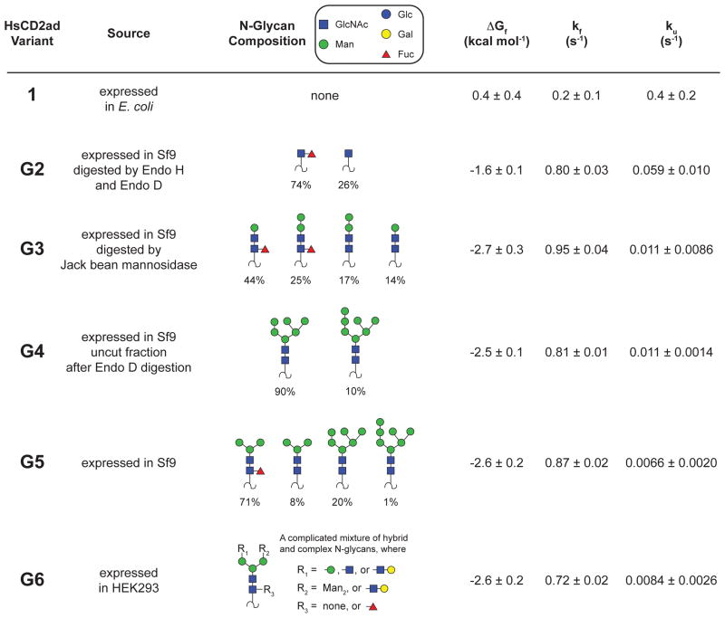
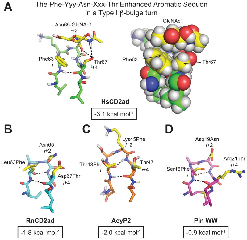
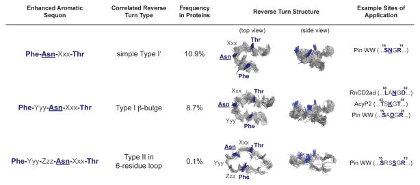
N-glycosylation can increase the rate of protein folding, enhance thermodynamic stability, and slow protein unfolding; however, the molecular basis for these effects is incompletely understood. Without clear engineering guidelines, attempts to use N-glycosylation as an approach for stabilizing proteins have resulted in unpredictable energetic consequences. Here, we review the recent development of three "enhanced aromatic sequons," which appear to facilitate stabilizing native-state interactions between Phe, Asn-GlcNAc and Thr when placed in an appropriate reverse turn context. It has proven to be straightforward to engineer a stabilizing enhanced aromatic sequon into glycosylation-naïve proteins that have not evolved to optimize specific protein-carbohydrate interactions. Incorporating these enhanced aromatic sequons into appropriate reverse turn types within proteins should enhance the well-known pharmacokinetic benefits of N-glycosylation-based stabilization by lowering the population of protease-susceptible unfolded and aggregation-prone misfolded states, thereby making such proteins more useful in research and pharmaceutical applications.


Figures

Figure 1
Figure 1
Cellular N-glycosylation allows a glycoprotein to enter the calnexin/calreticulin-assisted folding vs. degradation cycle in the ER, allowing the N-glycoprotein to fold and be secreted or to be targeted for degradation if attempts to fold the N-glycoprotein are unsuccessful.
Figure 2
Figure 2
The oligosaccharyl transferase enzyme transfers the Glc3Man9GlcNAc2 glycan en bloc to the side-chain amide nitrogen of an Asn residue within the consensus sequence Asn-Xxx-Thr/Ser, called the N-glycosylation “sequon”; Xxx is any amino acid but proline.
Figure 3
Figure 3
(A) Ribbon diagram of the adhesion domain of the human protein CD2, HsCD2ad (PDB code: 1GYA), which requires glycosylation at Asn65 to fold properly. The N-glycan and HsCD2ad side chains at positions 61, 63, and 67 are highlighted in yellow. Wyss et al. (ref 59) observed NOEs between the N-glycan and these side chains. (B) Ribbon diagram of the rat ortholog of HsCD2ad, RnCD2ad (PDB code: 1HNG), which is not glycosylated at Asn65, but is still able to fold properly. RnCD2ad side chains at positions 61, 63, and 67 are highlighted in yellow. (C) Comparison of the HsCD2ad amino acid sequence near the glycosylation site with the homologous sequence from RnCD2ad. All structures rendered in Pymol.
Figure 4
Figure 4
Folding free energies and folding and unfolding rates for HsCD2ad glycoforms generated via enzymatic remodeling (ref 65). Variants G2G6 are predominantly mixtures of the indicated glycoforms as determined by mass spectrometry. Only species with abundance >5% are shown, except for variant G2, where only species with relative abundance > 0.5 are shown (owing to the complexity of the heterogeneous mixture of glycans obtained upon expression of HsCD2ad in HEK293 cells).
Figure 5
Figure 5
The energetic contributions of N-glycan substructures to the change in HsCD2ad folding free energy (ΔΔGf), folding activation energy (ΔΔGf), and unfolding activation energy (ΔΔGu) upon N-glycosylation. Structure of glycosylated HsCD2ad (PDB code: 1GYA) rendered in Pymol.
Figure 6
Figure 6
Energy diagram view of the hypothesis that introduction of an N-glycosylation site into a glycosylation-naïve protein would restrict the conformational freedom of the denatured ensemble, thereby destabilizing it relative to the native state, resulting in a more negative free energy of folding (i.e., overall stabilization).
Figure 7
Figure 7
(A) Predicted and (B) observed energetic consequences of glycosylation (i.e., Asn to Asn-GlcNAc mutation) at each of the indicated positions within the WW domain of the human protein Pin 1 (WW). Positions where glycosylation is predicted or observed to stabilize WW are highlighted in green; positions where glycosylation is predicted or observed to destabilize WW are highlighted in red (ref. 52). WW domain structure rendered in Pymol (PDB code: 1PIN).
Figure 8
Figure 8
(A) Stick and space-filling representations of the Phe-Yyy-Asn-Xxx-Thr enhanced aromatic sequon in the type I β-turn of HsCD2ad (PDB code: 1GYA). We also installed the Phe-Yyy-Asn-Xxx-Thr enhanced aromatic sequon in similar reverse turns in (B) RnCD2ad (PDB code: 1HNG), (C) AcyP2 (PDB code: 1APS), and (D) WW (PDB code: 2F21) via the indicated mutations at the i, i+2, and i+4 positions (side chains at these positions in each structure are highlighted in yellow). The change in folding free energy (ΔΔGf) upon glycosylation of the Phe-Yyy-Asn-Xxx-Thr enhanced aromatic sequon in each structural context is shown below each structure in an outlined box (refs. 90, 93). All structures rendered in Pymol.
Figure 9
Figure 9
Folding free energies of WW-derived glycoproteins and their non-glycosylated counterparts along with the amino acid sequence of the five-residue reverse turn for each variant (ref. 93). The i, i+2, and i+4 positions where we installed the enhanced aromatic sequon are highlighted in bold font. Tabulated data are given as mean ± standard error at 65 °C for WW variants at 10 μM in 20 mM aqueous sodium phosphate, pH 7. N = Asn-GlcNAc.
Figure 10
Figure 10
(A) Triple mutant cycle formed by 5g-F,T and its derivatives (ref. 93). (B) Triple mutant cycle analysis of folding free energy data at 65 °C for 5g-F,T and its derivatives. Parameters are given as mean ± standard error. P values in parentheses indicate the probability that random sampling error accounts for the difference between zero and the observed value of the parameter.
Figure 11
Figure 11
We installed enhanced aromatic sequons (A) in a type I β-bulge turn (PDB code: 2F21) in WW variant 5 using the sequence Phe-Yyy-Asn-Xxx-Thr; (B) in a six-residue turn-within-a-turn (PDB code: 1PIN) in WW variant 6 using the sequence Phe-Zzz-Yyy-Asn-Xxx-Thr; and (C) in a type I′ β-turn (PDB code: 1ZCN) in WW variant 4 using the sequence Phe-Asn-Xxx-Thr, using the mutations indicated at each position. The stick representation of each reverse turn was rendered in Pymol, with hydrogen bonds shown as black dashed lines. Positions where we introduced mutations to install the enhanced aromatic sequons are highlighted in yellow. The change in folding free energy (ΔΔGf) upon glycosylation of each enhanced aromatic sequon is shown below each structure in a black outlined box.
Figure 12
Figure 12
Triple mutant cycle analysis of data from 4g-F,T, 5g-F,T, 6g-F,T, and their derivatives (ref. 93). The change in folding free energy (ΔΔGf,total) upon glycosylation of each enhanced aromatic sequon in its correlated reverse turn type can be attributed to energetic contributions from the Asn19 to Asn-GlcNAc mutation (ΔΔGN19N), the two-way interaction between Phe and Asn-GlcNAc, the two-way interaction between Asn-GlcNAc and Thr, and the three way interaction between Phe, Asn-GlcNAc, and Thr.
Figure 13
Figure 13
The enhanced aromatic sequons in their correlated reverse turn types. The first three columns list the sequence of each enhanced aromatic sequon, its correlated reverse turn type, and the frequency of that reverse turn type in the non-redundant protein structural database (<40% sequence homology, see ref. 95). In the fourth and fifth columns, the sequences are mapped onto an overlay of the main chains of members of each reverse turn family. The side chains where Phe, Asn(glycan), and Thr should be incorporated are highlighted in blue. The last column lists examples of proteins that we have studied that contain the reverse turn types that are compatible with the indicated enhanced aromatic sequons.

Similar articles

Cited by

References

    1. Kornfeld R, Kornfeld S. Annu Rev Biochem. 1985;54:631–664. - PubMed
    1. Yan A, Lennarz WJ. J Biol Chem. 2005;280:3121–3124. - PubMed
    1. Kelleher DJ, Gilmore R. Glycobiology. 2006;16:47R–62R. - PubMed
    1. Helenius A, Aebi M. Science. 2001;291:2364–2369. - PubMed
    1. Molinari M. Nat Chem Biol. 2007;3:313–320. - PubMed

Publication types

LinkOut - more resources