. 2012 Sep;30(9):2010-9.
doi: 10.1002/stem.1171.

Lysophosphatidic acid induces migration of human lung-resident mesenchymal stem cells through the β-catenin pathway

Affiliations

Lysophosphatidic acid induces migration of human lung-resident mesenchymal stem cells through the β-catenin pathway

Linda Badri et al. Stem Cells. 2012 Sep.

Abstract

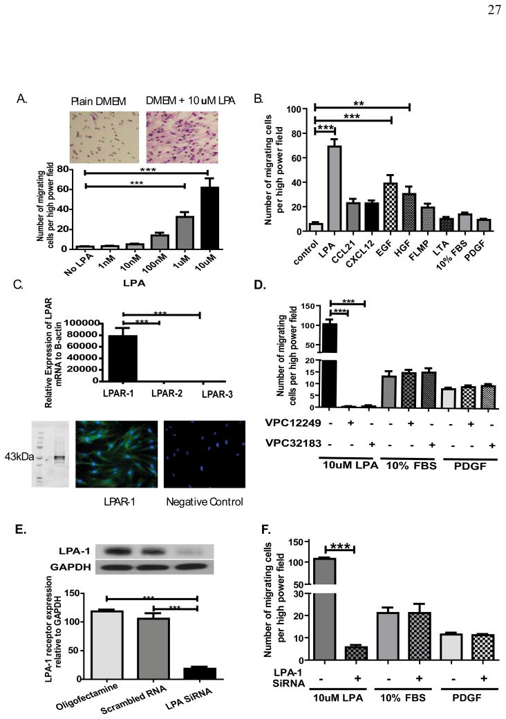
Mesenchymal stem cells (MSCs) have been demonstrated to reside in human adult organs. However, mechanisms of migration of these endogenous MSCs within their tissue of origin are not well understood. Here, we investigate migration of human adult lung-resident (LR) mesenchymal progenitor cells. We demonstrate that bioactive lipid lysophosphatidic acid (LPA) plays a principal role in the migration of human LR-MSCs through a signaling pathway involving LPA1-induced β-catenin activation. LR-MSCs isolated from human lung allografts and lungs of patients with scleroderma demonstrated a robust migratory response to LPA in vitro. Furthermore, LPA levels correlated with LR-MSC numbers in bronchoalveolar lavage (BAL), providing demonstration of the in vivo activity of LPA in human adult lungs. Migration of LR-MSCs was mediated via LPA1 receptor ligation and LPA1 silencing significantly abrogated the migratory response of LR-MSCs to LPA as well as human BAL. LPA treatment of LR-MSCs induced protein kinase C-mediated glycogen synthase kinase-3β phosphorylation, with resulting cytoplasmic accumulation and nuclear translocation of β-catenin. TCF/LEF dual luciferase gene reporter assay demonstrated a significant increase in transcriptional activity after LPA treatment. LR-MSC migration and increase in reporter gene activity in the presence of LPA were abolished by transfection with β-catenin small interfering RNA demonstrating that β-catenin is critical in mediating LPA-induced LR-MSC migration. These data delineate a novel signaling pathway through which ligation of a G protein-coupled receptor by a biologically relevant lipid mediator induces migration of human tissue-resident mesenchymal progenitors.


Figures

Figure 1
Figure 1. LPA induces migration of human lung-resident mesenchymal stem cells via LPA1 receptor
(A & B) LPA induces LR-MSC migration in a dose dependent manner. Human LR-MSCs were added to the upper chamber of the Transwell and allowed to migrate through the porous membrane into the lower chamber containing medium alone or medium with LPA. Migrated cells on the lower side of the membrane were stained with Hema3 stain and counted with Olympus BX41 light microscope using a ×20 objective. n=3 separate cell lines with 3 replicates of each condition and 5 counted high power fields for each replicate. Representative image from transwell demonstrating robust migration of LR-MSCs in response to LPA are shown above. LPA induced migration was compared to that induced by 10ng/ml CCL21, 30ng/ml CXCL12, 10ng/ml EGF, 20ng/ml HGF, 0.1uM FMLP, 1ug/ml LTA, 10% FBS, and 10nM PDGF, as shown in B. n=4 individual cell lines with 3 replicates of each condition and 5 counted high power fields for each replicate. (C) LPA receptors profile of human LR-MSCs. LPA1, LPA2 and LPA3 mRNA expression by real-time quantitative PCR in human LR-MSCs. n=4 LR-MSC lines. A representative image from western blot analysis and immunofluorescent staining demonstrating LPA1 protein expression in LR-MSCs is shown below. (D) Effect of LPA receptor antagonists on LPA-induced LR-MSC migration. LR-MSCs were pre-treated with competitive antagonists of the LPA1 and LPA3 receptors (1μM VPC12249 or 1μM VPC32183) for 30 min before migration assay. Data represent mean ± SEM from 3 independent experiments. (E & F) Effect of LPA1 siRNA on LPA-induced migration of human LR-MSCs. LR-MSCs were transfected with (20nM) validated LPA1 or scrambled siRNA for 24 h and serum starved for another 24 h. Protein was collected for western blot analysis to test transfection efficiency as shown in E (n=3). Effect of LPA1 siRNA on LR-MSC migration was studied using the migration assay. Data shown in F represent mean ± SEM from 3 independent experiments. *** P < 0.0001.
Figure 2
Figure 2. Demonstration of the in vivo role of LPA in human lung allografts and the role of β-catenin activation in mediating LPA induced lung-resident mesenchymal stem cell (LR-MSC) migration
(A) Correlation of LPA concentration with number of lung-resident mesenchymal stem cell in human bronchoalveolar lavage fluid. LR-MSCs were quantitated in BAL fluid from human lung transplant recipients by measuring number of mesenchymal CFUs per 2 ×10⁶ million cells. LPA concentration in supernatant of these BAL samples with high CFUs (CFU ≥ 10, n=10) and low CFUs (CFU< 10, n=10) was determined. **P = 0.004 (B) LPA accounts for chemotactic activity of BAL derived from human lung allografts. Ability of supernatant from samples with high and low CFUs to induce LR-MSC and its modulation by LPA receptor antagonist (VPC12249) was assessed by Transwell assay. Two LR-MSC lines (± VPC12449) were exposed to supernatant from 4 BAL samples with high CFUs (CFUs of 40, 13, 13 and 50) and 4 BAL samples with low CFUs (CFUs of 2, 0, 0, 0). Data is shown as mean ± SEM of migration induced by these BAL samples. Control signifies plain DMEM. *** p < 0.0001. (C) Effect of LPA treatment on β-catenin protein expression in human LR-MSCs. Serum starved LR-MSCs were treated with 10uM LPA for various time points. β-catenin protein expression in the cytoplasmic fraction was quantitated by western blot analysis. Data represent mean ± SEM from 4 individual LR- MSC lines. (D & E) Effect of β-catenin siRNA on LPA-induced LR-MSC migration. LR-MSCs were transfected with β-catenin siRNA (20nM) or scrambled siRNA for 24 hours. β-catenin protein was measured by western blot analysis in aliquots of transfected cells as shown in B. Effect of β-catenin silencing on LR-MSC migration in response to LPA and other control chemoattractants (10% FBS and PDGF) was studied using the Transwell migration assay. Data represent mean ± SEM from 4 individual experiments. ** p< 0.001, *** p < 0.0001.
Figure 3
Figure 3. LPA induces GSK3β phosphorylation and activates β-catenin pathway
(A) Effect of LPA treatment on GSK3β phosphorylation. Serum starved LR-MSCs were treated with 10μM LPA for 30 or 60 minutes. GSK3β phosphorylation was studied by western blot analysis using phospho-specific antibody (phospho-Ser-9) against GSK3β. n=4. (B) LPA induces nuclear translocation of β-catenin. Nuclear protein fractions of LR-MSCs treated with LPA were used to detect nuclear β-catenin accumulation by western blot analysis. Results were normalized to nuclear SAM 68 as a nuclear loading control. n = 4. (C) TCF/LEF/B-catenin transcription activation by LPA. LR-MSCs were transfected with TCF/LEF luciferase reporter construct (an inducible luciferase reporter), negative control (a non-inducible luciferase reporter used for background elimination) or positive control vector (a GFP expressing luciferase construct used to test transfection efficiency, data not shown) using Oligofectamine. In some conditions, LR-MSCs were also co-transfected with β-catenin siRNA (20nM). Transfected cells were then treated with either control vehicle or 10uM LPA for 2 hours. Reporter activity was assayed using dual luciferase assay system and data are shown as fold induction in relative light units (RLU). Experiments were done in triplicates. n=5 LR-MSC lines. (D& E) LPA1 receptor is the key receptor in LPA activation of β-catenin pathway. LR-MSCs were transiently transfected with (20nM) LPA1 siRNA for 24 hours before treatment with 10μM LPA. Total protein was collected at various time points after LPA treatment and analyzed for GSK3β phosphorylation by western blot analysis. β-catenin protein expression in nuclear fractions 1hr after LPA is shown. n=3.
Figure 4
Figure 4. LPA induced cPKC activation is essential for LPA-induced β-catenin pathway activation and migration
Serum starved LR-MSCs were pretreated with specific cPKC inhibitor (GÖ6976) for 30 min before treatment with 10μM LPA. The effect of cPKC inhibitor on GSK3β phosphorylation (A) and nuclear β-catenin expression (B) was studied by western blot analysis. Cell migration (C) was assayed in the presence of cPKC inhibitor. N=4. ** p< 0.001, *** p < 0.0001.
Figure 5
Figure 5. LPA causes migration of scleroderma LR-MSCs via an LPA1 mediated phosphorylation and inhibition of GSK-3β and subsequent β-catenin translocation
(A & B) LPS induces robust migration of scleroderma lung derived MSCs. Migration of LR-MSCs derived from patients with scleroderma in response to varying doses of LPA was assessed in vitro using transwell system. LPA induced migration was compared to that induced by 10% FBS, and 10nM PDGF as shown in B. (C) LPA1 is the predominant receptor on of scleroderma lung derived MSCs. mRNA expression of LPA receptors by real-time quantitative PCR in scleroderma LR-MSCs is shown (D to F) LPA1 receptor mediates LPA induced migration of scleroderma lung-derived MSCs. Effect of LPA receptor antagonists on LPA induced LR-MSC migration was studied by pre-treating cells with competitive antagonists of the LPA1 and LPA3 receptors (1μM VPC12249 or 1μM VPC32183) for 30 min before migration assay. Furthermore, effect of LPA1 siRNA on LPA-induced migration of human LR-MSCs was studied. (G & H) LPA induces GSK3β phosphorylation and nuclear translocation of β-catenin in scleroderma lung derived MSCs. Serum starved LR-MSCs were treated with 10μM LPA for 30 or 60 minutes. GSK3β phosphorylation was studied by western blot analysis using phospho-specific antibody (phospho-Ser-9) against GSK3β. Nuclear protein fractions of LR-MSCs treated with LPA were used to detect nuclear β-catenin accumulation by western blot analysis. Results were normalized to nuclear SAM 68 as a nuclear loading control. (I) LPA induced migration of scleroderma lung-derived MSCs is dependent on the β-catenin pathway. LR-MSCs were transfected with β-catenin siRNA (20nM) or scrambled siRNA for 24 hours. Effect of β-catenin silencing on LR-MSC migration in response to LPA was studied using the Transwell migration assay. N= 3 scleroderma cell lines. ** p < 0.001, *** p < 0.0001.

References

    1. Belperio JA, Weigt SS, Fishbein MC, et al. Chronic lung allograft rejection: mechanisms and therapy. Proc Am Thorac Soc. 2009;6:108–121. - PubMed
    1. Hoyles RK, Derrett-Smith EC, Khan K, et al. An essential role for resident fibroblasts in experimental lung fibrosis is defined by lineage-specific deletion of high-affinity type II transforming growth factor beta receptor. Am J Respir Crit Care Med. 2011;183:249–261. - PubMed
    1. Humphreys BD, Lin SL, Kobayashi A, et al. Fate tracing reveals the pericyte and not epithelial origin of myofibroblasts in kidney fibrosis. Am J Pathol. 2010;176:85–97. - PMC - PubMed
    1. Walker N, Badri L, Wettlaufer S, et al. Resident tissue-specific mesenchymal progenitor cells contribute to fibrogenesis in human lung allografts. Am J Pathol. 2011;178:2461–2469. - PMC - PubMed
    1. Bruno S, Bussolati B, Grange C, et al. Isolation and Characterization of Resident Mesenchymal Stem Cells in Human Glomeruli. Stem cells and development. 2009 - PubMed

Publication types

MeSH terms