. 2012 Aug 24;287(35):29873-86.
doi: 10.1074/jbc.M112.360768. Epub 2012 Jul 10.

Targeting serous epithelial ovarian cancer with designer zinc finger transcription factors

Affiliations

Targeting serous epithelial ovarian cancer with designer zinc finger transcription factors

Haydee Lara et al. J Biol Chem. .

Abstract

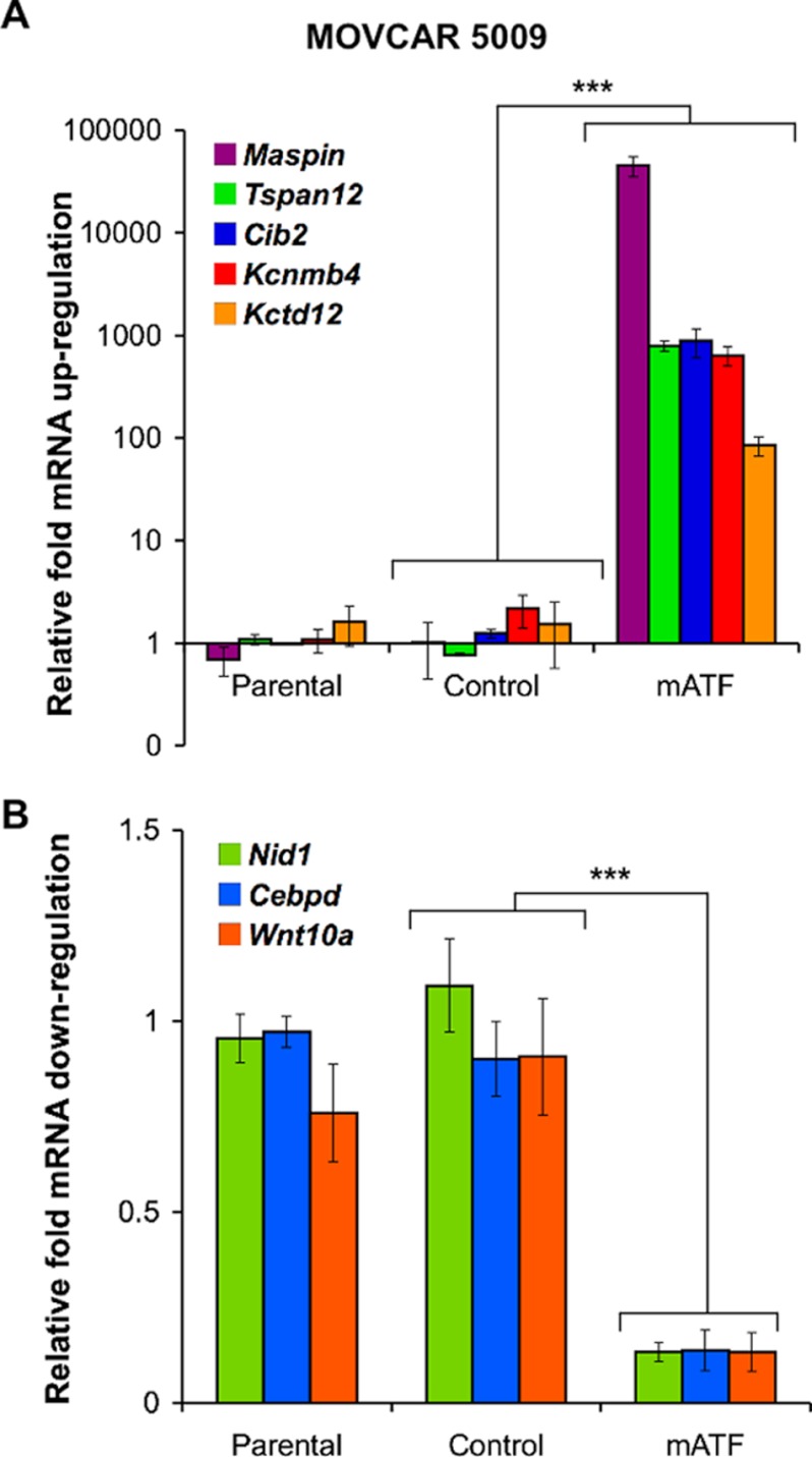
Ovarian cancer is the leading cause of death among gynecological malignancies. It is detected at late stages when the disease is spread through the abdominal cavity in a condition known as peritoneal carcinomatosis. Thus, there is an urgent need to develop novel therapeutic interventions to target advanced stages of ovarian cancer. Mammary serine protease inhibitor (Maspin) represents an important metastasis suppressor initially identified in breast cancer. Herein we have generated a sequence-specific zinc finger artificial transcription factor (ATF) to up-regulate the Maspin promoter in aggressive ovarian cancer cell lines and to interrogate the therapeutic potential of Maspin in ovarian cancer. We found that although Maspin was expressed in some primary ovarian tumors, the promoter was epigenetically silenced in cell lines derived from ascites. Transduction of the ATF in MOVCAR 5009 cells derived from ascitic cultures of a TgMISIIR-TAg mouse model of ovarian cancer resulted in tumor cell growth inhibition, impaired cell invasion, and severe disruption of actin cytoskeleton. Systemic delivery of lipid-protamine-RNA nanoparticles encapsulating a chemically modified ATF mRNA resulted in inhibition of ovarian cancer cell growth in nude mice accompanied with Maspin re-expression in the treated tumors. Gene expression microarrays of ATF-transduced cells revealed an exceptional specificity for the Maspin promoter. These analyses identified novel targets co-regulated with Maspin in human short-term cultures derived from ascites, such as TSPAN12, that could mediate the anti-metastatic phenotype of the ATF. Our work outlined the first targeted, non-viral delivery of ATFs into tumors with potential clinical applications for metastatic ovarian cancers.


Figures

FIGURE 1.
FIGURE 1.
The artificial transcription factor mATF reactivates Maspin expression in breast and ovarian cell lines derived from GEMMs. A, shown is a schematic representation of the multidomain structure of the mATF binding the Maspin promoter. The DNA binding domain, composed of six ZF motifs, was designed to bind an 18-bp sequence in the murine Maspin proximal promoter at −127 bp upstream the translation start site. The ZF proteins were linked to the VP64 transactivator domain (1). The hemagglutinin (HA) tag was engineered at the C terminus of the ZF construct (68). B, the mATF up-regulates Maspin mRNA expression in breast and ovarian cancer cell lines derived from GEMMs as assessed by qRT-PCR. The mATF and empty vector (control) were retrovirally transduced in the corresponding cell lines. Relative Maspin up-regulation in both transduced and parental cell lines are shown in the plot. Data were normalized to the parental MMTV-PYMT cell line. Student's t test was used to analyze the differences in gene expression between the parental and the transfected cells (*, p ≤ 0.05; **, p ≤ 0.01; ***, p ≤ 0.001). C, shown is an immunoblot analysis of Maspin up-regulation in cell lines derived from GEMMs transduced either with control or mATF. A densitometric analysis of the Western blot is shown. Data were normalized to the MMTV-PyMT cell line. Transgenic mouse strains and cell lines are indicated, and sources are described under “Experimental Procedures.”
FIGURE 2.
FIGURE 2.
The artificial transcription factor mATF binds the Maspin promoter in the MOVCAR 5009 cell line and reactivates silenced Maspin. A, retroviral delivery of mATF specifically reactivates Maspin, as assessed by qRT-PCR. VP64-SS refers to cells transduced with a vector carrying the transactivator domain but lacking the 6ZFs; ZF-VP64 indicates a scramble library of 3ZF domains linked to VP64 (69); Control refers to empty vector (pMX-IRES-GFP)-transduced cells. The parental cell line was treated with the epigenetic inhibitors: 5-aza-2′dC (5 μm), suberoylanilide hydroxamic acid (10 μm), and trichostatin A (100 nm) at saturating maximum doses. Data were normalized to the parental cell line. B, mATF physically binds its targeted site in the Maspin promoter. A ChIP assay was performed on the MOVCAR 5009 cell line retrovirally transfected with either control (empty vector) or mATF. α-RNA-polymerase II (RNAPII), IgG Control, and α-HA antibodies were used to immunoprecipitate the DNA-protein complexes. α-HA facilitated the immunoprecipitation of mATF. Densitometric analysis of the ChIP products loaded on agarose gels after PCR was carried out using the ImageJ software (NCBI, Bethesda, MD). Data were normalized to the signal of the input sample. Ab, antibody. C, retroviral transduction of mATF in MOVCAR 5009 cells results in reactivation of both cytoplasmic and nuclear Maspin. Immunofluorescence detected the nucleus (Hoechst 2258), the mATF expression (α-HA, red), and Maspin (green) in MOVCAR 5009 cells transduced with mATF, a Maspin cDNA-expressing retrovirus (18), and an empty retroviral vector or control.
FIGURE 3.
FIGURE 3.
mATF expression induces a decrease in tumor cell viability and invasion and promotes cytoskeleton disruption in the MOVCAR 5009 cell line. A, retroviral delivery of mATF decreases cell viability over control-transduced cells as determined by Cell Titer Glo assay. Student's t test was applied to analyze the difference in tumor cell survival between the control and mATF cells, with p < 0.001. Three different transductions were performed, each one monitored in triplicate wells. B, mATF decreases anchorage-independent growth of MOVCAR 5009 cells. Soft agar colony formation assays were performed on control and mATF-transduced cells. The arrow in the picture (left) points to a colony amplified on the left corner. Colony quantification was performed 20 days after the seeding. Three independent experiments were carried out, and Student's t test was applied to analyze the data; p = 0.0054. C, mATF-transduced cells abolished tumor formation potential, as evaluated by tumorsphere assay formation. Cells were retrovirally transduced with either mATF or control and seeded in low-attachment culture plates with spheroid media (43). After 10 days, representative pictures of the experiment are shown outlining the abolishment of spheroid formation in mATF transduced cells. (p = 0.0002). D, mATF transduction in MOVCAR 5009 cells inhibits the invasion potential of the cells relative to control, as assessed by Matrigel-invasion assays (10). A representative picture of the fixed cells after 24 h of invasion is indicated. Three independent experiments were performed, and Student's t test was applied to analyze the data; p = 0.0001. E, immunofluorescence staining of actin (green) and Maspin (red) on mATF and control-transduced cells is shown. To detect actin, Alexa 488-phalloidin was applied, and anti-Maspin-Alexa 594 was used as the secondary antibody for Maspin detection. Hoechst 33258 was used to stain the nuclei (blue).
FIGURE 4.
FIGURE 4.
Targeted LPR-AA nanoparticles encapsulating modified mATF mRNA results in functional production of the mATF and regulation of Maspin in the MOVCAR 5009 cell line. A, shown is a schematic illustration of the generation of LPR-AA nanoparticles. The LPR nanoparticle core was prepared mixing modified mRNA (e.g. EGFP, mATF, or control, which is the mATF lacking the 5′ cap and is unable to translate) plus protamine and liposome. The LPR core was pegylated by adding either DSPE-PEG2000-AA (targeted nanoparticle) or DSPE-PEG2000 (non-targeted). B, MOVCAR 5009 cells were transfected with either LPR-AA-EGFP (targeted) or LPR-EGFP (non-targeted) nanoparticles, and the fluorescence intensity (quantification of the total fluorescence intensity of EGFP+ events) was measured by flow cytometry 24 h post-transfection by gating the percentage % of EGFP-positive cells. Cells treated with nanoparticle controls not carrying EGFP and untreated cells were used to generate the negative gates. A representative 10× picture of the transfected cells with LPR-AA-EGFP (targeted) or LPR-EGFP (non-targeted) nanoparticles is shown. C, shown is immunofluorescence detection of the mATF (α-HA, green) in MOVCAR 5009 cells. Cells were transfected with either LPR-AA-mATF or control (uncapped mATF mRNA). The cells were stained 24 h after transfection. D, real-time detection of Maspin in MOVCAR 5009 cells using an anti-Maspin molecular beacon (MB) is shown. Targeted nanoparticles containing either a functional mATF or an inactive control were also loaded with an anti-Maspin MB. Red fluorescence emission due to the hybridization of the Maspin transcript with the MB was detected 24 h post-transfection.
FIGURE 5.
FIGURE 5.
Transfection of LPR-AA-mATF nanoparticles decreases the proliferation of MOVCAR 5009 cells in vitro and inhibits tumor growth in vivo. A, delivery of LPR-AA-mATF nanoparticles decreases survival of MOVCAR 5009 cells relative to control-treated targeted nanoparticles. Cell survival was determined by MTT cell proliferation assay on cells transfected with either LPR-AA-mATF or control. Student's t test was applied to analyze the difference on survival between mATF and control-transfected cells; p = 0.0002. Data represent the averages and S.E. of three independent experiments, each one done in triplicate wells. B, LPR-AA-mATF nanoparticles inhibit the growth of MOVCAR 5009 cells in subcutaneous allografts in nude mice. MOVCAR 5009 cells were inoculated in nude mice to induce subcutaneous tumors, and nanoparticles were injected intravenously at the time points indicated in the graph (arrows). Student's t test was applied to analyze the difference between control (n = 9) and mATF treatment (n = 9; p = 0.0220) and between vehicle and mATF (p = 0.0253). C, mATF and Maspin expression were detected on the treated tumors by Western blot. An anti-HA antibody was used to detect the ATF by Western blot.
FIGURE 6.
FIGURE 6.
mRNA quantification of targets up- and down-regulated by mATF in MOVCAR 5009 cells. Changes in mRNA expression of genes previously identified by gene expression microarrays (supplemental Table S1 and Table S2) were validated by qRT-PCR. A, genes differentially up-regulated by transduction of mATF in MOVCAR 5009 cells are shown. B, genes down-regulated by mATF are shown. Data were normalized to the parental MOVCAR 5009 cell line and represent the mean and S.D. of at least three independent experiments. Differences in gene expression were calculated with a student t test; ***, p ≤ 0.0001.
FIGURE 7.
FIGURE 7.
Down-regulation of Maspin and TSPAN12 in short term human ascites preparations from ovarian carcinoma patients. A, expression of Maspin and TSPAN12 in a panel of human ovarian primary tumors, ascites, and established cancer cell lines, as assessed by qRT-PCR is shown. Data were normalized to SR06, a preparation of normal ovary. UC064 and ASCUC064 represent tumor sample and ascites preparation, respectively, from the same patient. Tumor specimens, ascites preparations, and ovarian cell lines were homogenized and resuspended in Trizol for mRNA extraction and analyzed by quantitative qRT-PCR. B, shown is an immunoblot of Maspin and TSPAN12 on protein extracts from human ascites, the human cell line OVCAR-3, and parental, control, and mATF-transduced MOVCAR 5009 cells. C, down-regulation of Maspin and Tspan12 after Maspin siRNA knockdown in the mATF-transduced MOVCAR 5009 cell line as assessed by qRT-PCR is shown. Data were normalized to the cell line transfected with a control (anti-luciferase) siRNA (*, p ≤ 0.05; **, p ≤ 0.01; ***, p ≤ 0.001). D, shown is expression of Maspin and TSPAN12 by immunofluorescence in representative sections of epithelial ovarian cancer specimens (see also supplemental Fig. S5).

Similar articles

Cited by

References

    1. Jemal A., Bray F., Center M. M., Ferlay J., Ward E., Forman D. (2011) Global cancer statistics. CA Cancer J. Clin. 61, 69–90 - PubMed
    1. Rosen D. G., Yang G., Liu G., Mercado-Uribe I., Chang B., Xiao X. S., Zheng J., Xue F. X., Liu J. (2009) Ovarian cancer. Pathology, Biology, and Disease models. Front Biosci. 14, 2089–2102 - PMC - PubMed
    1. Havrilesky L., Darcy M., Hamdan H., Priore R. L., Leon J., Bell J., Berchuck A. (2003) Prognostic significance of p53 mutation and p53 overexpression in advanced epithelial ovarian cancer. A gynecologic oncology group study. J. Clin. Oncol. 21, 3814–3825 - PubMed
    1. Risch H. A., McLaughlin J. R., Cole D. E., Rosen B., Bradley L., Kwan E., Jack E., Vesprini D. J., Kuperstein G., Abrahamson J. L., Fan I., Wong B., Narod S. A. (2001) Prevalence and penetrance of germline BRCA1 and BRCA2 mutations in a population series of 649 women with ovarian cancer. Am. J. Hum. Genet. 68, 700–710 - PMC - PubMed
    1. Liu J., Matulonis U. A. (2010) New advances in ovarian cancer. Oncology 24, 721–728 - PubMed

Publication types

MeSH terms

Associated data