. 2012 Aug 31;287(36):30073-83.
doi: 10.1074/jbc.M111.338574. Epub 2012 Jul 10.

Cathepsin B is secreted apically from Xenopus 2F3 cells and cleaves the epithelial sodium channel (ENaC) to increase its activity

Affiliations

Cathepsin B is secreted apically from Xenopus 2F3 cells and cleaves the epithelial sodium channel (ENaC) to increase its activity

Abdel A Alli et al. J Biol Chem. .

Abstract

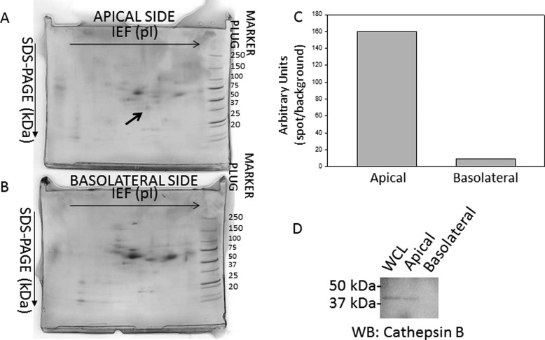
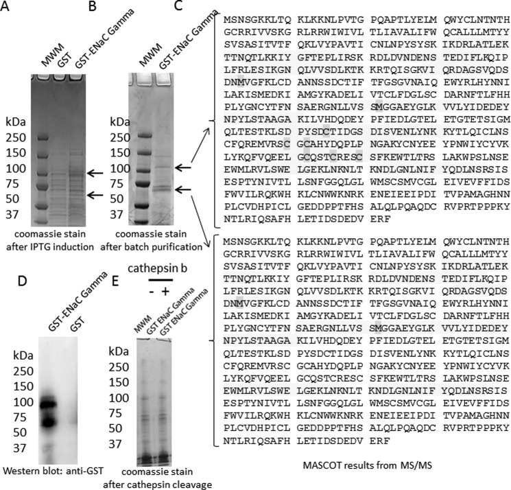
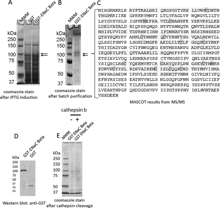
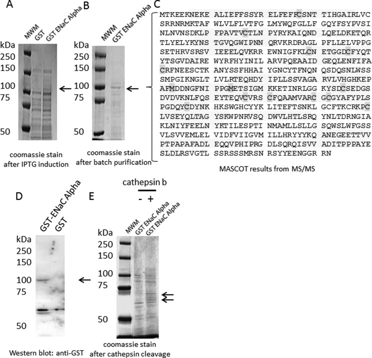
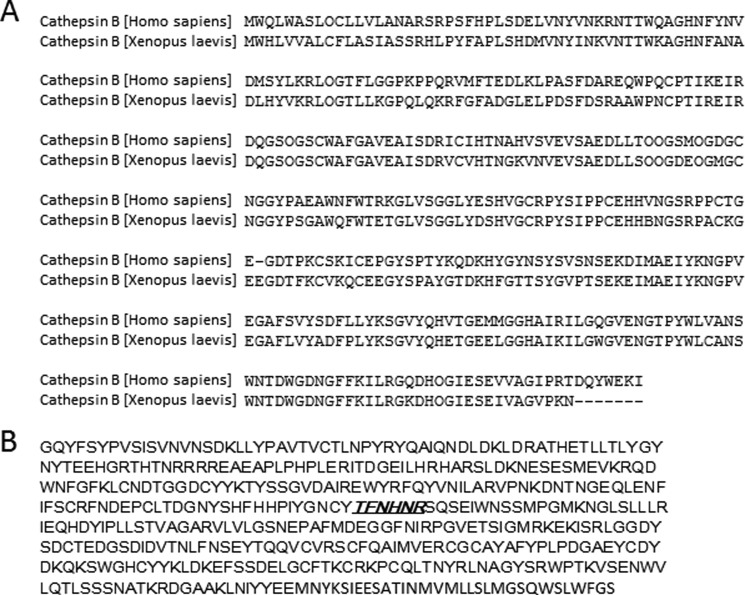
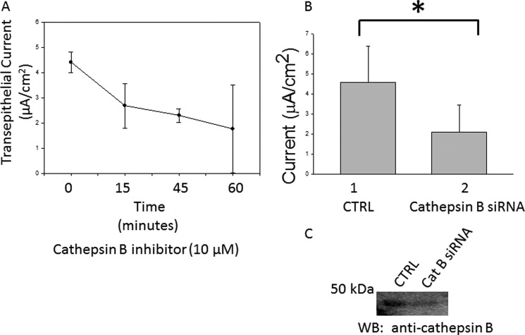
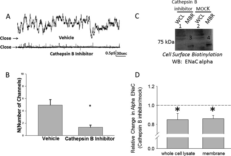
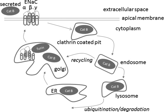
The epithelial sodium channel (ENaC) plays an important role in regulating sodium balance, extracellular volume, and blood pressure. Evidence suggests the α and γ subunits of ENaC are cleaved during assembly before they are inserted into the apical membranes of epithelial cells, and maximal activity of ENaC depends on cleavage of the extracellular loops of α and γ subunits. Here, we report that Xenopus 2F3 cells apically express the cysteine protease cathepsin B, as indicated by two-dimensional gel electrophoresis and mass spectrometry analysis. Recombinant GST ENaC α, β, and γ subunit fusion proteins were expressed in Escherichia coli and then purified and recovered from bacterial inclusion bodies. In vitro cleavage studies revealed the full-length ENaC α subunit fusion protein was cleaved by active cathepsin B but not the full-length β or γ subunit fusion proteins. Both single channel patch clamp studies and short circuit current experiments show ENaC activity decreases with the application of a cathepsin B inhibitor directly onto the apical side of 2F3 cells. We suggest a role for the proteolytic cleavage of ENaC by cathepsin B, and we suggest two possible mechanisms by which cathepsin B could regulate ENaC. Cathepsin B may cleave ENaC extracellularly after being secreted or intracellularly, while ENaC is present in the Golgi or in recycling endosomes.


Figures

FIGURE 1.
FIGURE 1.
Two-dimensional gel electrophoresis analysis of proteins located on the apical and basolateral side of 2F3 cells. Coomassie-stained gel of proteins from media collected and concentrated from the apical side (A) or basolateral side (B) of Xenopus 2F3 cells that were subcultured on permeable supports and allowed to form tight junctions and generate a measurable transepithelial resistance and voltage. Some proteins were found to be exclusively on the apical or basolateral side. The protein spot in A, labeled with an arrow, was excised and identified as cathepsin B after mass spectrometry protein sequencing. C, densitometric analysis of the protein spot indicated by an arrow in A. D, shown is a representative Western blot of cathepsin B expression in the cell lysate (whole cell lysate (WCL)) and in the media collected and concentrated from the apical and basolateral side of Xenopus 2F3 cells. IEF, isoelectric focusing.
FIGURE 2.
FIGURE 2.
Characterization and proteolytic analysis of full-length ENaC γ subunit. A, a Coomassie-stained gel shows the expression of the entire γ subunit of ENaC as a GST fusion protein (GST-ENaC γ) after isopropyl 1-thio-β-d-galactopyranoside (IPTG) induction. B, a Coomassie-stained gel shows the double band corresponding to GST-ENaC γ or ENaC γ after batch purification. C, MASCOT mass spectrometry peptide sequence coverage of several signature peptides corresponding to Xenopus ENaC γ subunit is shown. The shaded residues represent oxidation of methionine residues or alkylation of cysteine residues. D, shown is Western blot analysis of GST-ENaC using a peroxidase-conjugated antibody against GST to detect a 100-kDa immunoreactive band corresponding to GST-ENaC γ. E, a Coomassie-stained gel indicates GST-ENaC γ is not cleaved by cathepsin B in vitro. MWM, molecular weight markers.
FIGURE 3.
FIGURE 3.
Characterization and proteolytic analysis of full-length ENaC β subunit. A, a Coomassie-stained gel shows the expression of the entire β subunit of ENaC as a GST fusion protein (GST-ENaC β) after isopropyl 1-thio-β-d-galactopyranoside (IPTG) induction. B, a Coomassie-stained gel shows the double band corresponding to GST-ENaC β after batch purification. C, MASCOT mass spectrometry peptide sequence coverage of several signature peptides corresponding to Xenopus ENaC β subunit is shown. The shaded residues represent oxidation of methionine residues or alkylation of cysteine residues. D, shown is Western blot analysis of GST-ENaC using a peroxidase-conjugated antibody against GST to detect a 100-kDa immunoreactive band corresponding to GST-ENaC β. E, a Coomassie-stained gel indicates GST-ENaC β is not cleaved by cathepsin B in vitro. MWM, molecular weight markers.
FIGURE 4.
FIGURE 4.
Characterization and proteolytic analysis of full-length ENaC α subunit. A, a Coomassie-stained gel shows the expression of the entire α subunit of ENaC as a GST fusion protein (GST-ENaC α) after isopropyl 1-thio-β-d-galactopyranoside (IPTG) induction. B, a Coomassie-stained gel shows a band corresponding to GST-ENaC α after batch purification. C, shown is MASCOT mass spectrometry peptide sequence coverage of several signature peptides corresponding to the Xenopus ENaC α subunit. The shaded residues represent oxidation of methionine residues or alkylation of cysteine residues. D, shown is a Western blot analysis of GST-ENaC using a peroxidase-conjugated antibody against GST to detect a 100-kDa immunoreactive band corresponding to GST-ENaC α. E, shown is a Coomassie-stained gel indicating GST-ENaC α is cleaved by cathepsin B in vitro. MWM, molecular weight markers.
FIGURE 5.
FIGURE 5.
Sequence analysis of cathepsin B and ENaC. A, shown is multiple sequence alignment of H. sapiens and X. laevis cathepsin B. The amino acid sequence of cathepsin B is highly conserved between H. sapiens and X. laevis, as it is for several other species. B, shown is the predicted cleavage site of cathepsin B in the extracellular loop of Xenopus ENaC α subunit. Based on known cleavage sites of other substrates of cathepsin B, the amino acid region underlined and in bold italics is the predicted cathepsin B cleavage site within the extracellular loop of the α subunit of ENaC.
FIGURE 6.
FIGURE 6.
Measurements of amiloride-sensitive transepithelial current in 2F3 cells. A, transepithelial sodium current decreased with time after treating Xenopus 2F3 cells with 10 μm cathepsin B inhibitor. B, transepithelial sodium current decreased after siRNA-mediated knockdown of cathepsin B in Xenopus 2F3 cells. Values are presented as the means ± S.E., n = 3 for each data point. *, p < 0.05. CTRL, control. C, a representative Western blot (WB) shows a decrease in cathepsin B at the protein level after siRNA-mediated knockdown as presented in B.
FIGURE 7.
FIGURE 7.
Single channel recording from Xenopus 2F3 cells showing an effect on ENaC activity after application of a cathepsin B inhibitor. A, shown are representative traces of Xenopus 2F3 cells pretreated with vehicle (DMSO) or cathepsin B inhibitor (CA-074) for 1 h before acquiring a patch. B, results of eight independent patch clamp studies show the cathepsin B inhibitor significantly decreased the number of active channels in a patch of Xenopus 2F3 cells. *, p < 0.001. Empty patches were included in the calculation of N, but the inhibitor did not affect Po. C, a representative Western blot (WB) of three independent experiments shows a decrease in ENaC α protein in the cell lysate and at the membrane after treating Xenopus 2F3 cells with the cathepsin B inhibitor. WCL represents whole cell lysate, and MBR represents membrane fractions. D, a densitometric analysis of immunoreactive bands in C shows a statistically significant decrease in ENaC α protein in either the whole cell lysate or membrane fractions after treatment with the cathepsin B inhibitor relative to treatment with the vehicle (MOCK). n = 3; *, p < 0.05.
FIGURE 8.
FIGURE 8.
Proposed model depicting the cathepsin B cleavage-dependent activation of ENaC. The cysteine protease cathepsin B (Cat B) is synthesized as a latent preproenzyme and post-translationally processed in the rough endoplasmic reticulum, Golgi, endosomal, and lysosomal compartments. Cathepsin B is secreted or found in the cytoplasm of certain epithelial cells. Unlike other cysteine proteases, cathepsin B exhibits both endopeptidase and exopeptidase activities. Cathepsin B is shown to potentially cleave the extracellular loop of the α subunit of ENaC either within the Golgi, extracellularly, or within endosomes.

References

    1. Bertog M., Cuffe J. E., Pradervand S., Hummler E., Hartner A., Porst M., Hilgers K. F., Rossier B. C., Korbmacher C. (2008) Aldosterone responsiveness of the epithelial sodium channel (ENaC) in colon is increased in a mouse model for Liddle's syndrome. J. Physiol. 586, 459–475 - PMC - PubMed
    1. Lu C., Pribanic S., Debonneville A., Jiang C., Rotin D. (2007) The PY motif of ENaC, mutated in Liddle syndrome, regulates channel internalization, sorting, and mobilization from subapical pool. Traffic 8, 1246–1264 - PubMed
    1. Konstas A. A., Mavrelos D., Korbmacher C. (2000) Conservation of pH sensitivity in the epithelial sodium channel (ENaC) with Liddle's syndrome mutation. Pflugers Arch. 441, 341–350 - PubMed
    1. Prince L. S., Welsh M. J. (1999) Effect of subunit composition and Liddle's syndrome mutations on biosynthesis of ENaC. Am. J. Physiol. 276, C1346–C1351 - PubMed
    1. Schild L. (1996) The ENaC channel as the primary determinant of two human diseases. Liddle syndrome and pseudohypoaldosteronism. Nephrologie 17, 395–400 - PubMed

Publication types

MeSH terms