. 2012 Jul 24;109(30):12064-9.
doi: 10.1073/pnas.1207210109. Epub 2012 Jul 10.

Tmem100, an ALK1 receptor signaling-dependent gene essential for arterial endothelium differentiation and vascular morphogenesis

Affiliations

Tmem100, an ALK1 receptor signaling-dependent gene essential for arterial endothelium differentiation and vascular morphogenesis

Satoshi Somekawa et al. Proc Natl Acad Sci U S A. .

Abstract

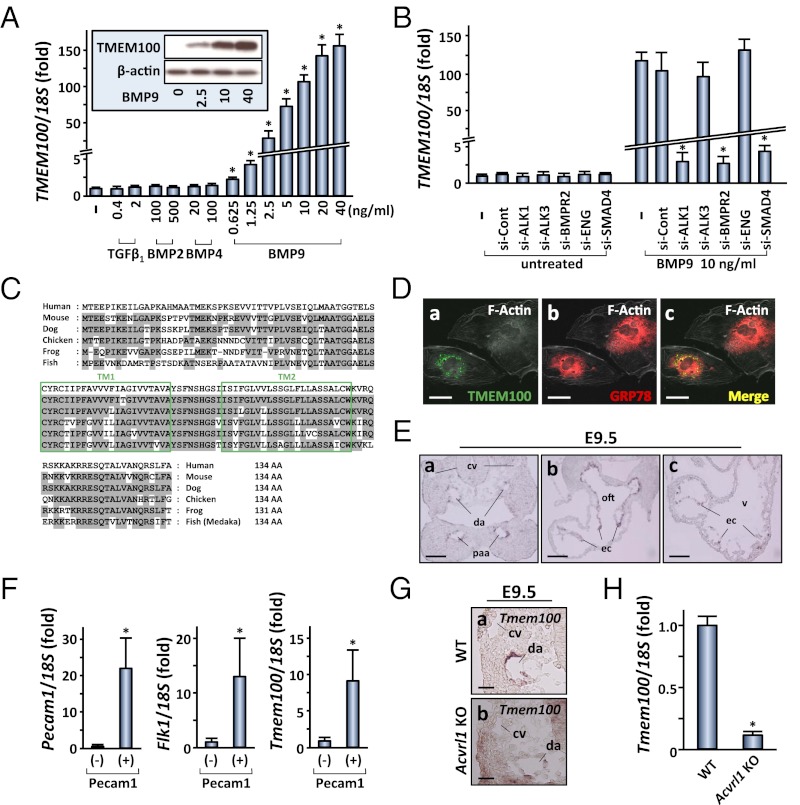
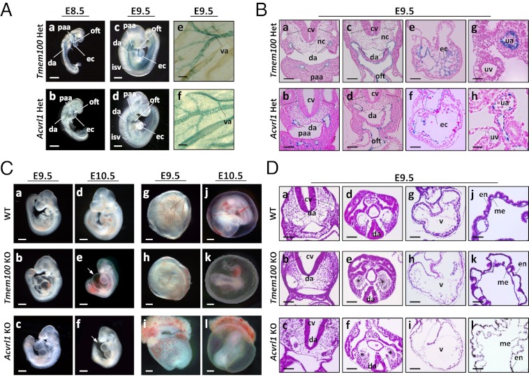
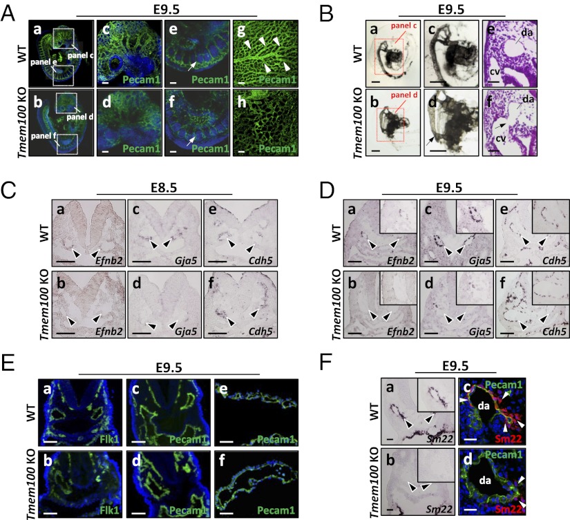
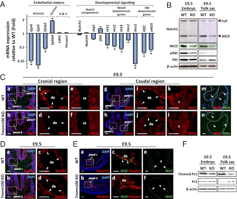
Members of the transforming growth factor-β superfamily play essential roles in various aspects of embryonic development and physiological organ function. Among them, bone morphogenetic protein (BMP) 9 and BMP10 regulate embryonic vascular development by activating their endothelial receptor ALK1 (activin receptor-like kinase 1, also called Acvrl1). ALK1-mediated intracellular signaling is implicated in the etiologies of human diseases, but their downstream functional proteins are largely unknown. In this study, we identified Tmem100, a gene encoding a previously uncharacterized intracellular transmembrane protein, to be an embryonic endothelium-enriched gene activated by BMP9 and BMP10 through the ALK1 receptor. Tmem100 null mice showed embryonic lethality due to impaired differentiation of arterial endothelium and defects of vascular morphogenesis, which phenocopied most of the vascular abnormalities observed with the Acvrl1/Alk1 deficiency. The activity of Notch- and Akt-mediated signaling, which is essential for vascular development, was down-regulated in Tmem100 null mice. Cre-mediated deletion of Tmem100 in endothelial cells was sufficient to recapitulate the null phenotypes. These data indicated that TMEM100 may play indispensable roles downstream of BMP9/BMP10-ALK1 signaling during endothelial differentiation and vascular morphogenesis.


Conflict of interest statement

The authors declare no conflict of interest.

Figures

Fig. 1.
Fig. 1.
Tmem100: an arterial endothelium-enriched gene downstream of BMP9/BMP10-ALK1 signaling. (A) BMP9 markedly increases TMEM100 mRNA expression at 24 h in a dose-dependent manner. Quantitative RT-PCR analysis is shown. Fold increase relative to the untreated cells is shown with SD. (Inset) Western blot result shows a dose-dependent increase in TMEM100 protein expression. −, untreated. (B) BMP9-induced TMEM100 mRNA expression is inhibited by pretreatment with ACVRL1/ALK1 siRNA (si-ALK1), BMPR2 siRNA (si-BMPR2), or SMAD4 siRNA (si-SMAD4), but not with BMPR1A/ALK3 siRNA (si-ALK3), Endoglin siRNA (si-ENG), or control siRNA (si-Cont). Quantitative RT-PCR is shown. −, untreated. (C) Amino acid sequences of TMEM100 from various species. Boxes represent two putative transmembrane (TM) domains. Residues conserved with the human sequence are shaded in gray. (D) TMEM100 protein predominantly resides in the perinuclear region overlapping endoplasmic reticulum marked by anti-GRP78/HSPA5 antibody. Immunocytochemistry of HUAEC transfected with a TMEM100-FLAG expression plasmid is shown. (E) Tmem100 mRNA is expressed specifically in arterial endothelial cells of pharyngeal arch artery (paa) and dorsal aorta (da) as well as in endocardium (ec), but not in cardinal vein (cv), at E9.5. In situ hybridization is shown. oft, outflow tract; v, ventricle. (F) Tmem100 expression is enriched in endothelial cells also at earlier developmental stages. Shown is FACS sorting of Pecam1-positive cells from E8.5 embryos followed by quantitative RT-PCR. Pecam1 and Kdr/Flk1 expression was examined to confirm proper selection of the endothelial population. (G) Tmem100 expression in dorsal aorta is reduced in Acvrl1/Alk1 null embryos at E9.5. In situ hybridization is shown. (H) Tmem100 expression significantly decreases in the yolk sac of Acvrl1/Alk1 null mice at E9.5. Quantitative RT-PCR is shown. In A, B, F, and H, asterisks indicate data with statistical significance (*P < 0.05). (Scale bars: D, 10 μm; E, 50 μm; and G, 20 μm.)
Fig. 2.
Fig. 2.
Tmem100 and Acvrl1/Alk1: arterial expression of knock-in lacZ reporter and angiogenesis defects in null embryos. (A and B) Knock-in lacZ reporter activity is detected in major arteries and endocardium in both Tmem100 and Acvrl1/Alk1 heterozygous mice. Sections counterstained with nuclear fast red are shown in B. (C) Tmem100 null embryos die in utero, showing cardiac dysmorphogenesis and enlargement at E9.5, massive pericardial effusion (arrow) and severe growth retardation at E10.5, and absence of large vitelline vessels in the yolk sac at E10.5. These phenotypes are also observed in Acvrl1/Alk1 null embryos. (D) Tmem100 null and Acvrl1/Alk1 null embryos have remarkably similar defects of cardiovascular morphogenesis. Paired dorsal aortas show marked dilatation and narrowing, and clumps of blood cells are frequently seen in the caudal region (asterisk). Mural layers surrounding the aorta and myocardial wall are thinner and ventricular trabeculation is not formed well. The yolk sac shows detachment of endodermal (en) and mesodermal (me) layers and abnormal vessel dilatation. H&E staining at E9.5 is shown. cv, cardinal vein; da, dorsal aorta; isv, intersomitic vessel; nc, notochord; oft, outflow tract; paa, pharyngeal arch artery; ua, umbilical artery; uv, umbilical vein; v, ventricle; va, vitelline artery. (Scale bars: A, a–d, and C, 200 μm; A, e and f, B, a–f, and D, a–i, 50 μm; and B, g and h, and D, j–l, 20 μm.)
Fig. 3.
Fig. 3.
Impairment of vascular remodeling and arterial endothelium differentiation in Tmem100 null embryos. (A) Impaired vascular remodeling is visualized by whole-mount Pecam1 immunostaining in the Tmem100 null embryos (b, d, and f) and yolk sac (h). Arrows indicate intersomitic vessels. Arrowheads indicate highly organized vascular branches in the wild-type yolk sac. a–f are shown with nuclear DAPI stain. (B) Abnormal vascular connections (arrow) between dorsal aorta and cardinal veins are observed in null embryos. Shown is microangiography by India ink injection and H&E staining. (C and D) Expression of arterial endothelium marker genes Efnb2 and Gja5/Cx40 diminishes in dorsal aorta (arrowheads) of Tmem100 null embryos at E8.5 (C) and E9.5 (D), whereas maintenance of Cdh5 expression indicates existence of endothelial layers. In situ hybridization is shown. (E) Flk1 and Pecam1 expression is preserved in dorsal aorta of Tmem100 null embryo (b and d) and yolk sac vasculature (f). Immunohistochemistry with nuclear DAPI stain is shown. (F) Expression of Sm22/Tagln mRNA and SM22 protein in the mural layers of dorsal aorta is significantly reduced in Tmem100 null embryos (arrowheads). In situ hybridization (a and b) and immunohistochemistry with nuclear DAPI stain (c and d) are shown. Insets in D and F show magnified views of dorsal aorta. (Scale bars: A, C, D, and E, a–d, 50 μm; B, a–d, 200 μm; and B, e and f, E, e and f, and F, 20 μm.)
Fig. 4.
Fig. 4.
Notch- and Akt-mediated signaling pathways are down-regulated in Tmem100 null embryos and yolk sac. (A) Expression of arterial endothelium markers, Notch downstream genes, and Akt downstream genes markedly decreases in Tmem100 null yolk sac at E9.5. Expression of a venous endothelium marker, Ephb4, is significantly up-regulated. Quantitative RT-PCR is shown. (B) The amount of the intracellular domain of Notch receptor (NICD) and phosphorylated Akt significantly decreases in Tmem100 null embryos and yolk sac at E9.5, whereas that of full-length Notch1 receptor and total Akt remains unchanged. Western blot analysis is shown. (C) NICD expression markedly decreases in dorsal aorta (da) and endocardium (arrowheads in d, j, and n), but not in the neural tube (nt) and somites (s) (f and l), of E9.5 null embryos. Immunohistochemistry using a NICD-specific antibody is shown. a, b, g, h, m, and n are shown with nuclear DAPI stain. (D) Total amount of Notch receptors does not decrease in vascular endothelium of null embryos. Immunohistochemistry using an antibody recognizing full-length Notch receptors and NICD is shown. a and b are shown with nuclear DAPI stain. (E) Expression of Hrt2, a Notch downstream transcription factor, significantly decreases in the vasculature of Tmem100 null embryos (d), whereas its expression in cardiac muscle was unchanged (f). Immunohistochemistry is shown. a and b are shown with nuclear DAPI stain. (F) Cleaved Presenilin-1 (Ps1) decreases in null embryos and yolk sac at E9.5. Western blot analysis is shown. (Scale bars in C–E, 20 μm.)

References

    1. Potente M, Gerhardt H, Carmeliet P. Basic and therapeutic aspects of angiogenesis. Cell. 2011;146:873–887. - PubMed
    1. Eilken HM, Adams RH. Dynamics of endothelial cell behavior in sprouting angiogenesis. Curr Opin Cell Biol. 2010;22:617–625. - PubMed
    1. David L, Feige JJ, Bailly S. Emerging role of bone morphogenetic proteins in angiogenesis. Cytokine Growth Factor Rev. 2009;20:203–212. - PubMed
    1. Pardali E, Goumans MJ, ten Dijke P. Signaling by members of the TGF-beta family in vascular morphogenesis and disease. Trends Cell Biol. 2010;20:556–567. - PubMed
    1. David L, Mallet C, Mazerbourg S, Feige JJ, Bailly S. Identification of BMP9 and BMP10 as functional activators of the orphan activin receptor-like kinase 1 (ALK1) in endothelial cells. Blood. 2007;109:1953–1961. - PubMed

Publication types

MeSH terms