. 2012 Jul 6:6:101.
doi: 10.3389/fnins.2012.00101. eCollection 2012.

Decision points: the factors influencing the decision to feed in the medicinal leech

Affiliations

Decision points: the factors influencing the decision to feed in the medicinal leech

Quentin Gaudry et al. Front Neurosci. .

Abstract

The decision to feed is a complex task that requires making several small independent choices. Am I hungry? Where do I look for food? Is there something better I'd rather be doing? When should I stop? With all of these questions, it is no wonder that decisions about feeding depend on several sensory modalities and that the influences of these sensory systems would be evident throughout the nervous system. The leech is uniquely well suited for studying these complicated questions due to its relatively simple nervous system, its exceptionally well-characterized behaviors and neural circuits, and the ease with which one can employ semi-intact preparations to study the link between physiology and decision-making. We will begin this review by discussing the cellular substrates that govern the decision to initiate and to terminate a bout of feeding. We will then discuss how feeding temporarily blocks competing behaviors from being expressed while the animal continues to feed. Then we will review what is currently known about how feeding affects long-term behavioral choices of the leech. Finally, we conclude with a short discussion of the advantages of the leech's decision-making circuit's design and how this design might be applicable to all decision circuits.

Keywords: behavioral choice; decision-making; distributed; feeding; leech; modular; sensory gating; serotonin.


Figures

Figure 1
Figure 1
Diet and not phylogeny determine leech behavioral choice. (A) The phylogenic relationship of leeches used for this study (Gaudry et al., 2010) is based on a comparison of morphological and molecular features (Borda and Siddall, 2004). An asterisk implies an ancestral state of unknown feeding preference. The most parsimonious explanation of these relationships is that the sanguivorous feeding strategy evolved three different times among these species from a carnivorous ancestor. The numbers following each species is used to reference that species in (B,C). (B) Canonical Correspondence Analysis (CCA) biplot showing the relationship between species, stimulus location, and behavioral output in the non-feeding state. Species and stimulus location serve as predictors and the magnitude of their vectors denotes the influence they have on the raw variables (behavioral outputs). The predictor vectors point toward the behaviors they are most strongly correlated with. The clustering of all species at the middle means that all species responded to all stimuli in similar ways. Thus species has little predictive power over the resulting behavior while stimulus location is a good indicator of which behavior will be elicited in response to stimulation in the non-feeding state. Coloring and numberings as in (A) where brown refers to carnivorous species and red refers sanguivorous species. (C) CCA results for the same group of leeches as in (B) but during the feeding state. The carnivorous leech vectors shown in brown point toward active behaviors [shortening, swimming, crawling, and back sucker release (BSR)] whereas the sanguivorous leech vectors shown in red point in the direction of local responses [Bend, Tense, local bend (LB)] or no response (NR). The results indicate that the diet of the leeches (regardless of their phylogenetic relationship) is the best predictor of stimulus response during feeding. (More details about CCA are found in Gaudry et al., 2010).
Figure 2
Figure 2
Evidence for presynaptic inhibition of pressure mechanosensory P cells. (A) Effects of feeding on EPSP amplitudes and PPR at the P cell-to-AP neuron synapse. Inserts show overlapping pairs of traces from one such experiment, sampled from pre-feeding, feeding, and post-feeding times. In each pair, the black trace is the first EPSP and the gray trace is the second EPSP in response to a P cell spike triggered 500 ms after the first one. Scale bars represent 2 mV and 50 ms (from Gaudry and Kristan, 2009). (B) Recording from a P cell in the leech head brain while blood serum was applied to the isolated lip of a semi-intact preparation similar to Groome et al. (1995). Diagonal dashes denote a break in the sample trace corresponding to ∼3 min. Fast vertical deflections in voltage trace are artifacts of switching the solution on at the lip of the preparation. (C) P cells of the cephalic or head brain are hyperpolarized when blood serum is applied. As controls we show that neurons capable of triggering swimming (Tr1) remain unaffected while the serotonergic motor effector LL cell depolarizes as described previously by others. *p < 0.05, N = 5 leeches. (D) A schematic diagram of semi-intact feeding preparation showing the sites of intracellular stimulation and extracellular recordings. Leeches were fed on warmed bovine serum. Dorsal posterior nerves (DP) contain the axon of a dorsal excitor motor neuron (DE-3) and spiking indicated the dorsal contraction phase of each swim cycle. (E) Depolarization of cell 204 with a 2-nA current elicited a swim pattern in the DP recorded in ganglion 15. Traces were recorded while the anterior end of the leech was feeding from the serum tube. The vertical scale bar represents 50 mV and the horizontal scale bar represents 5 s. Cell 204 spikes are small (∼5 mV) and are obscured by the relatively large depolarization caused by an inability to completely offset the electrodes resistance while passing large currents. (F) DP nerve recordings made anterior and posterior to the impaled 204 cell in (E). Scale bar represents 500 ms. (Data from Gaudry and Kristan, 2009).
Figure 3
Figure 3
Feeding has long-term effects on other leech behaviors. (A) Schematic diagram of the semi-intact preparation used to test the impact of distention on swimming. The nerve cord was severed between ganglia 2 and 3, ganglia 3 through 5 were dissected free of the body, and extracellular recordings were made from the DP nerves of ganglion 3 or 4. Saline solution was injected via a syringe into the gut to vary the amount of distention in the intact part of the animal. (B) Sample trace of a DP nerve recording showing the motor neuron bursts that define swimming. The horizontal bar above inset trace corresponds to 1 s. Between each pair of bursts, the intact portion of the leech swam one complete cycle. The large stimulus artifact at time zero shows when we stimulated the body wall electrically. Motor activity that precedes the stimulus is from contact made from the stimulating electrode onto the body wall before the electrical stimulus was delivered. The inset shows an expanded view of the swim bursts between 20 and 25 s within the swim episode. (C) The effect of induced distention on the number of swim cycles observed within 1 min of stimulation. The x-axis is a logarithmic scale because this relationship appeared to be exponential. The black line is the linear regression for these data points. The dashed gray line shows best fit derived from intact active feeding preparations. [(A–C) from Gaudry and Kristan, .] (D) Swimming speed measured following a bout of feeding. Leeches were fed and then stimulated to swim. The speed of each swim episode was calculated and leeches were tested for up to 10 days following feeding. Red lines represent the 95% confidence interval for post-feeding data. The horizontal black line and gray shaded area show the mean pre-feeding values and 95% confidence interval of the mean. (Modified from Claflin et al., .) (E) Preferred temperature of leeches before and up to 10 days following feeding. Leeches were acclimated to 21°C, fed, and then tested on subsequent days. The dashed line indicates the acclimation temperature (Ta). (A) is significantly different from the pre-feeding (PF) preferred temperature (ANCOVA, planned contrasts, Dunnett’s procedure, p < 0.05; n = 7 for PF, 3, 27 and 51 h, n = 6 for 123 and 243 h); (B) is significantly different from Ta (one-sample, two-tailed, t-test, p < 0.05 after applying Dunnett’s correction). Error bars indicate 1 SEM. (Data from Petersen et al., .)
Figure 4
Figure 4
Sensory receptors and targets involved in the leech’s decision to feed. (A) Sensory receptors implicated in feeding based on behavioral experimentation. Chemosensory and thermal receptors on the dorsal lip are used to determine whether to attempt to feed on a potential food source. Additional chemosensory receptors sample the food in the gut and determine whether feeding will continue or cease. Visual and mechanosensory receptors located in the body wall allow the leech to orient into water waves to find their point of origin and thus likely prey. Stretch receptors in the gut of the leech serve to terminate feeding once a full meal has been ingested. (B) Diagram summarizing the multiple ways that leech feeding is known to inhibit the swimming circuit. The circles represent cell populations; the letters and numbers inside the circles indicate one identified neuron from that population type. The lines ending in bars represent excitatory connections, and those ending in solid black circles represent inhibitory connections. The diagram shows the excitatory, feedforward nature of the circuit but does not show the inhibitory interactions among the CPG neurons and between particular motor neurons. The inhibition from ingestion arises from an unknown source, probably chemical sensory pathways; it inhibits the P cell terminals via an unidentified serotonergic neuron. The actions of distention likely originate from stretch receptors in the body wall and target either the gating neurons or CPG neurons. The inhibition of cell 204 is speculative but consistent with an increase in swim period and a cessation of swimming behavior. Because leech stretch receptors hyperpolarize during stretch, the excitation of cell 208 may reflect the removal of inhibition rather than direct excitation. The swim circuit connections have been identified previously (Kristan et al., 2005). P, pressure mechanosensory P cell; Tr1, trigger neuron 1; 204, gating neuron 204; 208, CPG neuron 208; 3, dorsal longitudinal muscle excitatory motor neuron 3; SR, stretch receptors; CR, chemosensory receptor; speculated to encode distention; ?, potential connections.

Similar articles

Cited by

References

    1. Baltzley M. J., Gaudry Q., Kristan W. B. (2010). Species-specific behavioral patterns correlate with differences in synaptic connections between homologous mechanosensory neurons. J. Comp. Physiol. A 196, 181–19710.1007/s00359-010-0503-y - DOI - PMC - PubMed
    1. Blackshaw S. E., Thompson S. W. (1988). Hyperpolarizing responses to stretch in sensory neurones innervating leech body wall muscle. J. Physiol. (Lond.) 396, 121–137 - PMC - PubMed
    1. Bontrager L. R., Jones D. M., Sievert L. M. (2006). Influence of meal size on postprandial thermophily in cornsnakes (Elapheguttata). Trans. Kans. Acad. Sci. 109, 184–19010.1660/0022-8443(2006)109[184:IOMSOP]2.0.CO;2 - DOI
    1. Borda E., Siddall M. E. (2004). Arhynchobdellida (Annelida: Oligochaeta: Hirudinida): phylogenetic relationships and evolution. Mol. Phylogenet. Evol. 30, 213–22510.1016/j.ympev.2003.09.002 - DOI - PubMed
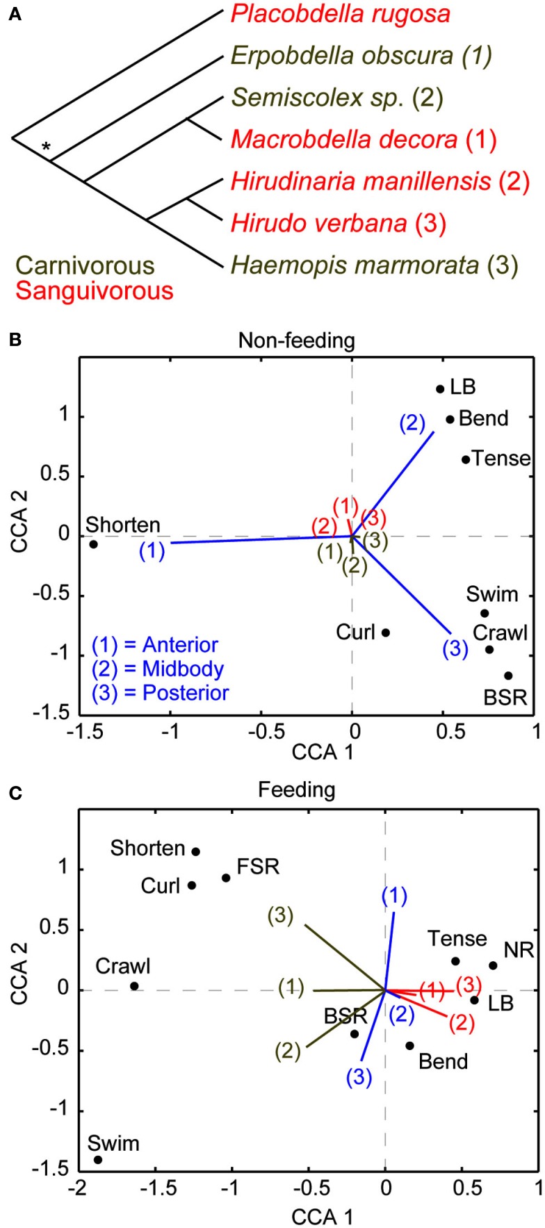
    1. Braak Ter C. J. F. (1986). Canonical correspondence analysis: a new eigenvector technique for multivariate direct gradient analysis. Ecology 67, 1167–117910.2307/1938672 - DOI

LinkOut - more resources