

Management of the Schneiderian membrane perforation during the maxillary sinus elevation procedure: a case report
- PMID: 22783452
- PMCID: PMC3392662
Management of the Schneiderian membrane perforation during the maxillary sinus elevation procedure: a case report
Abstract
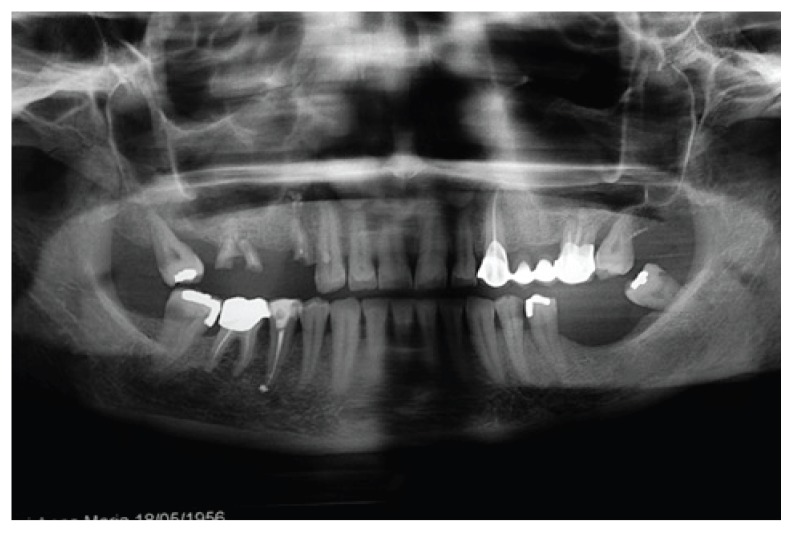
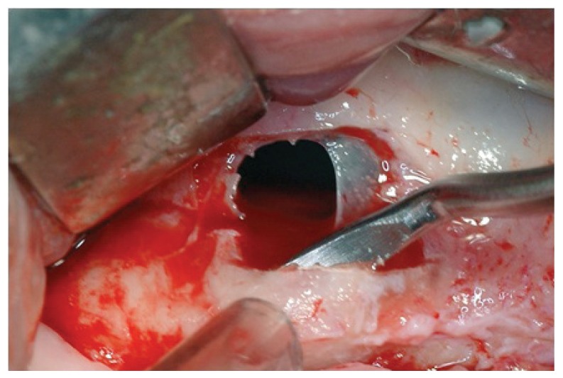
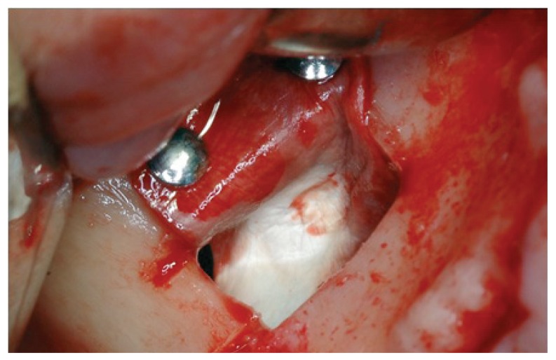
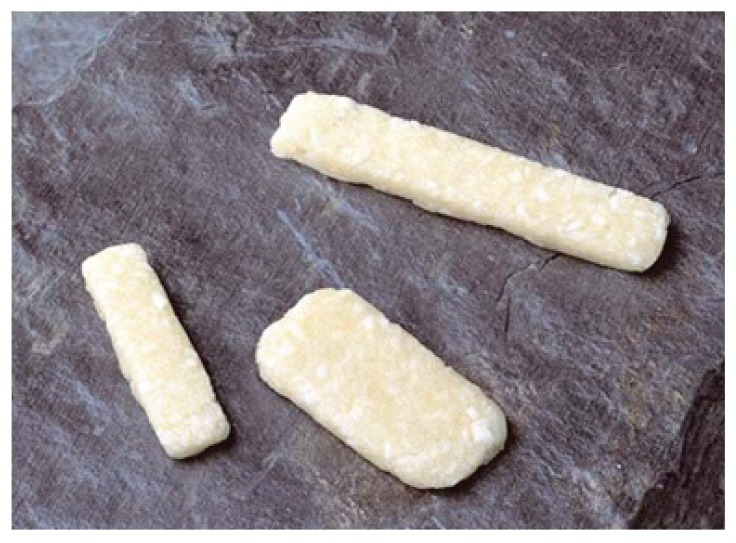
The maxillary sinus elevation is a standard and predictable procedure allowing the realization of dental implant rehabilitation in patients with severe bone atrophy in the lateral-posterior areas of the maxilla. Despite the presence of validated surgical methods and the broad availability of biomaterials, the procedures aimed at increasing the bone volume by lateral antrostomy still entail complications with different degrees of relevance. The prosthetic and surgical outcome is based on a successful coping with these aspects. The perforation of the Schneiderian membrane is one of the most frequent events for which a variety of protocols and approaches have been suggested by different authors. In this work is presented a case study in which a technique to repair the sinus mucosa laceration occurring during a maxillary sinus elevation procedure has been successfully adopted.
Keywords: Schneiderian membrane; maxillary sinus augmentation; sinus lift complications; underwood septa.
Figures
Similar articles
-
Effect of Schneiderian membrane perforation on sinus lift graft outcome using two different donor sites: a retrospective study of 105 maxillary sinus elevation procedures.GMS Interdiscip Plast Reconstr Surg DGPW. 2016 Mar 2;5:Doc11. doi: 10.3205/iprs000090. eCollection 2016. GMS Interdiscip Plast Reconstr Surg DGPW. 2016. PMID: 26955510 Free PMC article.
-
The effect of piezoelectric use on open sinus lift perforation: a retrospective evaluation of 56 consecutively treated cases from private practices.J Periodontol. 2010 Jan;81(1):167-71. doi: 10.1902/jop.2009.090190. J Periodontol. 2010. PMID: 20059429
-
The clinical and radiographic outcomes of Schneiderian membrane perforation without repair in sinus elevation surgery.Clin Implant Dent Relat Res. 2019 Oct;21(5):931-937. doi: 10.1111/cid.12752. Epub 2019 Apr 4. Clin Implant Dent Relat Res. 2019. PMID: 30950206
-
Maxillary Sinus Lift Procedures: An Overview of Current Techniques, Presurgical Evaluation, and Complications.Cureus. 2023 Nov 28;15(11):e49553. doi: 10.7759/cureus.49553. eCollection 2023 Nov. Cureus. 2023. PMID: 38156177 Free PMC article. Review.
-
Influence of Schneiderian Membrane Perforation on Implant Survival Rate: Systematic Review and Meta-Analysis.J Clin Med. 2024 Jun 27;13(13):3751. doi: 10.3390/jcm13133751. J Clin Med. 2024. PMID: 38999315 Free PMC article. Review.
Cited by
-
Management of Schneiderian membrane perforations during maxillary sinus floor augmentation with lateral approach in relation to subsequent implant survival rates: a systematic review and meta-analysis.Int J Implant Dent. 2021 Jul 12;7(1):91. doi: 10.1186/s40729-021-00346-7. Int J Implant Dent. 2021. PMID: 34250560 Free PMC article.
-
Endoscopic management of the schneiderian membrane perforation during transcrestal sinus augmentation: a case report.Oral Implantol (Rome). 2016 Nov 16;9(4):157-163. doi: 10.11138/orl/2016.9.4.157. eCollection 2016 Oct-Dec. Oral Implantol (Rome). 2016. PMID: 28042444 Free PMC article.
-
Tissue-Engineered Grafts from Human Decellularized Extracellular Matrices: A Systematic Review and Future Perspectives.Int J Mol Sci. 2018 Dec 18;19(12):4117. doi: 10.3390/ijms19124117. Int J Mol Sci. 2018. PMID: 30567407 Free PMC article.
References
-
- Froum SJ, Wallace SS, Elian N, Cho SC, Tarnow DP. Comparison of mineralized cancellous bone allograft (Puros) and anorganic bovine bone matrix (Bio-Oss) for sinus augmentation: histomorphometry at 26 to 32 weeks after grafting. Int J Periodontics Restorative Dent. 2006 Dec;26(6):543–51. - PubMed
-
- Shulman LB, Jensen OT. Sinus Graft Consensus Conference. Introduction. Int J Oral Maxillofac Implants. 1998;13(suppl):5, 6. - PubMed
-
- Esposito M, Grusovin MG, Rees J, Karasoulos D, Felice P, Alissa R, Worthington HV, Coulthard P. Effectiveness of sinus lift procedures for dental implant rehabilitation: a Cochrane systematic review. Eur J Oral Implantol. 2010;3(1):7–26. - PubMed
-
- Chiapasco M, Zaniboni M. Methods to treat the edentulous posterior maxilla: implants with sinus grafting. J Oral Maxillofac Surg. 2009;67(4):867–71. - PubMed
LinkOut - more resources
Full Text Sources