. 2012 Aug 14;51(32):6266-88.
doi: 10.1021/bi300010w. Epub 2012 Jul 31.

Membrane protein structure determination using crystallography and lipidic mesophases: recent advances and successes

Affiliations

Membrane protein structure determination using crystallography and lipidic mesophases: recent advances and successes

Martin Caffrey et al. Biochemistry. .

Abstract

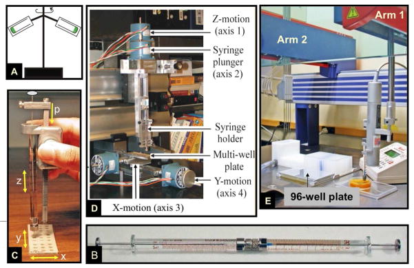
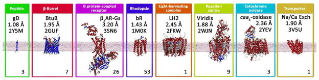
The crystal structure of the β(2)-adrenergic receptor in complex with an agonist and its cognate G protein has just recently been determined. It is now possible to explore in molecular detail the means by which this paradigmatic transmembrane receptor binds agonist, communicates the impulse or signaling event across the membrane, and sets in motion a series of G protein-directed intracellular responses. The structure was determined using crystals of the ternary complex grown in a rationally designed lipidic mesophase by the so-called in meso method. The method is proving to be particularly useful in the G protein-coupled receptor field where the structures of 13 distinct receptor types have been determined in the past 5 years. In addition to receptors, the method has proven to be useful with a wide variety of integral membrane protein classes that include bacterial and eukaryotic rhodopsins, light-harvesting complex II (LHII), photosynthetic reaction centers, cytochrome oxidases, β-barrels, an exchanger, and an integral membrane peptide. This attests to the versatility and range of the method and supports the view that the in meso method should be included in the arsenal of the serious membrane structural biologist. For this to happen, however, the reluctance to adopt it attributable, in part, to the anticipated difficulties associated with handling the sticky, viscous cubic mesophase in which crystals grow must be overcome. Harvesting and collecting diffraction data with the mesophase-grown crystals are also viewed with some trepidation. It is acknowledged that there are challenges associated with the method. Over the years, we have endeavored to establish how the method works at a molecular level and to make it user-friendly. To these ends, tools for handling the mesophase in the pico- to nanoliter volume range have been developed for highly efficient crystallization screening in manual and robotic modes. Methods have been implemented for evaluating the functional activity of membrane proteins reconstituted into the bilayer of the cubic phase as a prelude to crystallogenesis. Glass crystallization plates that provide unparalleled optical quality and sensitivity to nascent crystals have been built. Lipid and precipitant screens have been designed for a more rational approach to crystallogenesis such that the method can now be applied to an even wider variety of membrane protein types. In this work, these assorted advances are outlined along with a summary of the membrane proteins that have yielded to the method. The prospects for and the challenges that must be overcome to further develop the method are described.


Figures

Figure 1
Figure 1
Cartoon representation of a bicontinuous lipidic cubic mesophase. At its simplest, the cubic phase is formed by homogenizing lipid, typically monoolein (9.9 MAG), and water in approximately equal parts at 20 °C. An expanded view of the lipid component that forms the continuous curved bilayer is shown at the bottom of the figure. Water channels, on either side of the bilayer, that interpenetrate but never contact one another as they permeate the mesophase, are colored blue and red for clarity. The lattice parameter of the cubic phase, in this case of space group Im3m, obtained using small-angle X-ray scattering, is indicated.,
Figure 2
Figure 2
Temperature-composition phase diagrams for (A) 7.7 MAG and (B) 9.9 MAG, two monoacylglycerols that have proven to be particularly useful hosting lipids for the in meso crystallization of membrane proteins, complexes and peptides (Table 1). Cartoon representations of the different solid (Lc), liquid (FI) and liquid crystalline phases (Lα, HII, cubic-Pn3m, cubic-Ia3d) accessed in the temperature and composition range studied are shown along the top of the figure. The phase diagrams were constructed based on small-angle X-ray scattering measurements.,
Figure 3
Figure 3
The tools and supplies used to set up in meso crystallization trials in manual mode. Full details of the in meso method are available in print and in an online video. Key: A. Laboratory notebook. B. Temperature-composition phase diagram. C. Milli-Q water. D. Methanol. E. Paper towels. F. Pipeting devices covering volumes in the microliter range. G. Hamilton syringes (removable needle type, gas-tight) of varying sizes (10 and 100 μL usually). H. Narrow bore coupler. I. Repeat dispenser. J. Screwdriver. K. Glass slides and cover slips. L. Perforated double stick tape. M. Tweezers. N. Coupled syringes loaded with lipid and proteins solution, as indicated.
Figure 4
Figure 4
Crystals of membrane proteins in mesophases prepared with different hosting lipids. Examples of proteins and peptides are shown that either did not produce crystals or the crystals that grew were of lesser diffraction quality in the benchmark lipid, 9.9 MAG (monoolein), compared to the identified MAG. Diffraction quality (in brackets) is identified as is crystal growth temperature if other than 20 °C. Images for the gramicidin and β2AR-Gs complex are from references and , respectively. In all cases, the protein is colorless but the crystals are clearly visible in the hosting mesophase in wells of the glass sandwich crystallization plate.
Figure 5
Figure 5
Cartoon representation of the events proposed to take place during the crystallization of an integral membrane protein from the lipidic cubic mesophase. The process begins with the protein reconstituted into the curved bilayer of the “bicontinuous” cubic phase (tan). Added “precipitants” shift the equilibrium away from stability in the cubic membrane. This leads to phase separation wherein protein molecules diffuse from the bicontinuous bilayered reservoir of the cubic phase into a sheet-like or lamellar domain (A) and locally concentrate therein in a process that progresses to nucleation and crystal growth (B). Co-crystallization of the protein with native or additive lipid (cholesterol) is shown in this illustration. As much as possible, the dimensions of the lipid (tan oval with tail), detergent (pink oval with tail), cholesterol (purple), protein (blue and green; β2-adrenergic receptor-T4 lysozyme fusion; PDB code 2RH1), bilayer, and aqueous channels (dark blue) have been drawn to scale. The lipid bilayer is ~40 Å thick. Figure 5 is from reference .
Figure 6
Figure 6
Layered or Type I packing is observed in all crystals of membrane proteins produced to date by the in meso method. In the case of the β2-adrenoreceptor-Gs protein complex shown here (PDB code 3SN6), the transmembrane receptor (tan) drives the layering process by the proposed mechanism outlined in Figure 4. The approximate location of the bilayer supporting the receptor is indicated by the paired horizontal white lines.
Figure 7
Figure 7
Intrinsic tryptophan fluorescence quenching curve of gramicidin D in the cubic phase of hydrated monoolein. Bromo-MAG is the quenching lipid and its concentration is expressed as mole% in monoolein. Flourescence data were corrected for background fluorescence from buffer and lipid and for the inner filter effect, and were normalized to the quencher-free (Fc,0) value. The quenching profile is consistent with the peptide being reconstituted into the bilayer of the cubic phase. Data from reference .
Figure 8
Figure 8
Monitoring the γ-phosphoryl group transfer activity of diacylglycerol kinase (DgkA) reconstituted into the bilayer of the cubic phase by a coupled enzyme assay method (A) in a muli-well plate. Protein-laden mesophase (shaded grey in (B)) is positioned on the wall of the well where it remains in place throughout the assay as a consequence of its intrinsic viscosity and stickiness. The mesophase is bathed in buffer (blue) containing the water-soluble ingredients of the coupled assay (ATP, ADP, pyruvate, lactate, phosphoenolpyruvate (PEP), NADH/NAD+, pyruvate kinase (PK) and lactate dehydrogenase (LDH)). Water soluble substrate, ATP, diffuses into the nanoporous mesophase for use by DgkA in synthesizing phosphatidic acid from diacylglycerol both of which reside in and are confined to the bilayer of the mesophase. Water soluble product, ADP, diffuses out of the mesophase into the bathing solution where it is used by the coupled enzyme assay system to regenerate ATP. The coupling process that involves PK and LDH leads to a drop in the concentration of NADH which, in turn, is monitored continuously in situ in a multi-plate reader by a reduction in absorbance at 340 nm of the bathing solution (C). The slope of the progress curve (C) provides a measure of the initial velocity, Vo, as indicated.
Figure 9
Figure 9
Approaches and equipment used to set up in meso crystallization trials since the method was introduced in the mid-nineties. The original method used repeated centrifugation in a fixed angle rotor (A) to effect lipid and protein solution homogenization and cubic phase formation. The coupled syringe mixing device (B) was introduced in 1998 as a more practical and efficient means to generate and to dispense conveniently nanoliter volumes of protein-laden mesophase for use in in meso crystallization trials. Manual dispensing of the protein-laden mesophase prepared in the coupled syringe mixing device was greatly facilitated by the repeat dispenser (C). The x, y and z motions executed in dispensing mesophase manually, as in (C), inspired the building of a prototype robot consisting of a series of motorized orthogonal translation stages connected to a computer under LabView control (D). The success of the prototype in ‘automatically’ setting up crystallization plates in which membrane protein crystals grew was proof-of-concept and enough to secure funding with which to build in-house a custom-designed in meso crystallization robot (E). Variations on the original robot shown in (E) are now available commercially. The instrument shown in the figure comes equipped with a 4-tip liquid handling dispensing arm. An 8-tip version of the instrument is available commercially.
Figure 10
Figure 10
Temperature-induced changes in the lipid and aqueous channel dimensions of the cubic phase. Temperature dependence of the lipid length in the cubic phase (A) and of fully hydrated, cubic-Pn3m phase water channel radius (B) for three monoacylglycerols. Lipid identity is reported in the N.T notation. Data are from references , and .
Figure 11
Figure 11
Structure of the β2-adrenoreceptor solved with crystals grown by the in meso method using monoolein doped with cholesterol as an additive lipid. Cholesterol molecules (space filling model, asterisks) are part of the crystal structure. White horizontal lines mark the approximate location of the membrane/aqueous interface with respect to the receptor (green model, PDB code 2RH1).
Figure 12
Figure 12
A gallery of membrane protein structures solved using crystals grown by the lipidic cubic phase or in meso method. A single representative structure within a membrane protein class is shown along with diffraction resolution and hyperlinked PDB identifier. The figure at the bottom of each panel refers to the number of record entries in the PDB for that particular membrane protein class. Thus, within the peptide class there are 3 records for gramicidin D at different resolutions. Within the β-barrel class there are 7 entries, 1 each for BtuB, OpcA, Intimin, and Invasin, and 3 for OmpF. Within the G protein-coupled receptor class there are 26 records: 1 for the β2-adrenoreceptor-Gs complex, 7 for the β2-adrenorecptor, 3 for the A2a-adenosine receptor, 5 for the CXCR4 receptor, 2 for the sphingosine 1-phosphate subtype 1 receptor, and 1 each for the D3 dopamine receptor, the H1 histamine receptor, the M2 muscarinic receptor, the M3. muscarinic receptor, the δ-opioid receptor, the κ-opioid receptor, the μ-opioid receptor, and the nociceptin receptor. Within the non-GPCR rhodopsin class there are 53 records with 38 for bacteriorhodopsin, 3 for halorhodopsin, 6 for sensory rhodopsin II, 3 for the sensory rhodopsin II/transducer complex, and 1 each for sensory rhodopsin from Nostoc sp. Pcc 7120 and for rhodopin from Acetabularia acetabulum. Within the light harvesting complex class there is a single entry for LHII. Within the photosynthetic reaction center class there are 5 and 4 entries for the reaction centers from Blastochloris viridis and Rhodobacter sphaeroides, respectivly. Within the cytochrome oxidase class there are 2 entries for ba3 and 1 for caa3. Within the exchanger group there is a single entry for the Na+-Ca2+ exchanger. As of June 2012, the total count for in meso structures in the PDB is 103. Hyperlinks: 2Y5M; 2GUF; 3SN6; 1M0K; 2FKW; 2WJN; 2YEV; 3V5U
Chart 1
Chart 1
Issues to consider when undertaking an in meso crystallogenesis study assuming little prior knowledge about the crystallization potential of the membrane protein target. Items with an asterisk should be given priority.

References

    1. Caffrey M. Membrane protein crystallization. J Struct Biol. 2003;142:108–132. - PubMed
    1. Michel H. Crystallization of membrane-proteins. Trends Biochem Sci. 1983;8:56–59.
    1. Chen YJ, Pornillos O, Lieu S, Ma C, Chen AP, Chang G. X-ray structure of EmrE supports dual topology model. Proc Natl Acad Sci U S A. 2007;104:18999–19004. - PMC - PubMed
    1. Wada T, Shimono K, Kikukawa T, Hato M, Shinya N, Kim SY, Kimura-Someya T, Shirouzu M, Tamogami J, Miyauchi S, Jung KH, Kamo N, Yokoyama S. Crystal structure of the eukaryotic light-driven proton-pumping rhodopsin, Acetabularia rhodopsin II, from marine alga. J Mol Biol. 2011;411:986–998. - PubMed
    1. Lahiri S, Brehs M, Olschewski D, Becker CFW. Total chemical synthesis of an integral membrane enzyme: Diacylglycerol kinase from Escherichia coli. Angewandte Chemie-International Edition. 2011;50:3988–3992. - PubMed

Publication types