

doi: 10.3109/17435390.2012.711862.
Epub 2012 Aug 20.
Effect of modifying quantum dot surface charge on airway epithelial cell uptake in vitro
Affiliations
- PMID: 22783847
- PMCID: PMC3737271
- DOI: 10.3109/17435390.2012.711862
Item in Clipboard
Effect of modifying quantum dot surface charge on airway epithelial cell uptake in vitro
Nanotoxicology.
2013 Sep.
Abstract
The respiratory system is one of the portals of entry into the body, and hence inhalation of engineered nanomaterials is an important route of exposure. The broad range of physicochemical properties that influence biological responses necessitate the systematic study to contribute to understanding occupational exposure. Here, we report on the influence of nanoparticle charge and dose on human airway epithelial cells, and show that this platform can be used to evaluate consequences of exposure to engineered nanomaterials.
Figures
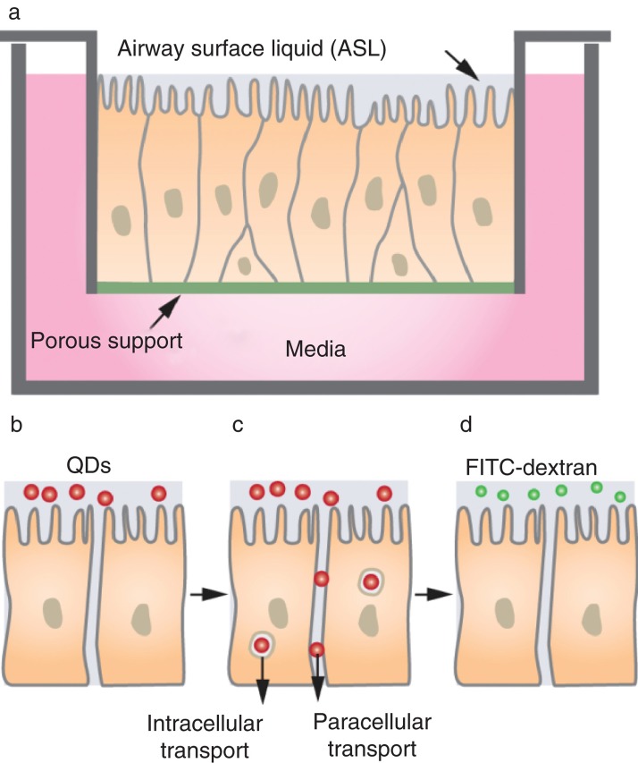
Schematic illustration of the platform used to study the response of the human airway epithelium to nanoparticles. (A) Human airway epithelial cells are grown on an insert such that the apical (top) surface is exposed to air and the basolateral (bottom) surface to media for 6 weeks prior to study to allow the cells to differentiate. The epithelial resistance across the monolayer is determined using a current source. (B) Quantum dots functionalised with positive or negative charge are introduced at the apical surface. (C) QDs can traverse the airway epithelium via paracellular or transcellular pathways. (D After exposures of airway epithelial cells to QDs, cells are washed and then 4 kDa FITC-dextran is placed on the apical surface and incubated for 20 min, after which the basolateral media is sampled to assess FITC concentration as a permeability assessment.
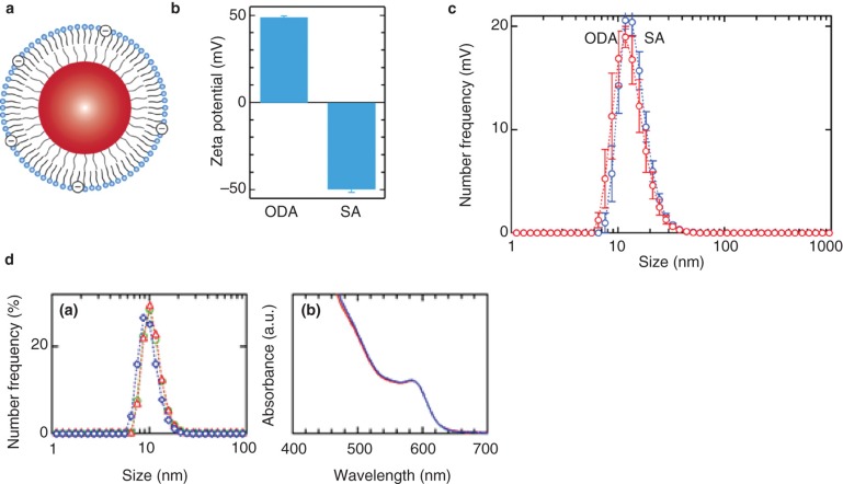
(A) Schematic illustration of lipid functionalised quantum dots. The outer leaflet contains 20 mol% Octadecylamine (positive charge) or stearic acid (negative charge). (B) The zeta potential was 49 mV for ODA-modified QDs and -50 mV for SA-modified QDs. (C) Particle size distributions for ODA-modified QDs (red) and SA-modified QDs (blue) (Green 2004). (D) (left) Size distributions and (right) absorbance spectra for ODA-functionalised QDs: (green) QDs in distilled water, (blue) QDs immediately after transferring to media, and (red) QDs after 12 suspension in media.
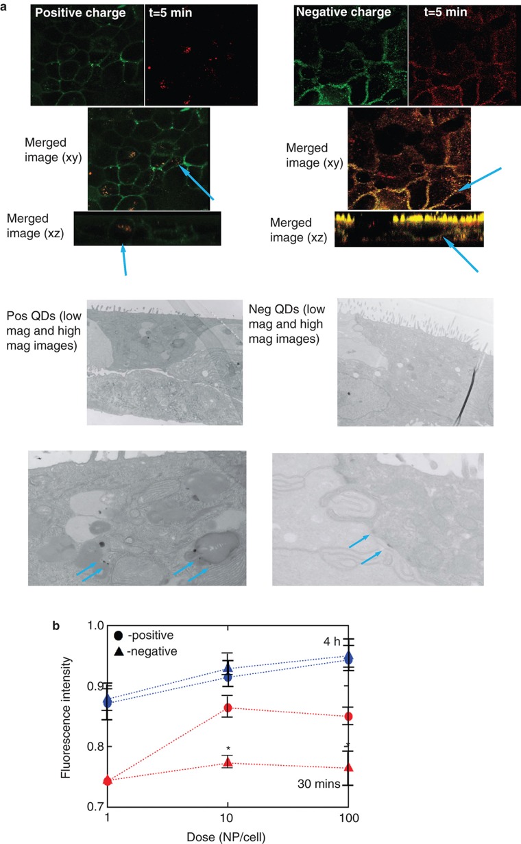
(A) After 5 min exposure, QDs penetrate the epithelium. Fluorescence images for airway epithelial cells 5 min after incubating with a dose of 10 NP/cell. (Green) Cell membrane, (red) QDs. After 5 min exposure, the majority of the negatively charged QDs are located at the cell–cell junctions, whereas the majority of the positively charged QDs are located with the cells. (B) By measuring the fluorescence in the basolateral media we can assess QD transport across the epithelial barrier. Both negatively and positively charged particles cross the epithelial barrier. (Bars represent standard error (SE), n = 3 for each dose; Anova one-way (p < 0.025)).
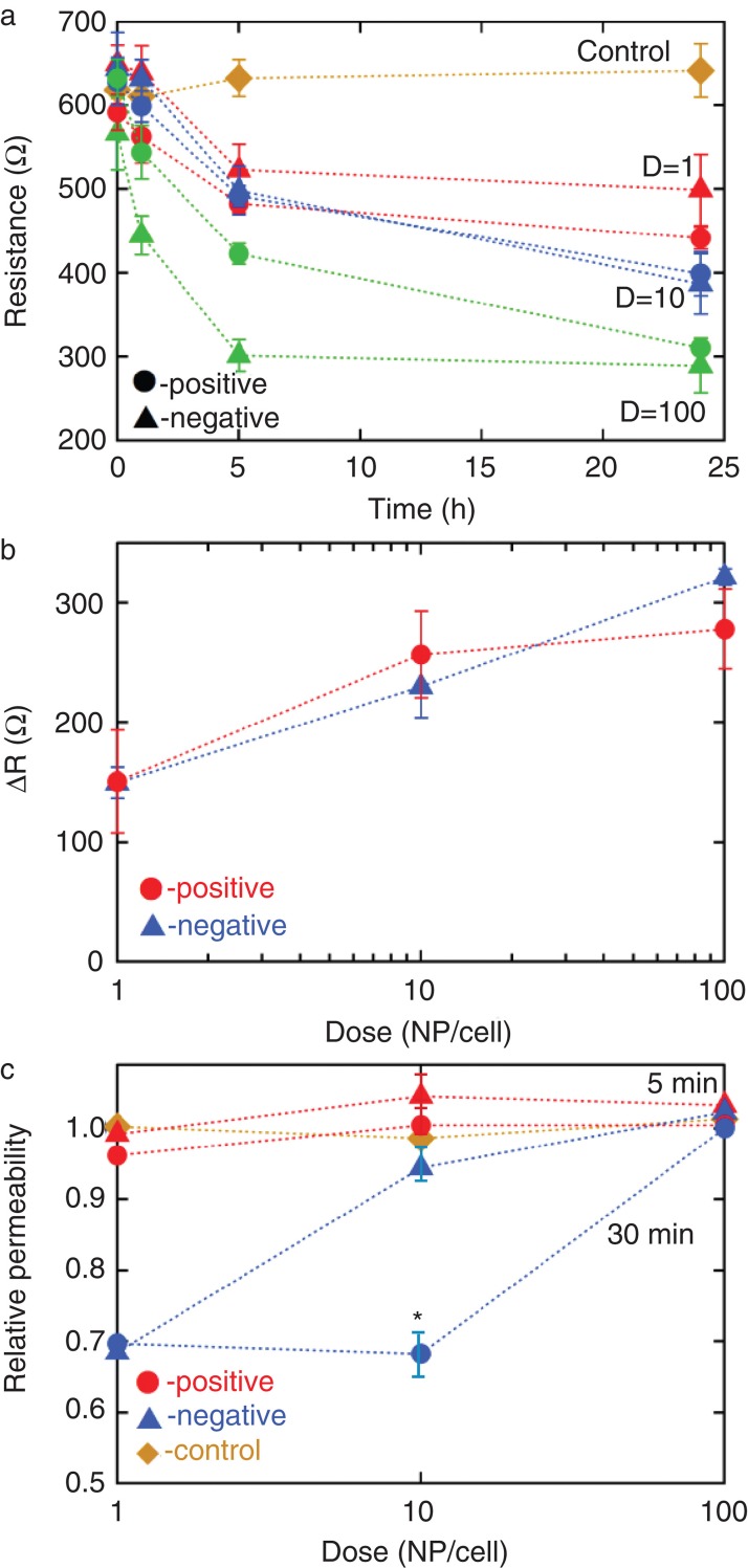
Nanoparticle exposure results in altered airway epithelial barrier function. (A) TER versus time for airway epithelial cells incubated with 1, 10 or 100 NP/cell. Both positively and negatively charged QDs decrease TER in a dose-responsive fashion. The control represents incubation with buffer only. (Bars represent standard error (SE) with at least three independent experiments) (B) The change in TER after 24 h increases exponentially with dose, independent of charge. (C) After exposure to QD, the 4 kD FITC-dextran was placed in the apical chamber and after incubating for 20 min (37oC) the basolateral media was sampled to assess fluorescence. In response to 30 min of exposure to a very low exposure of QD (in the order of 1 NP/cell), there was a significant decrease in paracellular permeability. (Bars represent standard error (SE), n = 3 per condition; Anova one-way (p < 0.025)).
Similar articles
-
Translocation of PEGylated quantum dots across rat alveolar epithelial cell monolayers.Int J Nanomedicine. 2011;6:2849-57. doi: 10.2147/IJN.S26051. Epub 2011 Nov 10. Int J Nanomedicine. 2011. PMID: 22131830 Free PMC article.
-
Selenium Redox Reactivity on Colloidal CdSe Quantum Dot Surfaces.J Am Chem Soc. 2016 Sep 7;138(35):11105-8. doi: 10.1021/jacs.6b06548. Epub 2016 Aug 24. J Am Chem Soc. 2016. PMID: 27518320 Free PMC article.
-
Functionalization-dependent induction of cellular survival pathways by CdSe quantum dots in primary normal human bronchial epithelial cells.ACS Nano. 2013 Oct 22;7(10):8397-411. doi: 10.1021/nn305532k. Epub 2013 Sep 17. ACS Nano. 2013. PMID: 24007210
-
Cell type-dependent changes in CdSe/ZnS quantum dot uptake and toxic endpoints.Toxicol Sci. 2015 Apr;144(2):246-58. doi: 10.1093/toxsci/kfv002. Epub 2015 Jan 19. Toxicol Sci. 2015. PMID: 25601991 Free PMC article.
-
Effect of Nanoparticle Surface Coating on Cell Toxicity and Mitochondria Uptake.J Biomed Nanotechnol. 2017 Feb;13(2):155-66. doi: 10.1166/jbn.2017.2337. J Biomed Nanotechnol. 2017. PMID: 29377103 Free PMC article.
Cited by
-
Quantifying engineered nanomaterial toxicity: comparison of common cytotoxicity and gene expression measurements.J Nanobiotechnology. 2017 Nov 9;15(1):79. doi: 10.1186/s12951-017-0312-3. J Nanobiotechnology. 2017. PMID: 29121949 Free PMC article.
-
Acute exposure to silica nanoparticles aggravate airway inflammation: different effects according to surface characteristics.Exp Mol Med. 2015 Jul 17;47(7):e173. doi: 10.1038/emm.2015.50. Exp Mol Med. 2015. PMID: 26183169 Free PMC article.
-
Role of air pollutants in airway epithelial barrier dysfunction in asthma and COPD.Eur Respir Rev. 2022 Mar 23;31(163):210112. doi: 10.1183/16000617.0112-2021. Print 2022 Mar 31. Eur Respir Rev. 2022. PMID: 35321933 Free PMC article. Review.
References
-
- Anderson JM, Van Itallie CM. Tight junctions and the molecular basis for regulation of paracellular permeability. Am J Physiol. 1995;269(4 Pt 1):G467–G475. - PubMed
-
- Colegio OR, Van Itallie C, Rahner C, Anderson JM. Claudin extracellular domains determine paracellular charge selectivity and resistance but not tight junction fibril architecture. Am J Physiol Cell Physiol. 2003;284(6):C1346–C1354. - PubMed
-
- Dausend J, Musyanovych A, Dass M, Walther P, Schrezenmeier H, Landfester K, et al. Uptake mechanism of oppositely charged fluorescent nanoparticles in HeLa cells. Macromol Biosci. 2008;8(12): 1135–1143. - PubMed
Publication types
MeSH terms
Substances
Grants and funding
LinkOut - more resources
Full Text Sources