. 2012 Jul 12;367(2):115-23.
doi: 10.1056/NEJMoa1110740.

Potassium channel KIR4.1 as an immune target in multiple sclerosis

Affiliations

Potassium channel KIR4.1 as an immune target in multiple sclerosis

Rajneesh Srivastava et al. N Engl J Med. .

Abstract

Background: Multiple sclerosis is a chronic inflammatory demyelinating disease of the central nervous system. Many findings suggest that the disease has an autoimmune pathogenesis; the target of the immune response is not yet known.

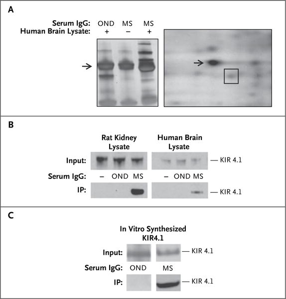
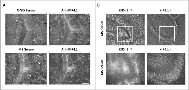
Methods: We screened serum IgG from persons with multiple sclerosis to identify antibodies that are capable of binding to brain tissue and observed specific binding of IgG to glial cells in a subgroup of patients. Using a proteomic approach focusing on membrane proteins, we identified the ATP-sensitive inward rectifying potassium channel KIR4.1 as the target of the IgG antibodies. We used a multifaceted validation strategy to confirm KIR4.1 as a target of the autoantibody response in multiple sclerosis and to show its potential pathogenicity in vivo.

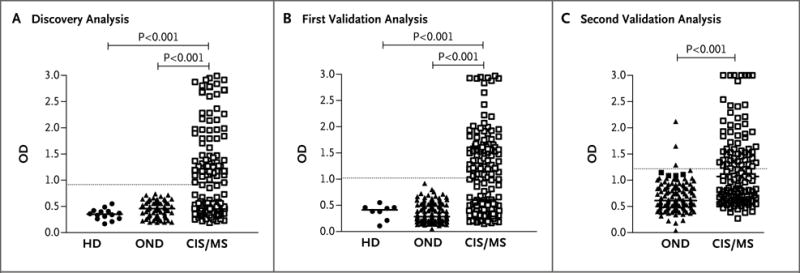
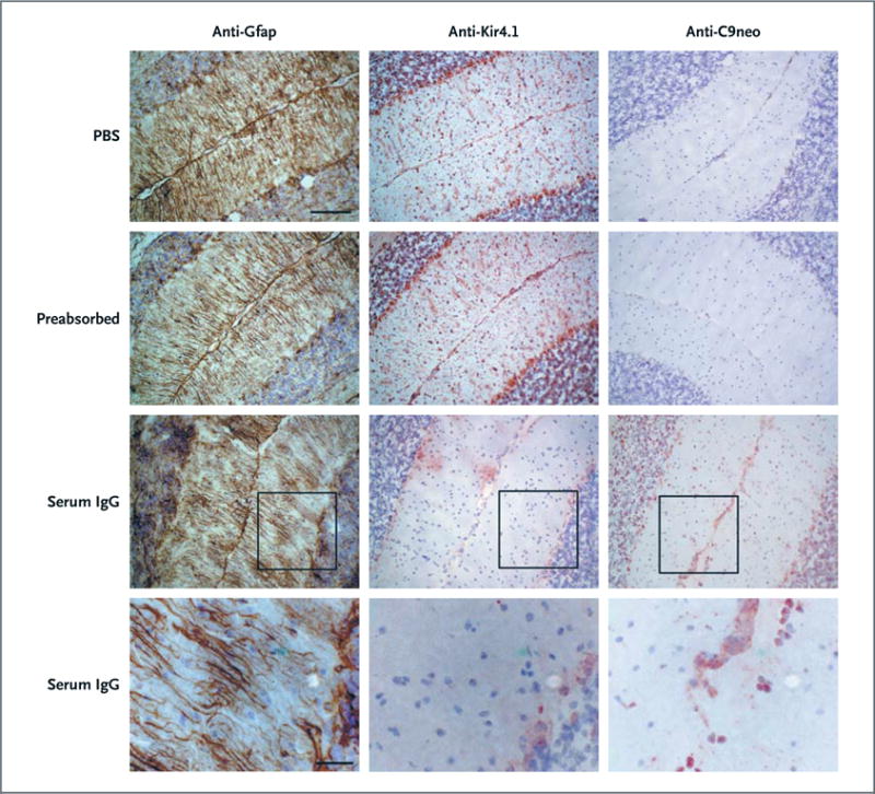
Results: Serum levels of antibodies to KIR4.1 were higher in persons with multiple sclerosis than in persons with other neurologic diseases and healthy donors (P<0.001 for both comparisons). We replicated this finding in two independent groups of persons with multiple sclerosis or other neurologic diseases (P<0.001 for both comparisons). Analysis of the combined data sets indicated the presence of serum antibodies to KIR4.1 in 186 of 397 persons with multiple sclerosis (46.9%), in 3 of 329 persons with other neurologic diseases (0.9%), and in none of the 59 healthy donors. These antibodies bound to the first extracellular loop of KIR4.1. Injection of KIR4.1 serum IgG into the cisternae magnae of mice led to a profound loss of KIR4.1 expression, altered expression of glial fibrillary acidic protein in astrocytes, and activation of the complement cascade at sites of KIR4.1 expression in the cerebellum.

Conclusions: KIR4.1 is a target of the autoantibody response in a subgroup of persons with multiple sclerosis. (Funded by the German Ministry for Education and Research and Deutsche Forschungsgemeinschaft.).


Figures

Figure 1
Figure 1. Identification of KIR4.1 as a Target of Serum IgG in Multiple Sclerosis (MS)
Panel A, left, shows a one-dimensional SDS-PAGE of human brain lysate precipitated with IgG from the pooled serum of 12 patients with MS and 12 with other neurologic diseases (OND). (For the middle lane [marked –], no brain lysate was used in the precipitation.) Unique bands (third lane) above and below the IgG heavy chain band (arrow) are observed after immunoprecipitation with purified IgG from the pooled serum of patients with MS. Panel A, right, shows a two-dimensional electrophoresis of brain antigens obtained after immunoprecipitation with serum IgG from patients with MS. The spot containing the KIR4.1 protein, identified by means of matrix-assisted laser desorption–tandem mass spectrometry, is outlined with a square. The arrow shows the IgG heavy chain spot. Panel B shows KIR4.1 detection by Western blot analysis in two different immunoprecipitation (IP) assays, as indicated. IP assays were performed with serum IgG from patients with OND and from patients with MS on membrane-protein– enriched fractions of rat kidney and human brain tissue lysates. The lanes on the left (marked –) of the blots are negative controls (no IgG used for IP). Panel C shows a Western blot analysis of KIR4.1 immunoprecipitation from in vitro–translated human KIR4.1 protein with serum IgG from patients with OND and from patients with MS.
Figure 2
Figure 2. Validation of KIR4.1 as the Target of the Serum IgG Reactivity in Patients with MS
Panel A shows double immunofluorescence labeling revealing colocalization of monoclonal anti-KIR4.1 with serum IgG from a patient with MS in cerebellar sections of rat brain. Staining with serum of a patient with other neurologic diseases (OND) is shown as a control (scale bar, 200 μm for all parts of Panel A). Panel B shows immunofluorescence labeling of cerebellar sections of wild-type (left) and Kir4.1−/− (right) mice with purified serum IgG from a patient with MS (scale bar, 100 μm for the upper panels and 50 μm for the lower panels). KIR4.1-antibody–negative serum specimens did not stain central nervous system (CNS) tissue from wild-type mice or Kir4.1−/− mice (data not shown).
Figure 3
Figure 3. High-Titer Serum Reactivity to the KIR4.1 Protein in a Subgroup of Patients with MS
A protein-based enzyme-linked immunosorbent assay (ELISA) was used to detect anti-KIR4.1 serum autoantibodies. Purified recombinant KIR4.1 from HEK293 cells was covalently coupled to ELISA plates. Serum antibody binding to KIR4.1 was determined in healthy donors (HD), in patients with other neurologic diseases (OND), and in patients with MS or a clinically isolated syndrome (CIS) for the discovery series (Panel A), the first validation series (Panel B), and the second validation series (Panel C). For the first two series, KIR4.1 antibody titers of healthy persons, of patients with OND, and of patients with MS or a CIS were compared with the use of the Kruskal–Wallis test of one-way analysis of variance followed by Dunn’s multiple comparison test, for which P values are shown. In the third series, KIR4.1 antibody titers in patients with OND and those with MS or a CIS were compared with the use of the Mann–Whitney t-test. The threshold for anti-KIR4.1 antibody positivity (5 SD above the mean optical density [OD] for healthy persons) is indicated by a dashed horizontal line. For further details, see the Supplementary Appendix.
Figure 4
Figure 4. KIR4.1-Specific MS Serum IgG Antibodies and Loss of Kir4.1 Staining, Disruption of Gfap Architecture, and Activation of Complement in Vivo in Mice
Phosphate-buffered saline (PBS; first row), serum IgG from a patient with MS depleted of KIR4.1-specific antibodies (preabsorbed, second row), or serum IgG with preserved anti-KIR4.1 reactivity (third and fourth rows) was injected into the cisternae magnae of C57BL/6 mice together with human complement. Twenty-four hours after injection, the mice were killed and brain sections were assessed for glial fibrillary acidic protein (Gfap) expression (left column), Kir4.1 expression (middle column), and C9neo reactivity (right column) by means of immunohistochemical analysis. The monoclonal anti-Kir4.1 antibody, which was used to visualize Kir4.1 expression, binds the intracellular domain of the protein and does not compete with the KIR4.1 reactive serum IgG for the same epitope of the KIR4.1 protein (Fig. S7 in the Supplementary Appendix and data not shown). Moreover, the lack of Kir4.1 immunoreactivity in mice that were injected with serum IgG with preserved anti-KIR4.1 specific antibodies is not caused by masking of the Kir4.1 antigen by nonspecific serum IgG antibodies, because the monoclonal antibody used to detect glial Kir4.1 expression was not blocked when it was applied together with serum IgG that had been preabsorbed with KIR4.1 (see second row) (scale bars, 50 μm in the upper three rows and 20 μm in the bottom row).

Comment in

References

    1. Noseworthy JH, Lucchinetti C, Rodriguez M, Weinshenker BG. Multiple sclerosis. N Engl J Med. 2000;343:938–52. - PubMed
    1. Compston A, Coles A. Multiple sclerosis. Lancet. 2008;372:1502–17. - PubMed
    1. Ascherio A, Munger KL. Environmental risk factors for multiple sclerosis. Part I: the role of infection. Ann Neurol. 2007;61:288–99. - PubMed
    1. The International Multiple Sclerosis Genetics Consortium. Risk alleles for multiple sclerosis identified by a genome-wide study. N Engl J Med. 2007;357:851–62. - PubMed
    1. McFarland HF, Martin R. Multiple sclerosis: a complicated picture of autoimmunity. Nat Immunol. 2007;8:913–9. - PubMed

Publication types