Review
. 2012 Oct;12(10):1347-60.
doi: 10.1517/14712598.2012.707181. Epub 2012 Jul 12.

Advances in the formation, use and understanding of multi-cellular spheroids

Affiliations
Review

Advances in the formation, use and understanding of multi-cellular spheroids

Toni-Marie Achilli et al. Expert Opin Biol Ther. 2012 Oct.

Abstract

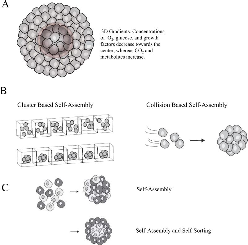
Introduction: Developing in vitro models for studying cell biology and cell physiology is of great importance to the fields of biotechnology, cancer research, drug discovery, toxicity testing, as well as the emerging fields of tissue engineering and regenerative medicine. Traditional two-dimensional (2D) methods of mammalian cell culture have several limitations and it is increasingly recognized that cells grown in a three-dimensional (3D) environment more closely represent normal cellular function due to the increased cell-to-cell interactions, and by mimicking the in vivo architecture of natural organs and tissues.

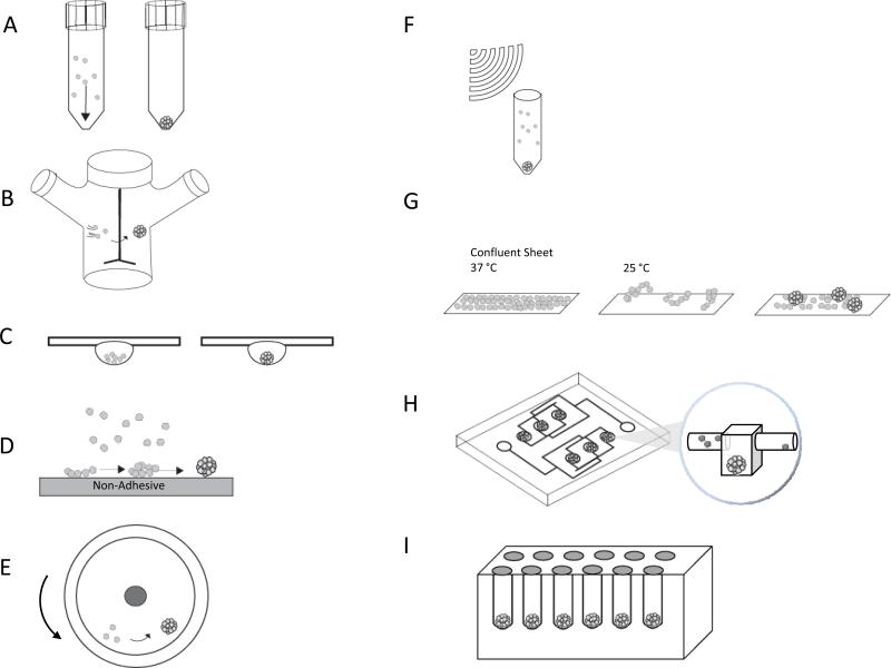
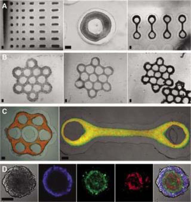
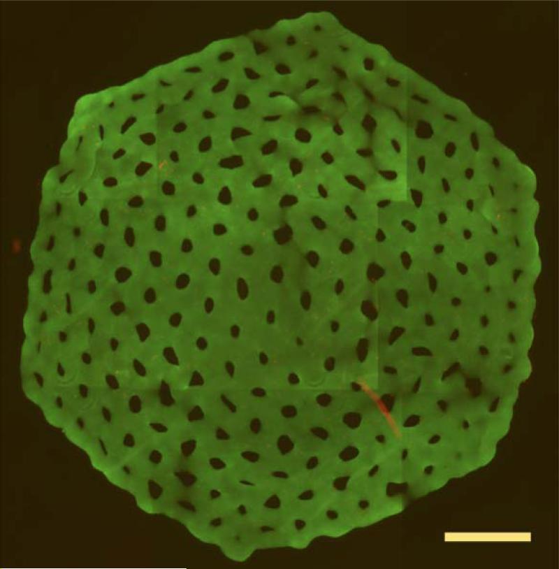
Areas covered: In this review, we discuss the methods to form 3D multi-cellular spheroids, the advantages and limitations of these methods, and assays used to characterize the function of spheroids. The use of spheroids has led to many advances in basic cell sciences, including understanding cancer cell interactions, creating models for drug discovery and cancer metastasis, and they are being investigated as basic units for engineering tissue constructs. As so, this review will focus on contributions made to each of these fields using spheroid models.

Expert opinion: Multi-cellular spheroids are rich in biological content and mimic better the in vivo environment than 2D cell culture. New technologies to form and analyze spheroids are rapidly increasing their adoption and expanding their applications.


Figures

Figure 1
Figure 1
(A) The concentric organization of proliferating and necrotic cells in multicellular spheroids is influenced by the molecular gradients of soluble factors which are established by convection and diffusion within the spheroid microenvironment. (B) There are two main categories of spheroid bio-fabrication, cluster based and collision based self-assembly. In cluster based self-assembly, monodispersed cells are segregated into compartments, settle, and aggregate to form spheroids. In collision based self-assembly, suspended cells collide to form spheroids. (C) When certain mixed cell populations self-assemble, the specific pattern of segregation that occurs is referred to as self-sorting.
Figure 2
Figure 2
Methods of spheroid bio-fabrication (A) Pellet Culture (B) Spinner Culture (C) Hanging Drop (D) Liquid Overlay (E) Rotating Wall Vessel (F) External Force (G) Cell Sheets (H) Microfluidics (I) Nonadhesive Hydrogel Micro-molds
Figure 3
Figure 3
Micro-molded hydrogels can be used to direct the self-assembly of a number of shapes including rodes, toroids, loop-ended dogbones, and honeycombs. (A) Microtissues with complex geometries were self-assembled from various cell types: rat hepatoma (H35) rods (left), MCF-7 toroid (middle), and H35 loop-ended dog bones (right). (B) MCF-7 cells seeded into honeycomb recesses (left), formed a branched microtissue by 1 day (middle), and compacted slightly but retained the honeycomb shape for at least 3 days after removal from the mold (right). Mixed-cell populations produced multilayered microtissues with controlled shape. (C) Images of microtissues with normal human fibroblasts (NHF) (red) cores coated by an outer layer of H35 (green, left) or HUVEC (green, right). (D) Confocal microscopy images of self-sorting of three cell types within a 2-day-old tritypic spheroid. (E) The eight orbital honeycomb of NHG (6×10⁶ cells) was stained with a live/dead assay after 24 hours to show viability. Scale bars are 200 μm (panels A-C), 100 μm (panel D), and 1800 μm (panel E). Figure permissions Biotechniques, and Biofabrication.
Figure 3
Figure 3
Micro-molded hydrogels can be used to direct the self-assembly of a number of shapes including rodes, toroids, loop-ended dogbones, and honeycombs. (A) Microtissues with complex geometries were self-assembled from various cell types: rat hepatoma (H35) rods (left), MCF-7 toroid (middle), and H35 loop-ended dog bones (right). (B) MCF-7 cells seeded into honeycomb recesses (left), formed a branched microtissue by 1 day (middle), and compacted slightly but retained the honeycomb shape for at least 3 days after removal from the mold (right). Mixed-cell populations produced multilayered microtissues with controlled shape. (C) Images of microtissues with normal human fibroblasts (NHF) (red) cores coated by an outer layer of H35 (green, left) or HUVEC (green, right). (D) Confocal microscopy images of self-sorting of three cell types within a 2-day-old tritypic spheroid. (E) The eight orbital honeycomb of NHG (6×10⁶ cells) was stained with a live/dead assay after 24 hours to show viability. Scale bars are 200 μm (panels A-C), 100 μm (panel D), and 1800 μm (panel E). Figure permissions Biotechniques, and Biofabrication.

References

    1. Pampaloni F, Reynaud EG, Stelzer EH. The third dimension bridges the gap between cell culture and live tissue. Nat Rev Mol Cell Biol. 2007;810:839–45. - PubMed
    1. Kleinman HK, Philp D, Hoffman MP. Role of the extracellular matrix in morphogenesis. Curr Opin Biotechnol. 2003;145:526–32. - PubMed
    1. Duguay D, Foty RA, Steinberg MS. Cadherin-mediated cell adhesion and tissue segregation: qualitative and quantitative determinants. Dev Biol. 2003;2532:309–23. - PubMed
    1. Foty RA, Pfleger CM, Forgacs G, et al. Surface tensions of embryonic tissues predict their mutual envelopment behavior. Development. 1996;1225:1611–20. - PubMed
    1. Moscona A, Moscona H. The dissociation and aggregation of cells from organ rudiments of the early chick embryo. J Anat. 1952;863:287–301. [One of the original papers showing the self-assembly and self-sorting of cells.] - PMC - PubMed

Publication types