

Longitudinal MRI contrast enhanced monitoring of early tumour development with manganese chloride (MnCl2) and superparamagnetic iron oxide nanoparticles (SPIOs) in a CT1258 based in vivo model of prostate cancer
- PMID: 22784304
- PMCID: PMC3520113
- DOI: 10.1186/1471-2407-12-284
Longitudinal MRI contrast enhanced monitoring of early tumour development with manganese chloride (MnCl2) and superparamagnetic iron oxide nanoparticles (SPIOs) in a CT1258 based in vivo model of prostate cancer
Abstract
Background: Cell lines represent a key tool in cancer research allowing the generation of neoplasias which resemble initial tumours in in-vivo animal models. The characterisation of early tumour development is of major interest in order to evaluate the efficacy of therapeutic agents. Magnetic resonance imaging (MRI) based in-vivo characterisation allows visualisation and characterisation of tumour development in early stages prior to manual palpation. Contrast agents for MRI such as superparamagnetic iron oxide nanoparticles (SPIOs) and manganese chloride (MnCl2) represent powerful tools for the in-vivo characterisation of early stage tumours. In this experimental study, we labelled prostate cancer cells with MnCl2 or SPIOs in vitro and used 1 T MRI for tracing labelled cells in-vitro and 7 T MRI for tracking in an in-vivo animal model.
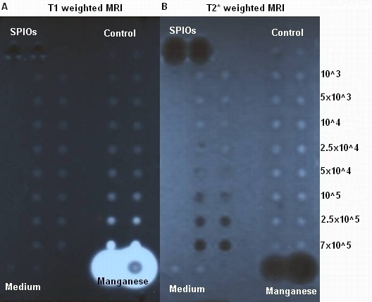
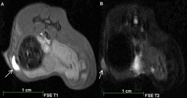
Methods: Labelling of prostate cancer cells CT1258 was established in-vitro with MnCl2 and SPIOs. In-vitro detection of labelled cells in an agar phantom was carried out through 1 T MRI while in-vivo detection was performed using 7 T MRI after subcutaneous (s.c.) injection of labelled cells into NOD-Scid mice (n = 20). The animals were scanned in regular intervals until euthanization. The respective tumour volumes were analysed and corresponding tumour masses were subjected to histologic examination.
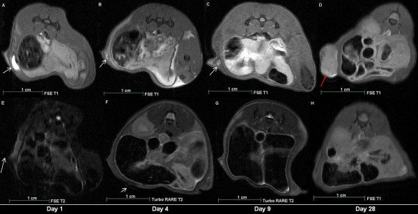
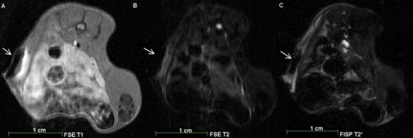
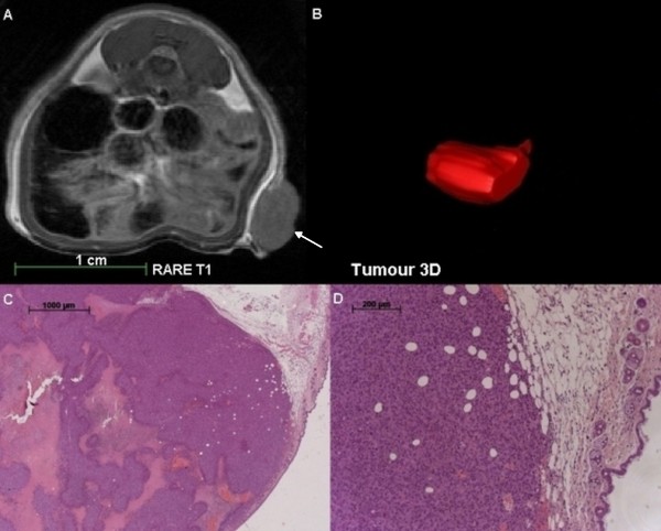
Results: MnCl2in-vitro labelling resulted in no significant metabolic effects on proliferation and cell vitality. In-vitro detection-limit accounted 105 cells for MnCl2 as well as for SPIOs labelling. In-vivo 7 T MRI scans allowed detection of 103 and 104 cells. In-vivo MnCl2 labelled cells were detectable from days 4-16 while SPIO labelling allowed detection until 4 days after s.c. injection. MnCl2 labelled cells were highly tumourigenic in NOD-Scid mice and the tumour volume development was characterised in a time dependent manner. The amount of injected cells correlated with tumour size development and disease progression. Histological analysis of the induced tumour masses demonstrated characteristic morphologies of prostate adenocarcinoma.
Conclusions: To the best of our knowledge, this is the first study reporting direct in-vitro MnCl2 labelling and 7 T based in-vivo MRI tracing of cancer cells in a model of prostate cancer. MnCl2 labelling was found to be suitable for in-vivo tracing allowing long detection periods. The labelled cells kept their highly tumourigenic potential in-vivo. Tumour volume development was visualised prior to manual palpation allowing tumour characterisation in early stages of the disease.
Figures
Similar articles
-
Monitoring of in vivo function of superparamagnetic iron oxide labelled murine dendritic cells during anti-tumour vaccination.PLoS One. 2011;6(5):e19662. doi: 10.1371/journal.pone.0019662. Epub 2011 May 27. PLoS One. 2011. PMID: 21637760 Free PMC article.
-
Imaging monocytes with iron oxide nanoparticles targeted towards the monocyte integrin MAC-1 (CD11b/CD18) does not result in improved atherosclerotic plaque detection by in vivo MRI.Contrast Media Mol Imaging. 2010 Sep-Oct;5(5):268-75. doi: 10.1002/cmmi.384. Contrast Media Mol Imaging. 2010. PMID: 20973112
-
Current limitations of molecular magnetic resonance imaging for tumors as evaluated with high-relaxivity CD105-specific iron oxide nanoparticles.Invest Radiol. 2012 Jul;47(7):383-91. doi: 10.1097/RLI.0b013e31824c5a57. Invest Radiol. 2012. PMID: 22659596
-
Multiparametric MRI in detection and staging of prostate cancer.Dan Med J. 2017 Feb;64(2):B5327. Dan Med J. 2017. PMID: 28157066 Review.
-
Recent advances in superparamagnetic iron oxide nanoparticles for cellular imaging and targeted therapy research.Curr Pharm Des. 2013;19(37):6575-93. doi: 10.2174/1381612811319370003. Curr Pharm Des. 2013. PMID: 23621536 Free PMC article. Review.
Cited by
-
Establishment and characterization of stable red, far-red (fR) and near infra-red (NIR) transfected canine prostate cancer cell lines.Cancer Cell Int. 2020 Apr 29;20:139. doi: 10.1186/s12935-020-01211-0. eCollection 2020. Cancer Cell Int. 2020. PMID: 32368185 Free PMC article.
-
Generation and characterisation of a canine EGFP-HMGA2 prostate cancer in vitro model.PLoS One. 2014 Jun 10;9(6):e98788. doi: 10.1371/journal.pone.0098788. eCollection 2014. PLoS One. 2014. PMID: 24914948 Free PMC article.
-
The role of manganese-based MRI contrast agents for cancer theranostics: Where do we stand in 2025?Theranostics. 2025 Mar 15;15(9):4147-4174. doi: 10.7150/thno.108705. eCollection 2025. Theranostics. 2025. PMID: 40213669 Free PMC article. Review.
-
Manganese-Based Nanotheranostics for Magnetic Resonance Imaging-Mediated Precise Cancer Management.Int J Nanomedicine. 2023 Oct 26;18:6077-6099. doi: 10.2147/IJN.S426311. eCollection 2023. Int J Nanomedicine. 2023. PMID: 37908669 Free PMC article. Review.
-
A brief review of cytotoxicity of nanoparticles on mesenchymal stem cells in regenerative medicine.Int J Nanomedicine. 2019 May 24;14:3875-3892. doi: 10.2147/IJN.S205574. eCollection 2019. Int J Nanomedicine. 2019. PMID: 31213807 Free PMC article. Review.
References
-
- Ferlay J, Shin HR, Bray F, Forman D, Mathers C, Parkin D. GLOBOCAN 2008, Cancer Incidence and Mortality Worldwide: IARC No. 10. Lyon. International Agency for Research on Cancer, France; 2010. Available from: http://globocaniarcfr 2008.
-
- Pienta KJ, Abate-Shen C, Agus DB, Attar RM, Chung LW, Greenberg NM, Hahn WC, Isaacs JT, Navone NM, Peehl DM, Simons JW, Solit DB, Soule HR, VanDyke TA, Weber MJ, Wu L, Vessella RL. The current state of preclinical prostate cancer animal models. Prostate. 2008;68(6):629–639. doi: 10.1002/pros.20726. - DOI - PMC - PubMed
Publication types
MeSH terms
Substances
LinkOut - more resources
Full Text Sources
Medical
