

Increased CSF osmolarity reversibly induces hydrocephalus in the normal rat brain
- PMID: 22784705
- PMCID: PMC3493274
- DOI: 10.1186/2045-8118-9-13
Increased CSF osmolarity reversibly induces hydrocephalus in the normal rat brain
Abstract
Background: Hydrocephalus is a central nervous system (CNS) disorder characterized by the abnormal accumulation of cerebrospinal fluid (CSF) in cerebral ventricles, resulting in their dilatation and associated brain tissue injury. The pathogenesis of hydrocephalus remains unclear; however, recent reports suggest the possible involvement of abnormal osmotic gradients. Here we explore the kinetics associated with manipulating CSF osmolarity on ventricle volume (VV) in the normal rat brain.
Methods: CSF was made hyper-osmotic by introducing 10KD dextran into the lateral ventricle, either by acute injection at different concentrations or by chronic infusion at a single concentration. The induction and withdrawal kinetics of dextran infusion on VV were explored in both contexts.
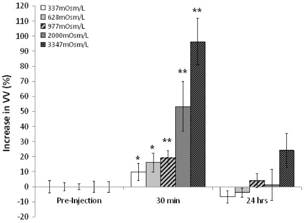
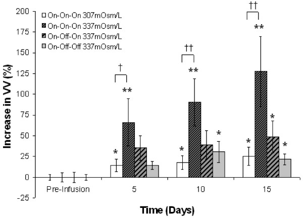
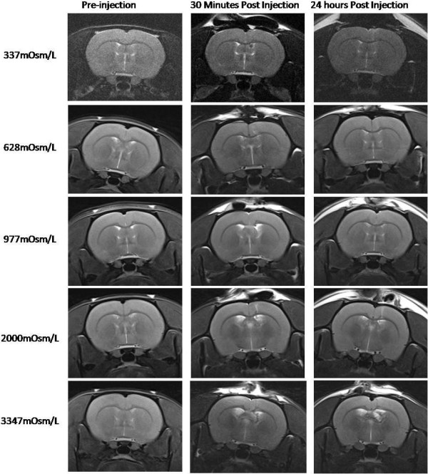
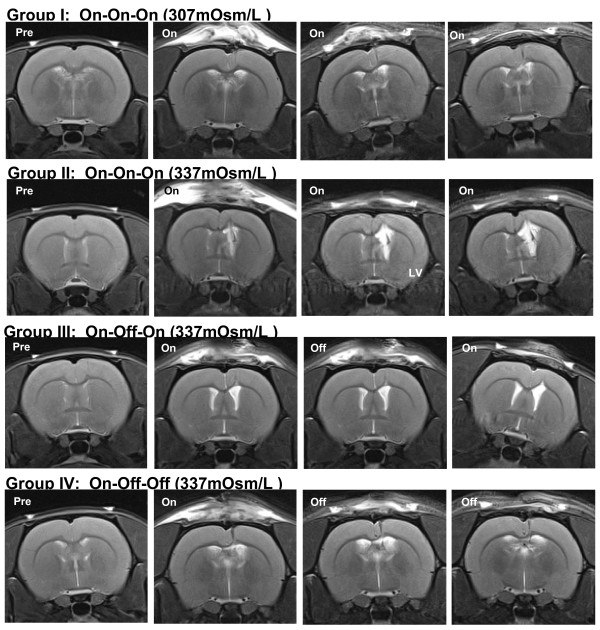
Results: Acute intraventricular injection of dextran caused a rapid increase in VV which completely reversed within 24 hours. These kinetics are seemingly independent of CSF osmolarity across a range spanning an order of magnitude; however, the magnitude of the transient increase in VV was proportional to CSF osmolarity. By contrast, continuous intraventricular infusion of dextran at a relatively low concentration caused a more gradual increase in VV which was very slow to reverse when infusion was suspended after five days.
Conclusion: We conclude that hyperosmolar CSF is sufficient to produce a proportional degree of hydrocephalus in the normal rat brain, and that this phenomenon exhibits hysteresis if CSF hyperosmolarity is persistent. Thus pathologically-induced increases in CSF osmolarity may be similarly associated with certain forms of clinical hydrocephalus. An improved understanding of this phenomenon and its kinetics may facilitate the development of novel therapies for the treatment of clinical hydrocephalus.
Figures
Similar articles
-
Intraventricular infusion of hyperosmolar dextran induces hydrocephalus: a novel animal model of hydrocephalus.Cerebrospinal Fluid Res. 2009 Dec 11;6:16. doi: 10.1186/1743-8454-6-16. Cerebrospinal Fluid Res. 2009. PMID: 20003330 Free PMC article.
-
Ventricle wall movements and cerebrospinal fluid flow in hydrocephalus.J Neurosurg. 2011 Jul;115(1):159-64. doi: 10.3171/2010.12.JNS10926. Epub 2011 Jan 28. J Neurosurg. 2011. PMID: 21275563
-
Biomechanical effects of hyper-dynamic cerebrospinal fluid flow through the cerebral aqueduct in idiopathic normal pressure hydrocephalus patients.J Biomech. 2023 Jul;156:111671. doi: 10.1016/j.jbiomech.2023.111671. Epub 2023 Jun 3. J Biomech. 2023. PMID: 37327645
-
A proposed role for efflux transporters in the pathogenesis of hydrocephalus.Croat Med J. 2014 Aug 28;55(4):366-76. doi: 10.3325/cmj.2014.55.366. Croat Med J. 2014. PMID: 25165050 Free PMC article. Review.
-
A unifying theory for the definition and classification of hydrocephalus.Childs Nerv Syst. 1994 Jan;10(1):2-12. doi: 10.1007/BF00313578. Childs Nerv Syst. 1994. PMID: 8194058 Review.
Cited by
-
Modulation of brain cation-Cl- cotransport via the SPAK kinase inhibitor ZT-1a.Nat Commun. 2020 Jan 7;11(1):78. doi: 10.1038/s41467-019-13851-6. Nat Commun. 2020. PMID: 31911626 Free PMC article.
-
Targeting choroid plexus epithelium as a novel therapeutic strategy for hydrocephalus.J Neuroinflammation. 2022 Jun 17;19(1):156. doi: 10.1186/s12974-022-02500-3. J Neuroinflammation. 2022. PMID: 35715859 Free PMC article. Review.
-
Cerebrospinal fluid biomarkers of infantile congenital hydrocephalus.PLoS One. 2017 Feb 17;12(2):e0172353. doi: 10.1371/journal.pone.0172353. eCollection 2017. PLoS One. 2017. PMID: 28212403 Free PMC article.
-
Experimental Spinal Stenosis in Cats: New Insight in Mechanisms of Hydrocephalus Development.Brain Pathol. 2016 Nov;26(6):701-712. doi: 10.1111/bpa.12337. Epub 2015 Dec 14. Brain Pathol. 2016. PMID: 26549012 Free PMC article.
-
Endoscopic Third Ventriculostomy and Cortical Biopsy in Patients With Normal Pressure Hydrocephalus.Cureus. 2022 Nov 15;14(11):e31523. doi: 10.7759/cureus.31523. eCollection 2022 Nov. Cureus. 2022. PMID: 36532929 Free PMC article.
References
-
- Fisher CM. Reversal of normal pressure hydrocephalus symptoms by subdural collections. Can J Neurol Sci. 2002;29:171–174. - PubMed
LinkOut - more resources
Full Text Sources
