. 2013 Jan;53(1-2):24-34.
doi: 10.1016/j.molimm.2012.06.001. Epub 2012 Jul 10.

Structural analysis of B-cell epitopes in antibody:protein complexes

Affiliations

Structural analysis of B-cell epitopes in antibody:protein complexes

Jens Vindahl Kringelum et al. Mol Immunol. 2013 Jan.

Abstract

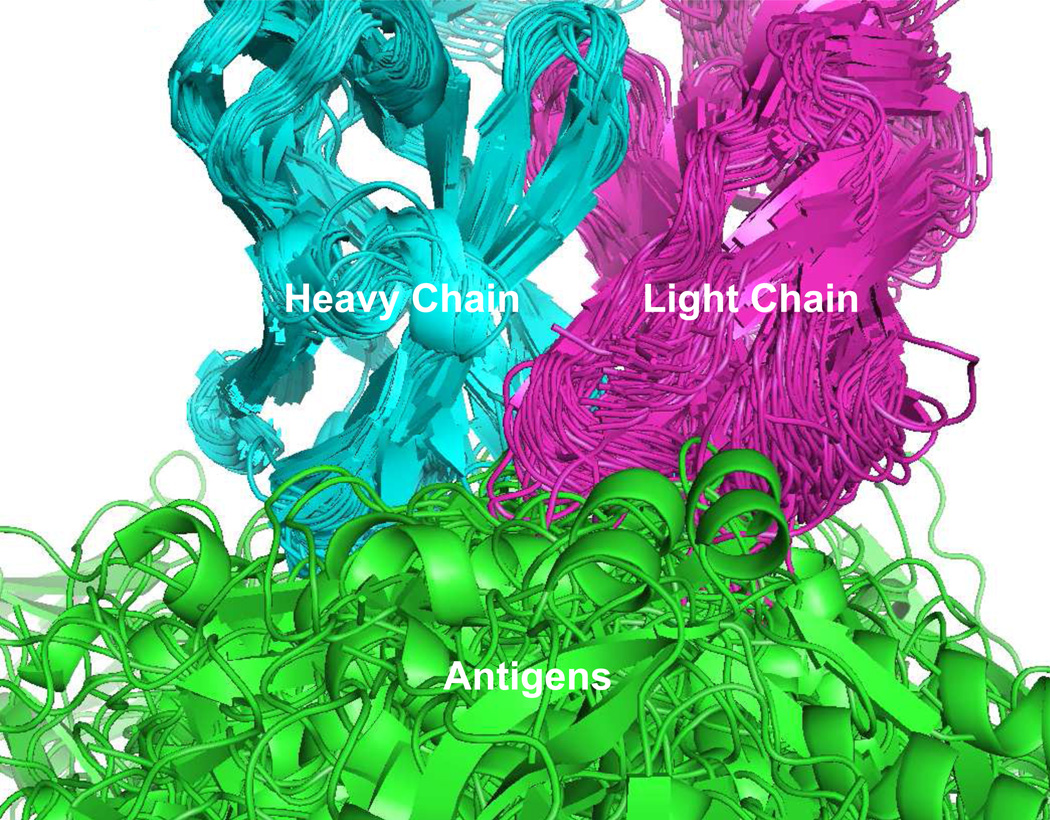
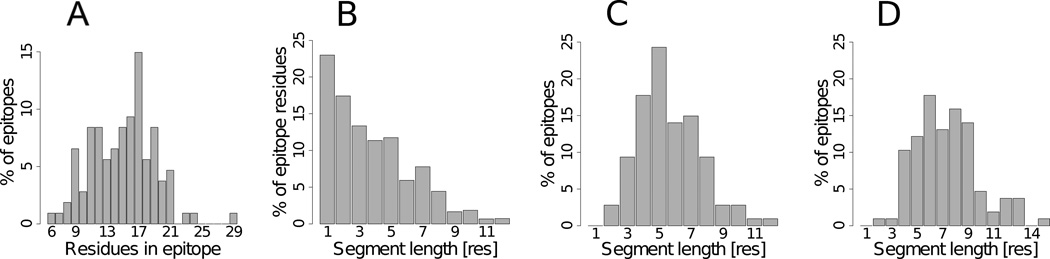
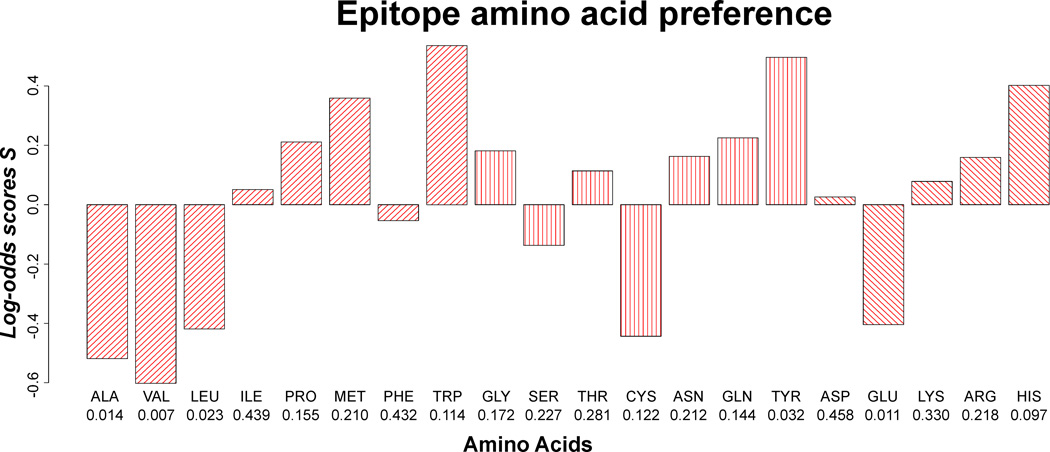
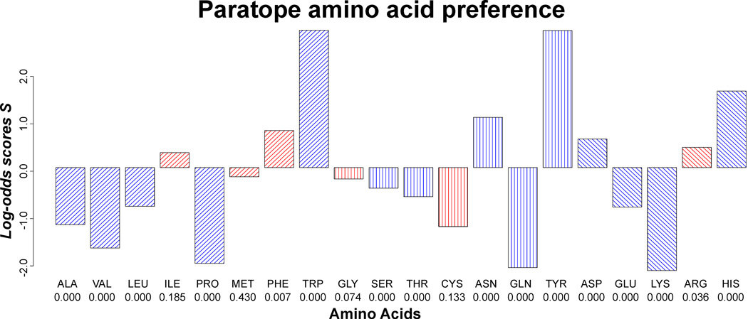
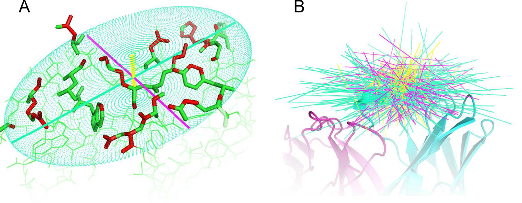
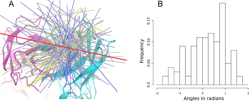
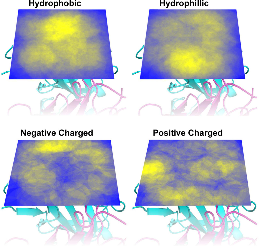
The binding of antigens to antibodies is one of the key events in an immune response against foreign molecules and is a critical element of several biomedical applications including vaccines and immunotherapeutics. For development of such applications, the identification of antibody binding sites (B-cell epitopes) is essential. However experimental epitope mapping is highly cost-intensive and computer-aided methods do in general have moderate performance. One major reason for this moderate performance is an incomplete understanding of what characterizes an epitope. To fill this gap, we here developed a novel framework for comparing and superimposing B-cell epitopes and applied it on a dataset of 107 non-similar antigen:antibody structures extracted from the PDB database. With the presented framework, we were able to describe the general B-cell epitope as a flat, oblong, oval shaped volume consisting of predominantly hydrophobic amino acids in the center flanked by charged residues. The average epitope was found to be made up of ∼15 residues with one linear stretch of 5 or more residues constituting more than half of the epitope size. Furthermore, the epitope area is predominantly constrained to a plane above the antibody tip, in which the epitope is orientated in a -30° to 60° angle relative to the light to heavy chain antibody direction. Contrary to previously findings, we did not find a significant deviation between the amino acid composition in epitopes and the composition of equally exposed parts of the antigen surface. Our results, in combination with previously findings, give a detailed picture of the B-cell epitope that may be used in development of improved B-cell prediction methods.


Figures

Fig. 1
Fig. 1. Alignment of data used in the study
Antibody:Antigen complexes were structural superimposed using the antibody heavy chain as template. For illustrative purpose the number of structures displayed in the figure is limited to 60.
Fig. 2
Fig. 2. Size and segmentation of discontinues epitopes used in the study
A) Distribution of epitope size. B) Distribution of epitope residues segmented by sequential stretches of residues. C) Distribution of maximum sequential stretches of residues in each epitope. D) Same as C, but allowing one non-binding residue in the sequential stretch.
Fig. 3
Fig. 3. Epitope amino acid composition
P-values are stated beneath each amino acid.
Fig. 4
Fig. 4. Paratope amino acid preferences
Log-odds scores significantly different from zero are colered blue, and unsignificant red. P-values are stated beneath each amino acid.
Fig. 5
Fig. 5. Illustration of principle components fitted to epitopes
A) The three principle components (axis of inertia) fitted to the antigen heavy atoms (marked red) in contact with the antibody. Primary axes are shown in green, secondary axes in purple and tertiary axes in yellow. B) Spatial orientation of principle components relative to the antibody. Refer to supplementary materials Figure S2 for an animation of B.
Fig. 6
Fig. 6. Directions of epitope relative to the antibody
A) Cartoon drawing represents the antibody and the red line indicates the antibody light to heavy chain direction. Directions of epitopes are represented by the first principle component fitted to the epitope (see methods) and colored from blue to yellow based on the number of other epitopes pointing in roughly the same direction (within an angle of 0.2 radian). Blue indicates that the epitope points in a less preferred direction and yellow that the epitope points in a preferred direction. B) Histogram of angles between epitope direction and the antibody light to heavy chain vector.
Fig. 7
Fig. 7. Density heatmaps of amino acid position in the epitopes plane above the antibody tip
Areas are colored on a scale from yellow (high density) to blue (low density) (see methods). The antibody structures display are included to enhance visualization.

References

    1. Andersen PH, Nielsen M, Lund O. Prediction of residues in discontinuous B-cell epitopes using protein 3D structures. Protein Sci. 2006;15:2558–2567. - PMC - PubMed
    1. Bhat TN, Bentley GA, Boulot G, Greene MI, Tello D, Dall’Acqua W, Souchon H, Schwarz FP, Mariuzza RA, Poljak RJ. Bound water molecules and conformational stabilization help mediate an antigen-antibody association. Proc. Natl. Acad. Sci. U.S.A. 1994;91:1089–1093. - PMC - PubMed
    1. Bickerton GR, Higueruelo AP, Blundell TL. Comprehensive, atomiclevel characterization of structurally characterized protein-protein interactions: the PICCOLO database. BMC Bioinformatics. 2011;12:313. - PMC - PubMed
    1. Bogan AA, Thorn KS. Anatomy of hot spots in protein interfaces. Journal of Molecular Biology. 1998;280:1–9. - PubMed
    1. Bordner AJ, Abagyan R. Statistical analysis and prediction of proteinprotein interfaces. Proteins. 2005;60:353–366. - PubMed

Publication types

MeSH terms

Substances