

Amyloid-β assessed by florbetapir F 18 PET and 18-month cognitive decline: a multicenter study
- PMID: 22786606
- PMCID: PMC3468774
- DOI: 10.1212/WNL.0b013e3182661f74
Amyloid-β assessed by florbetapir F 18 PET and 18-month cognitive decline: a multicenter study
Abstract
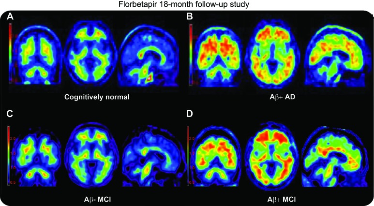
Objectives: Florbetapir F 18 PET can image amyloid-β (Aβ) aggregates in the brains of living subjects. We prospectively evaluated the prognostic utility of detecting Aβ pathology using florbetapir PET in subjects at risk for progressive cognitive decline.
Methods: A total of 151 subjects who previously participated in a multicenter florbetapir PET imaging study were recruited for longitudinal assessment. Subjects included 51 with recently diagnosed mild cognitive impairment (MCI), 69 cognitively normal controls (CN), and 31 with clinically diagnosed Alzheimer disease dementia (AD). PET images were visually scored as positive (Aβ+) or negative (Aβ-) for pathologic levels of β-amyloid aggregation, blind to diagnostic classification. Cerebral to cerebellar standardized uptake value ratios (SUVr) were determined from the baseline PET images. Subjects were followed for 18 months to evaluate changes in cognition and diagnostic status. Analysis of covariance and correlation analyses were conducted to evaluate the association between baseline PET amyloid status and subsequent cognitive decline.
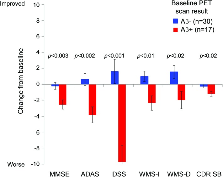
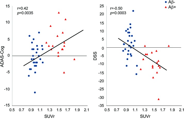
Results: In both MCI and CN, baseline Aβ+ scans were associated with greater clinical worsening on the Alzheimer's Disease Assessment Scale-Cognitive subscale (ADAS-Cog (p < 0.01) and Clinical Dementia Rating-sum of boxes (CDR-SB) (p < 0.02). In MCI Aβ+ scans were also associated with greater decline in memory, Digit Symbol Substitution (DSS), and Mini-Mental State Examination (MMSE) (p < 0.05). In MCI, higher baseline SUVr similarly correlated with greater subsequent decline on the ADAS-Cog (p < 0.01), CDR-SB (p < 0.03), a memory measure, DSS, and MMSE (p < 0.05). Aβ+ MCI tended to convert to AD dementia at a higher rate than Aβ- subjects (p < 0.10).
Conclusions: Florbetapir PET may help identify individuals at increased risk for progressive cognitive decline.
Figures
Comment in
-
Alzheimer disease: Florbetapir-a useful tool to image amyloid load and predict cognitive decline in Alzheimer disease.Nat Rev Neurol. 2012 Sep;8(9):471. doi: 10.1038/nrneurol.2012.162. Epub 2012 Aug 7. Nat Rev Neurol. 2012. PMID: 22868860 No abstract available.
References
-
- The National Institute on Aging and Reagan Institute Working Group on Diagnostic Criteria for the Neuropathological Assessment of Alzheimer's Disease. Neurobiol Aging 1997;18:S1–S2. - PubMed
Publication types
MeSH terms
Substances
Grants and funding
LinkOut - more resources
Full Text Sources
Other Literature Sources