

Diffusion tensor imaging in autism spectrum disorder: a review
- PMID: 22786754
- PMCID: PMC3474893
- DOI: 10.1002/aur.1243
Diffusion tensor imaging in autism spectrum disorder: a review
Abstract
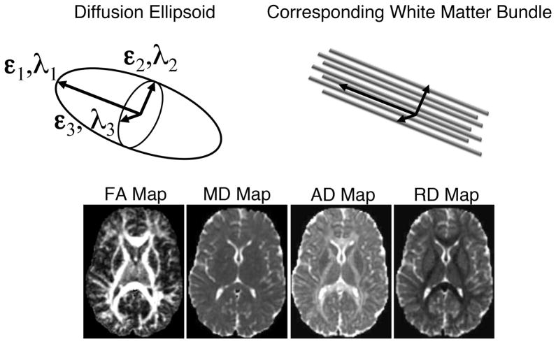
White matter tracts of the brain allow neurons and neuronal networks to communicate and function with high efficiency. The aim of this review is to briefly introduce diffusion tensor imaging methods that examine white matter tracts and then to give an overview of the studies that have investigated white matter integrity in the brains of individuals with autism spectrum disorder (ASD). From the 48 studies we reviewed, persons with ASD tended to have decreased fractional anisotropy and increased mean diffusivity in white matter tracts spanning many regions of the brain but most consistently in regions such as the corpus callosum, cingulum, and aspects of the temporal lobe. This decrease in fractional anisotropy was often accompanied by increased radial diffusivity. Additionally, the review suggests possible atypical lateralization in some white matter tracts of the brain and a possible atypical developmental trajectory of white matter microstructure in persons with ASD. Clinical implications and future research directions are discussed.
© 2012 International Society for Autism Research, Wiley Periodicals, Inc.
Figures
References
-
- Adluru N, Hinrichs C, Chung MK, Lee JE, Singh V, Bigler ED, Alexander AL. Classification in DTI using shapes of white matter tracts. Conference Proceedings: …Annual International Conference of the IEEE Engineering in Medicine and Biology Society. IEEE Engineering in Medicine and Biology Society. Conference; 2009; 2009. pp. 2719–2722. - DOI - PMC - PubMed
-
- Alexander AL, Hasan K, Kindlmann G, Parker DL, Tsuruda JS. A geometric analysis of diffusion tensor measurements of the human brain. Magnetic Resonance in Medicine: Official Journal of the Society of Magnetic Resonance in Medicine/Society of Magnetic Resonance in Medicine. 2000;44(2):283–291. - PubMed
-
- Alexander AL, Hasan KM, Lazar M, Tsuruda JS, Parker DL. Analysis of partial volume effects in diffusion-tensor MRI. Magnetic Resonance in Medicine: Official Journal of the Society of Magnetic Resonance in Medicine/Society of Magnetic Resonance in Medicine. 2001;45(5):770–780. - PubMed
Publication types
MeSH terms
Grants and funding
LinkOut - more resources
Full Text Sources
