

One-electron oxidation of an oxoiron(IV) complex to form an [O═FeV═NR]+ center
- PMID: 22786933
- PMCID: PMC3409744
- DOI: 10.1073/pnas.1206457109
One-electron oxidation of an oxoiron(IV) complex to form an [O═FeV═NR]+ center
Abstract
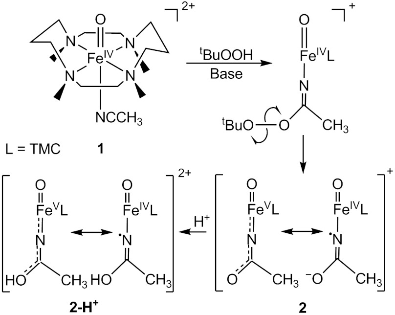
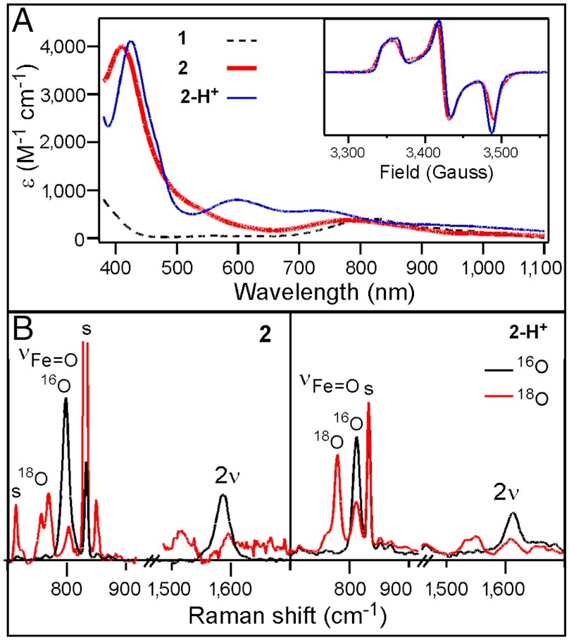
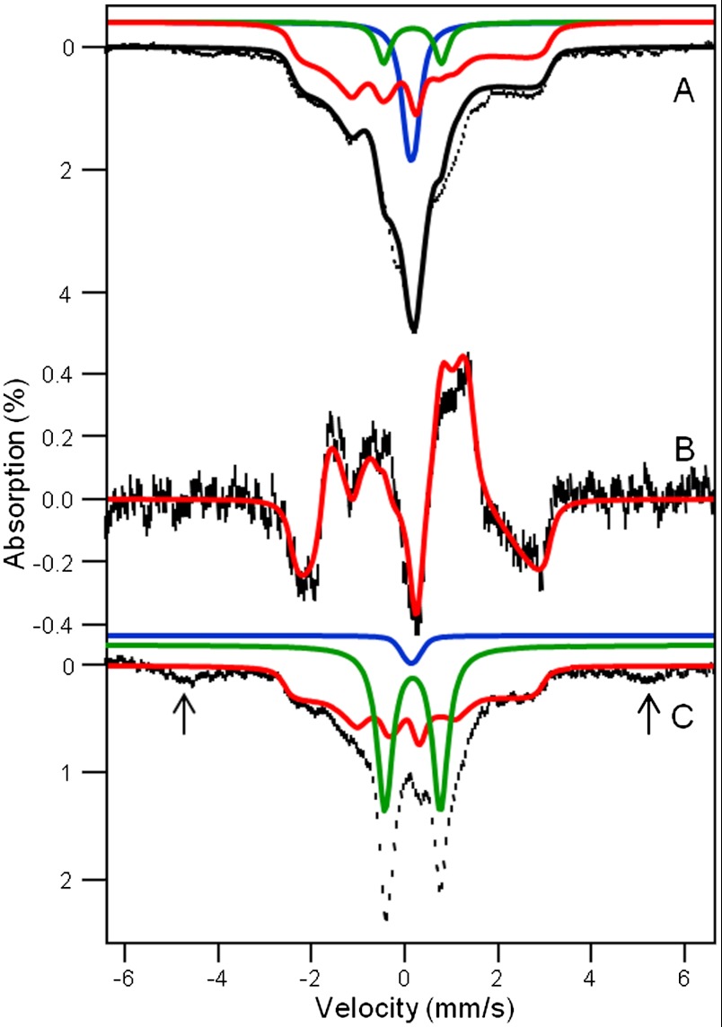
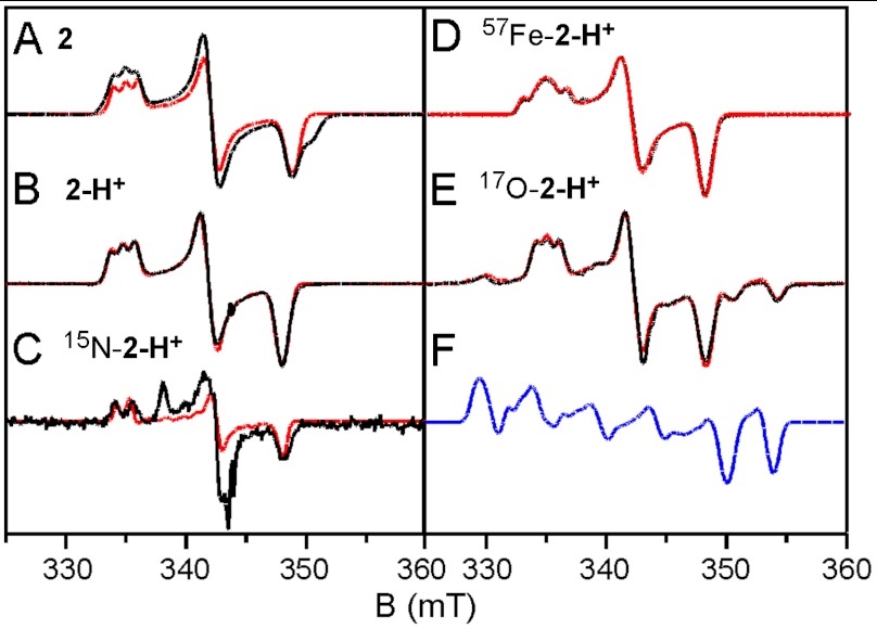
Oxoiron(V) species are postulated to be involved in the mechanisms of the arene cis-dihydroxylating Rieske dioxygenases and of bioinspired nonheme iron catalysts for alkane hydroxylation, olefin cis-dihydroxylation, and water oxidation. In an effort to obtain a synthetic oxoiron(V) complex, we report herein the one-electron oxidation of the S = 1 complex [Fe(IV)(O)(TMC)(NCCH(3))](2+) (1, where TMC is tetramethylcyclam) by treatment with tert -butyl hydroperoxide and strong base in acetonitrile to generate a metastable complex 2 at -44 °C, which has been characterized by UV-visible, resonance Raman, Mössbauer, and EPR methods. The defining spectroscopic characteristic of 2 is the unusual x/y anisotropy observed for the (57)Fe and (17)O A tensors associated with the high-valent Fe═O unit and for the (14)N A tensor of a ligand derived from acetonitrile. As shown by detailed density functional theory (DFT) calculations, the unusual x/y anisotropy observed can only arise from an iron center with substantially different spin populations in the d(xz) and d(yz) orbitals, which cannot correspond to an Fe(IV)═O unit but is fully consistent with an Fe(V) center, like that found for [Fe(V)(O)(TAML)](-) (where TAML is tetraamido macrocyclic ligand), the only well-characterized oxoiron(V) complex reported. Mass spectral analysis shows that the generation of 2 entails the addition of an oxygen atom to 1 and the loss of one positive charge. Taken together, the spectroscopic data and DFT calculations support the formulation of 2 as an iron(V) complex having axial oxo and acetylimido ligands, namely [Fe(V)(O)(TMC)(NC(O)CH(3))](+).
Conflict of interest statement
The authors declare no conflict of interest.
Figures
 ,
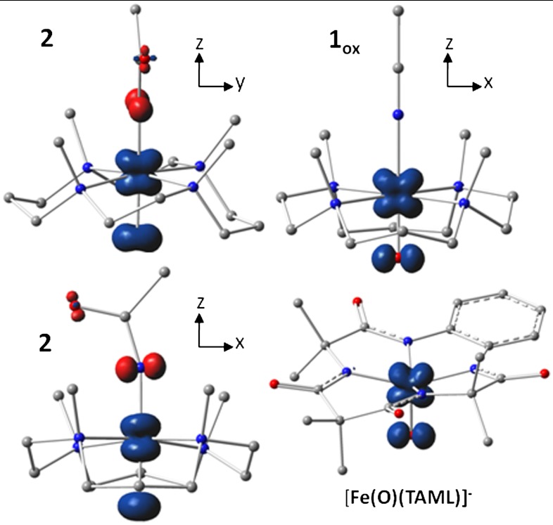
,  , and [Fe(O)(TAML)]-. The plot for
  , shown in two views, reveals the contours of the orbitals carrying spin density. From top to bottom, px (Nam), dyz (Fe), and py (17O). For
  , the TMC ligand has been rotated by approximately 90° around the Fe═O bond relative to the orientation shown for
  . Majority spin α in blue; minority spin β in red.
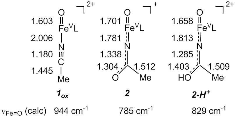
References
-
- Denisov IG, Makris TM, Sligar SG, Schlichting I. Structure and chemistry of cytochrome P450. Chem Rev. 2005;105:2253–2278. - PubMed
-
- Zilly FE, et al. Tuning a P450 enzyme for methane oxidation. Angew Chem Int Ed. 2011;50:2720–2724. - PubMed
-
- Kawakami N, Shoji O, Watanabe Y. Use of perfluorocarboxylic acids to trick cytochrome P450BM3 into initiating the hydroxylation of gaseous alkanes. Angew Chem Int Ed. 2011;50:5315–5318. - PubMed
-
- Rittle J, Green MT. Cytochrome P450 compound I: Capture, characterization, and C─H bond activation kinetics. Science. 2010;330:933–937. - PubMed
Publication types
MeSH terms
Substances
Grants and funding
LinkOut - more resources
Full Text Sources
Medical
Research Materials
