. 2012 Jul 11;32(28):9485-98.
doi: 10.1523/JNEUROSCI.0311-12.2012.

The mechanosensory structure of the hair cell requires clarin-1, a protein encoded by Usher syndrome III causative gene

Affiliations

The mechanosensory structure of the hair cell requires clarin-1, a protein encoded by Usher syndrome III causative gene

Ruishuang Geng et al. J Neurosci. .

Abstract

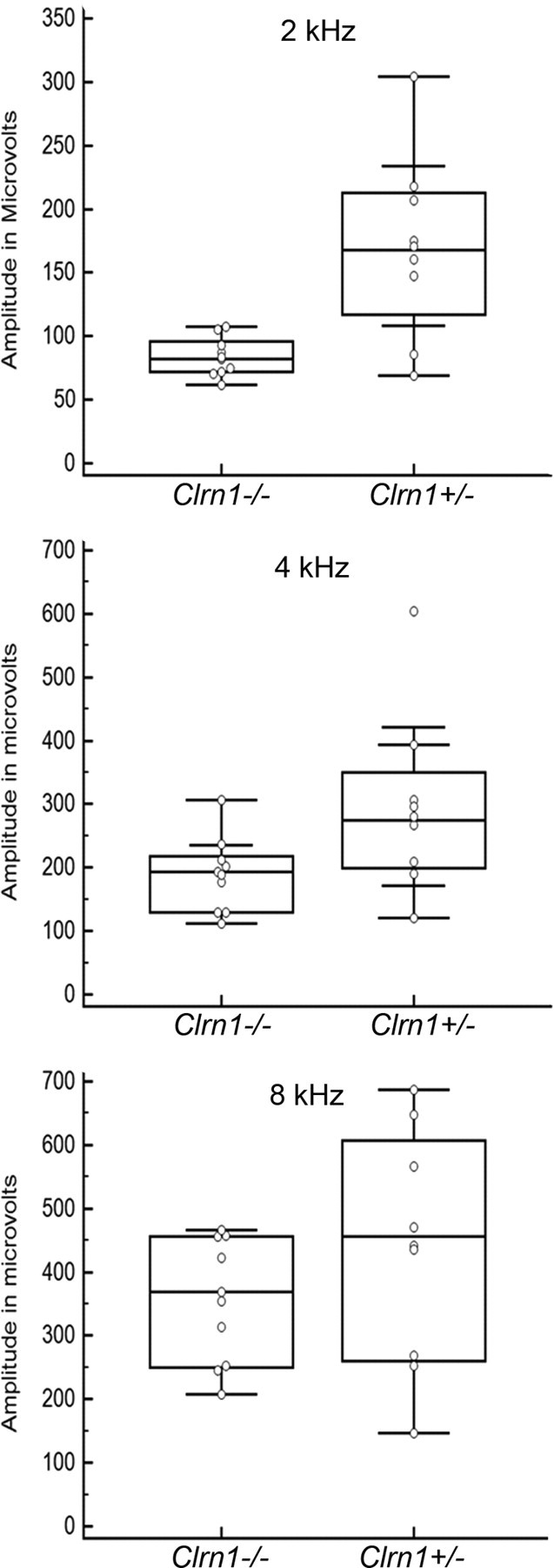
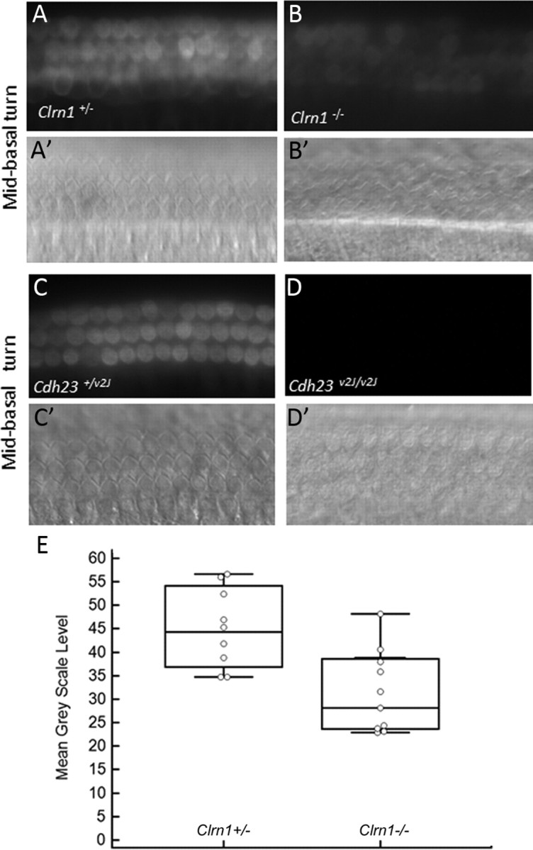
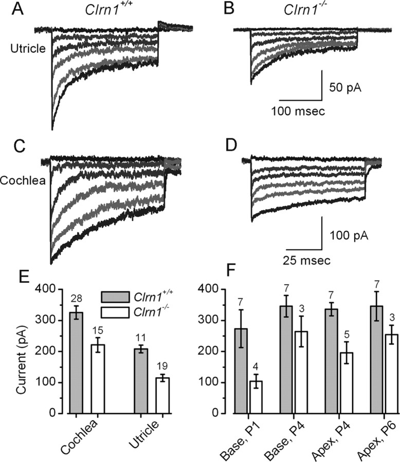
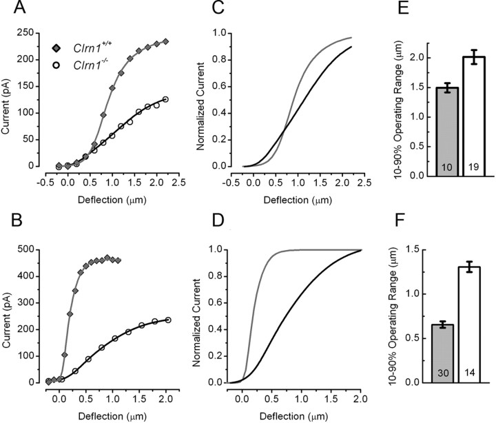
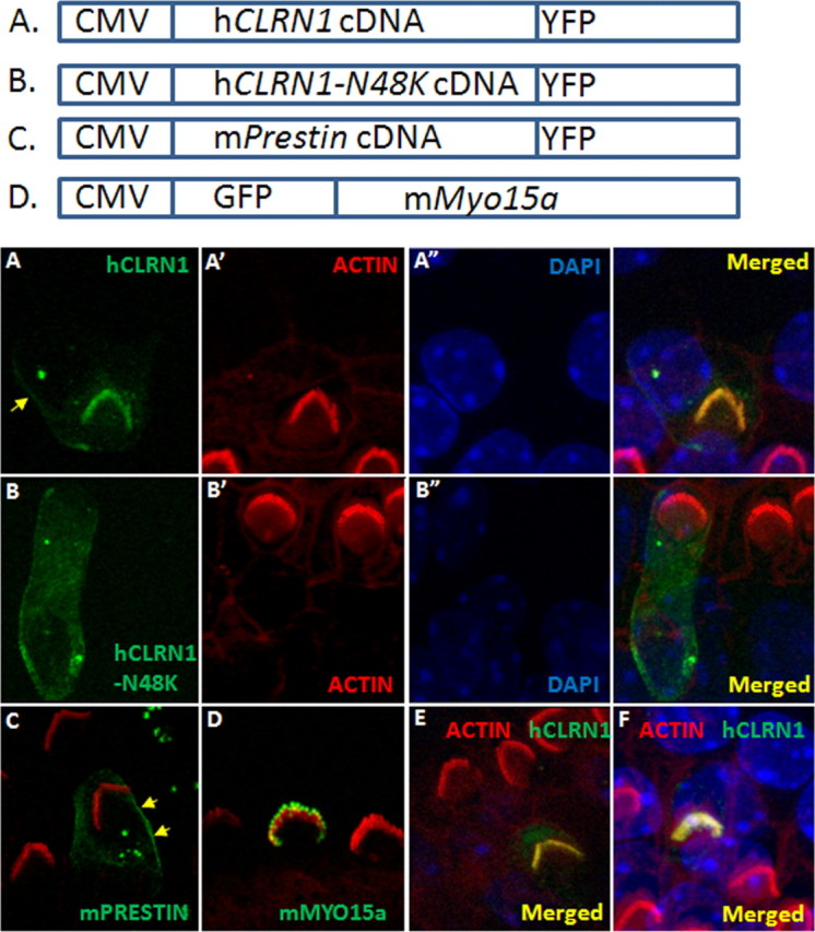
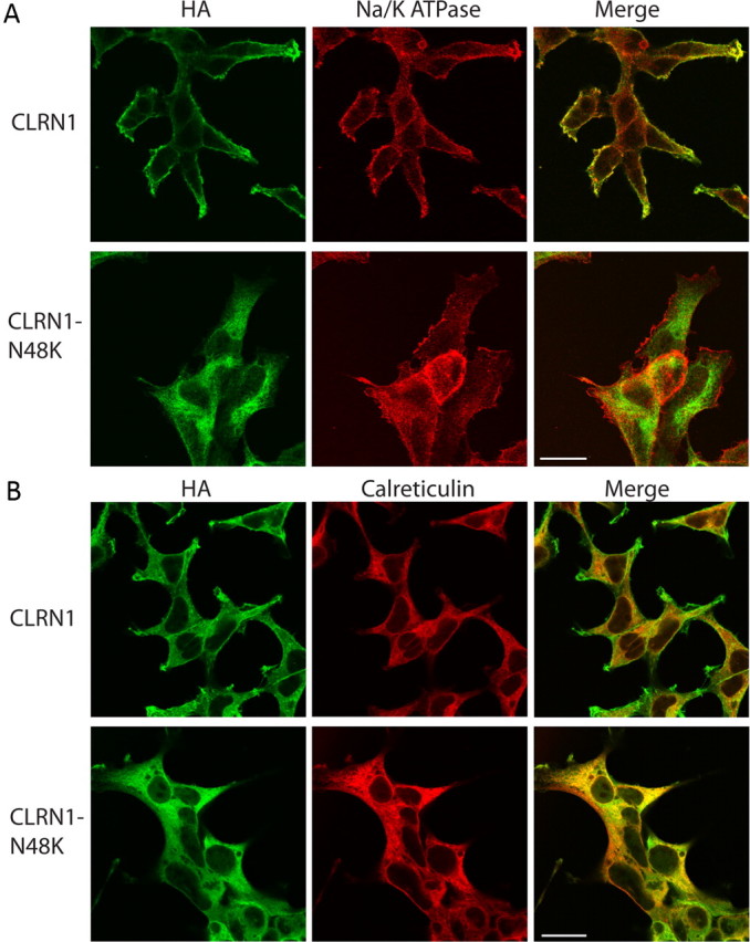
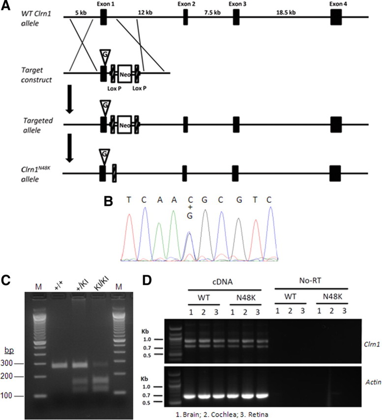
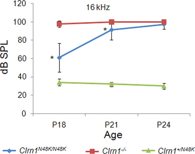
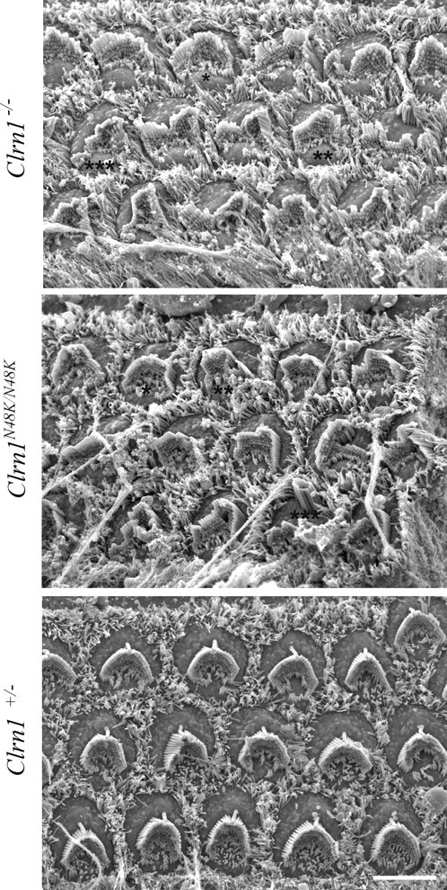
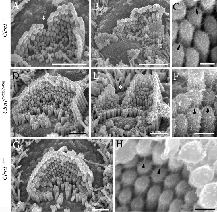
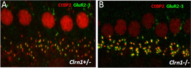
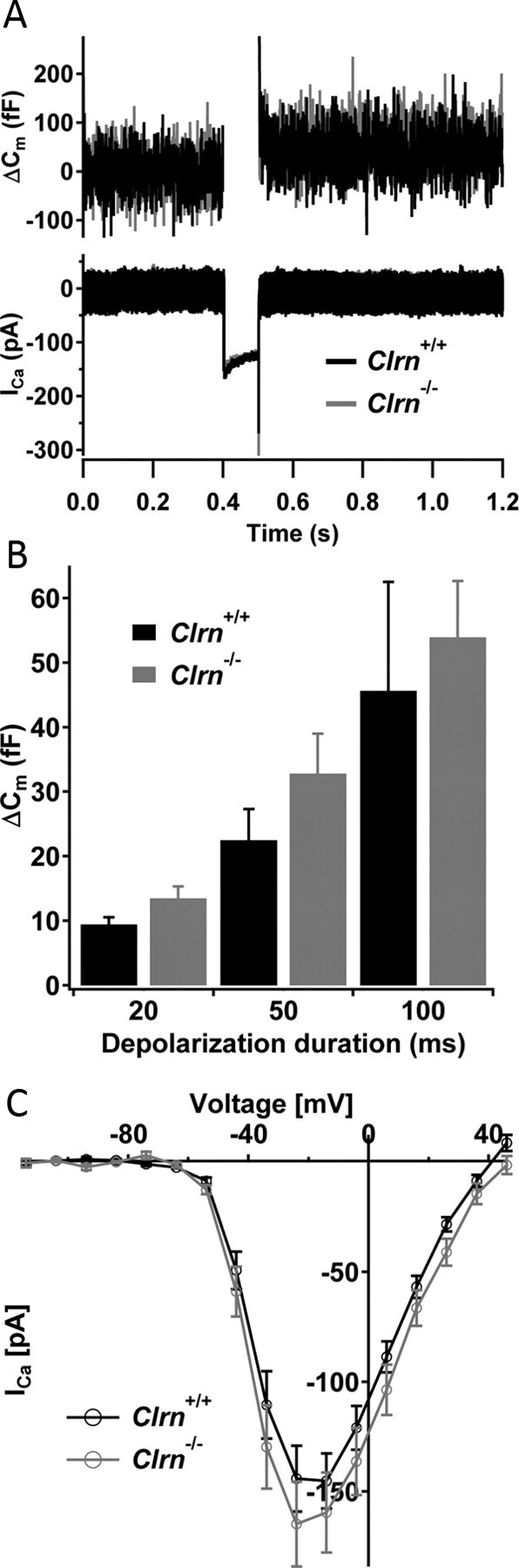
Mutation in the clarin-1 gene (Clrn1) results in loss of hearing and vision in humans (Usher syndrome III), but the role of clarin-1 in the sensory hair cells is unknown. Clarin-1 is predicted to be a four transmembrane domain protein similar to members of the tetraspanin family. Mice carrying null mutation in the clarin-1 gene (Clrn1(-/-)) show loss of hair cell function and a possible defect in ribbon synapse. We investigated the role of clarin-1 using various in vitro and in vivo approaches. We show by immunohistochemistry and patch-clamp recordings of Ca(2+) currents and membrane capacitance from inner hair cells that clarin-1 is not essential for formation or function of ribbon synapse. However, reduced cochlear microphonic potentials, FM1-43 [N-(3-triethylammoniumpropyl)-4-(4-(dibutylamino)styryl) pyridinium dibromide] loading, and transduction currents pointed to diminished cochlear hair bundle function in Clrn1(-/-) mice. Electron microscopy of cochlear hair cells revealed loss of some tall stereocilia and gaps in the v-shaped bundle, although tip links and staircase arrangement of stereocilia were not primarily affected by Clrn1(-/-) mutation. Human clarin-1 protein expressed in transfected mouse cochlear hair cells localized to the bundle; however, the pathogenic variant p.N48K failed to localize to the bundle. The mouse model generated to study the in vivo consequence of p.N48K in clarin-1 (Clrn1(N48K)) supports our in vitro and Clrn1(-/-) mouse data and the conclusion that CLRN1 is an essential hair bundle protein. Furthermore, the ear phenotype in the Clrn1(N48K) mouse suggests that it is a valuable model for ear disease in CLRN1(N48K), the most prevalent Usher syndrome III mutation in North America.


Figures

Figure 1.
Figure 1.
Cochlear microphonics. CM was recorded from Clrn1−/− or Clrn1+/− mice (P18–P19) at 2, 4, and 8 kHz. The graph shows the spread of the data at each frequency tested for Clrn1−/− (n = 9) and Clrn1+/− (n = 8) mice. The results show that, on average, the receptor (hair cell) potential was lower in the Clrn1−/− mice compared with the controls. Amplitude reduction in Clrn1−/− mice was statistically significant at 2 kHz (p = 0.01) and 4 kHz (p = 0.04) but not 8 kHz (p = 0.15).
Figure 2.
Figure 2.
FM1-43 uptake in Clrn1−/− and Cdh23−/− cochlear hair cells in culture at P3. Uptake in Clrn1−/− (B) is diminished compared with the control (A). A′ and B′ are DIC images of A and B showing the location of the hair cells. Hair cells from Cdh23v2J/v2J mice hair cells fail to load with FM1-43 (C, D). C′ and D′ are DIC images of C and D showing the location of the hair cells. When there is complete loss of hair cell function, no dye uptake is observed (no background). E, Quantitative analysis of FM1-43 dye loading in the basal coils of the Clrn1+/− controls (n = 8) and Clrn1−/− mutants (n = 9). The number of hair cells included to determine the mean grayscale levels per tissue is 24. The reduction in dye loading was significant (p = 0.0205). The error bars represent SD.
Figure 3.
Figure 3.
MET currents in Clrn1+/+ and Clrn1−/− hair cells. A–D, Representative families of MET currents evoked by rapid hair bundle deflections that ranged in amplitude from −0.5 to 2 μm. Currents were recorded in voltage-clamp mode (Vhold = −64 mV) from the indicated organs and genotypes between P1 and P6. The scale bars in B and D also apply to A and C, respectively. E, Mean maximal transduction currents measured from 73 hair cells as the difference between the smallest currents evoked by negative bundle deflection and the largest currents evoked by positive deflections. The number of cells for each of the four groups is indicated above. F, Mean maximal transduction currents plotted for different cochlear regions and ages. The number of cells for each group is indicated above.
Figure 4.
Figure 4.
I(X) relationships from P1–P6 hair cells excised from Clrn1+/+ and Clrn1−/− mice. Peak current is plotted as a function of the stimulus amplitude for utricle (A) and cochlea (B) cells, respectively. The data points were fitted with second-order Boltzmann equations. The data shown in A and B were normalized to the maximal current and replotted to illustrate the broader relationships in hair cells from the utricle (C) and cochlea (D) of Clrn1−/− mice. To quantify the sensitivity of the cells, we measured the 10–90% operating range for each of the 73 cells. The mean operating ranges are plotted on the bar graphs for utricle (E) and cochlea (F) hair cells. The number of cells in each group is indicated on the graphs.
Figure 5.
Figure 5.
CLRN1 localizes to the hair bundle. P3 wild-type mouse cochlear epithelia explants transfected with a specific construct (using gene gun) and neighboring nontransfected hair cells after 1 d in culture and ∼20 h after transfection. Expression is driven by the CMV promoter in all cases, and detection of the fluorescent protein marks transfected cells; the tissue was counterstained with phalloidin-conjugated Alexa Fluor 546 (red) and DAPI (blue). A, CLRN1–YFP (green); A′ and A″ are phalloidin and DAPI counterstained images of A. CLRN1–YFP predominantly localized to the bundle with a small amount of the fusion protein localizing to the plasma membrane in the soma (arrow). B, CLRN1N48K–YFP (green); B′ and B″ are phalloidin and DAPI counterstained images of B. C, Merged image showing Prestin–YFP (green)-expressing hair cell and phalloidin-labeled bundles on transfected and nontransfected cells. Arrows point to membrane localization of Prestin–YFP. D, GFP–Myo15a (green)-expressing hair cell and phalloidin-labeled bundles on transfected and nontransfected cells. GFP–Myo15a appears at the tip of the stereocilia as expected. E, F, YFP/phalloidin merged images from two separate (from A) rounds of transfection with CLRN1–YFP construct to show reproducibility of results shown in A.
Figure 6.
Figure 6.
Localization of CLRN1 and its CLRN1N48K mutant heterologously expressed in HEK293 cells. The HEK cell lines stably expressing either wild-type human CLRN1 fused to HA tag (top rows of A and B) or CLRN1N48K fused to HA tag (bottom rows of A and B) were treated with bortezomib (15 nm) for 16 h before fixed by 4% paraformaldehyde. A, The cells were stained by antibodies against HA tag (green) and Na/K ATPase (red), which localizes to the plasma membrane. CLRN1 localizes to the plasma membrane, whereas CLRN1N48K fails to reach the plasma membrane. B, The cells were stained by antibodies against HA tag (green) and calreticulin (red). The majority of CLRN1N48K is observed in the ER and the cytoplasm. Scale bars, 20 μm.
Figure 7.
Figure 7.
Generation of Clrn1N48K knock-in mouse. A, Targeting map for Clrn1N48K knock-in mice. Top, Mouse Clrn1 consists of four exons. Second row, The targeting vector contained 5.7 kb of 5′ and 2.2 kb of 3′ homologous sequence with C-to-G transversion at the codon 48. Third row, As a result of homologous recombination, codon 48 in exon 1 was changed to AAG, and the loxP flanked Neo gene was inserted after exon 1. Bottom row, Neo was removed from targeted locus by cre–loxP recombination. B, DNA sequence analysis confirms mutation at the target nucleotide in exon 1: both wild-type nucleotide C and mutant nucleotide G were observed in a cDNA sample from heterozygous knock-in mice (+/KI). C, The missense mutation introduced a BsaHI restriction site, which was used to distinguish the wild-type (+/+), heterozygous (+/KI), and homozygous (KI/KI) mutants. D, Mouse Clrn1 mRNA is expressed in brain, cochlea, and retina; the upper band (∼850 bp) and the lower band (∼750 bp) seen in the upper half is typical Clrn1 mRNA and alternative splicing expression pattern. Results show that Clrn1 mRNA expression is not affected by the missense mutation. WT, Wild type.
Figure 8.
Figure 8.
Assessment of hearing in Clrn1N48/K/N48K knock-in compared with the Clrn1−/− mice over time. The plot shows that mean ABR thresholds of Clrn1N48K/N48K mice (n = 25) are significantly elevated compared with the thresholds of Clrn1+/N48K mice (n = 6), which displayed wild-type thresholds at all time points tested. The plot also shows that ABR thresholds of Clrn1N48K/N48K mice are significantly lower compared with Clrn1−/− mice (n = 12) at P18 and P21; the difference is not significant at P24 because mice from both groups display profound hearing loss at P24. *p < 0.0001.
Figure 9.
Figure 9.
Scanning electron microscopy of the organ of Corti showing regions of ∼15 OHCs each from the Clrn1−/−, Clrn1N48K/N48K, and Clrn1+/− mice. Mild (*), moderate (**), and severe (***) bundle disruption can be seen in both the Clrn1−/− and Clrn1N48/K/N48K panels compared with the Clrn1+/− panel. However, the organ of Corti from the Clrn1−/− mouse showed a greater number of moderately to severely disturbed hair bundles compared with that of the Clrn1N48/K/N48K mouse. Scale bar, 5 μm.
Figure 10.
Figure 10.
Scanning electron microscopy of P3 clarin knock-out (top row), knock-in (middle row), and heterozygote (bottom row) organ of Corti. The Clrn1−/− and Clrn1N48K /N48K both show substantial but variable disruption of the bundle, the former to a greater degree than the latter as exemplified by B and E. The disruption includes splits in the bundle (*) and loss of some of the tallest stereocilia in particular. At a higher magnification (C, F), tip-link-like links (arrows) and abundant lateral links typical of this age are evident in both. The heterozygote shows normal bundle features (G) and links (H; tip-link-like links highlighted with arrows). Scale bars: A, 2 μm; B, 1.5 μm; D, E, G, 1 μm; C, F, H, 200 nm.
Figure 11.
Figure 11.
Light microscopy analysis of ribbon synapse in Clrn1−/− mice. Whole mounts of organs of Corti from P15 mice are double immunolabeled for CtBP2 and GluR2/3 and analyzed by confocal microscopy, followed by deconvolution and surface reconstruction. The difference in the number of double-labeled spots in Clrn1+/− (A) and Clrn1−/− (B) mice was not significant (for quantitative analysis, see Table 2).
Figure 12.
Figure 12.
Patch-clamp analysis of presynaptic IHC function. A, Representative exocytic membrane capacitance changes (ΔCm, top) and Ca2+ currents (ICa, bottom) in response to 50 ms depolarization to −14 mV in Clrn1−/− (gray) and Clrn1+/+ (black) IHCs. B, Current–voltage relationship for Cav1.3 Ba2+ currents in control (black, n = 12 IHCs) and mutant (n = 10 IHCs) IHCs. Currents were evoked by 10 ms step depolarizations from a resting potential of −84 mV to variable potentials, and their average of the last 2 ms of current (excluding the tail current) was plotted against the test potential. C, Average ΔCm responses of control IHCs (7, 8, and 10 IHCs for 20, 50, and 100 ms depolarizations) and mutant IHCs (5, 6, and 8 IHCs for 20, 50, and 100 ms depolarizations) to 50 ms depolarizations to −14 mV. All responses of B and C are given as grand averages (calculated from the means of the individual cells) ± SEM.

References

    1. Adato A, Kalinski H, Weil D, Chaib H, Korostishevsky M, Bonne-Tamir B. Possible interaction between USH1B and USH3 gene products as implied by apparent digenic deafness inheritance. Am J Hum Genet. 1999;65:261–265. - PMC - PubMed
    1. Adato A, Vreugde S, Joensuu T, Avidan N, Hamalainen R, Belenkiy O, Olender T, Bonne-Tamir B, Ben-Asher E, Espinos C, Millán JM, Lehesjoki AE, Flannery JG, Avraham KB, Pietrokovski S, Sankila EM, Beckmann JS, Lancet D. USH3A transcripts encode clarin-1, a four-transmembranedomain protein with a possible role in sensory synapses. Eur J Hum Genet. 2002;10:339–350. - PubMed
    1. Ahmed ZM, Goodyear R, Riazuddin S, Lagziel A, Legan PK, Behra M, Burgess SM, Lilley KS, Wilcox ER, Riazuddin S, Griffith AJ, Frolenkov GI, Belyantseva IA, Richardson GP, Friedman TB. The tip-link antigen, a protein associated with the transduction complex of sensory hair cells, is protocadherin-15. J Neurosci. 2006;26:7022–7034. - PMC - PubMed
    1. Alagramam KN, Murcia CL, Kwon HY, Pawlowski KS, Wright CG, Woychik RP. The mouse Ames waltzer hearing-loss mutant is caused by mutation of Pcdh15, a novel protocadherin gene. Nat Genet. 2001;27:99–102. - PubMed
    1. Alagramam KN, Stahl JS, Jones SM, Pawlowski KS, Wright CG. Characterization of vestibular dysfunction in the mouse model for Usher syndrome 1F. J Assoc Res Otolaryngol. 2005;6:106–118. - PMC - PubMed

Publication types

MeSH terms