. 2012 Jul 11;32(28):9588-600.
doi: 10.1523/JNEUROSCI.5977-11.2012.

Blood-brain barrier permeability is increased after acute adult stroke but not neonatal stroke in the rat

Affiliations

Blood-brain barrier permeability is increased after acute adult stroke but not neonatal stroke in the rat

David Fernández-López et al. J Neurosci. .

Abstract

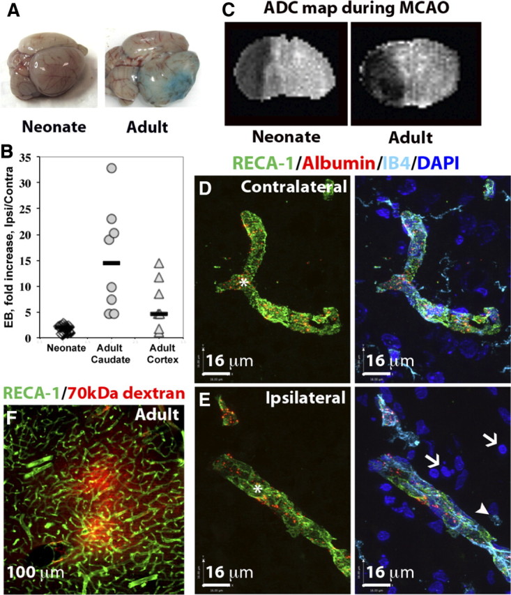
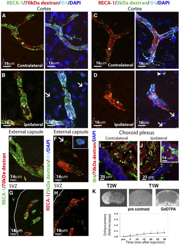
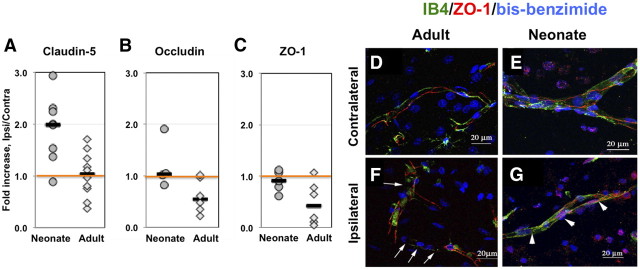
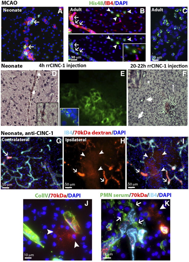
The immaturity of the CNS at birth greatly affects injury after stroke but the contribution of the blood-brain barrier (BBB) to the differential response to stroke in adults and neonates is poorly understood. We asked whether the structure and function of the BBB is disrupted differently in neonatal and adult rats by transient middle cerebral artery occlusion. In adult rats, albumin leakage into injured regions was markedly increased during 2-24 h reperfusion but leakage remained low in the neonates. Functional assays employing intravascular tracers in the neonates showed that BBB permeability to both large (70 kDa dextran) and small (3 kDa dextran), gadolinium (III)-diethyltriaminepentaacetic acid tracers remained largely undisturbed 24 h after reperfusion. The profoundly different functional integrity of the BBB was associated with the largely nonoverlapping patterns of regulated genes in endothelial cells purified from injured and uninjured adult and neonatal brain at 24 h (endothelial transcriptome, 31,042 total probe sets). Within significantly regulated 1266 probe sets in injured adults and 361 probe sets in neonates, changes in the gene expression of the basal lamina components, adhesion molecules, the tight junction protein occludin, and matrix metalloproteinase-9 were among the key differences. The protein expression of collagen-IV, laminin, claudin-5, occludin, and zonula occludens protein 1 was also better preserved in neonatal rats. Neutrophil infiltration remained low in acutely injured neonates but neutralization of cytokine-induced neutrophil chemoattractant-1 in the systemic circulation enhanced neutrophil infiltration, BBB permeability, and injury. The markedly more integrant BBB in neonatal brain than in adult brain after acute stroke may have major implications for the treatment of neonatal stroke.


Figures

Figure 1.
Figure 1.
Blood–brain barrier permeability is markedly increased in injured brain regions in the adult but is largely preserved in the neonate after acute MCAO. A, Representative whole brains showing Evans blue extravasation and accumulation in neonatal and adult brain 24 h after reperfusion. B, Quantification of intravenously administered Evans blue in the brain between 2 and 24 h after reperfusion. Evans blue accumulation is profoundly increased in the adult but not in the neonatal rat. Numbers show how many times greater the accumulation of Evans blue occurred in the injured region as compared to the contralateral region in the same rat. Shown are data for individual rats; horizontal bars indicate medians. C, Apparent diffusion coefficient (ADC) maps showing a similar extension of brain edema during MCAO in neonate and adult brains. D, E, The spatial distribution of intravenously administered Alexa-555-conjugated albumin in contralateral (D) and injured (E) cortical regions in neonates 24 h after reperfusion. Injured areas in the ipsilateral hemisphere (E) were identified by the presence of pyknotic nuclei (DAPI, white arrows) and round, ameboid-like IB4+/RECA-1 microglia/macrophages (white arrowheads). In the injured areas, intravenous tracers colocalized with brain vessels (RECA1+/IB4+, asterisk) and were not observed in the extravascular spaces or in phagocytic microglia/macrophages (white arrowheads). Green, RECA-1; turquoise, IB4; blue, DAPI. Sections are 12 μm thick. F, Extravasation of Alexa-555-conjugated albumin into the injured cortex of adult rats 24 h after reperfusion (section thickness, 50 μm).
Figure 2.
Figure 2.
Extravasation of intravascular tracers of different size is low in the injured regions of the neonate after acute MCAO. A–D, The distribution of TRITC-conjugated 70 kDa dextran (A, B) and fluorescein-conjugated 3 kDa dextran (C, D) was restricted to the vasculature (RECA-1+/IB4+ vessels, asterisks) in the contralateral (A, C) brain hemisphere. In the injured areas (B, D), intravenous tracers colocalized with brain vessels (RECA1+/IB4+, asterisks) and were not observed in the extravascular spaces. E, F, The distribution of 3 and 70 kDa dextran was restricted to the brain vasculature in the ipsilateral external capsule 24 h after reperfusion (E, F), although occasional 3 kDa signal was detected in microglia/macrophages located in this brain region (F, arrow and inset). G, H, No leakage of 70 kDa dextran (G) or 3 kDa dextran (H) was observed in the ipsilateral SVZ 24 h after reperfusion. I, J, Leakage of 70 kDa dextran was detected in the choroid plexus in both contralateral (I) and ipsilateral (J) ventricles. K, Coronal T1-weighted images showing contrast imaging of Gd-DTPA 24 h after neonatal MCAO. Contrast was increased in areas outside the brain (right) but was minimal in the brain up to 30 min after injection of Gd-DTPA (right vs middle, bottom graph). Injured areas were identified in the same animals by T2-weighted imaging (left). V, ventricle.
Figure 3.
Figure 3.
Differential effect of acute focal stroke on endothelial gene expression in adult and neonatal rats. The endothelial transcriptome data sets (31,042 probe sets) were obtained in endothelial cells purified by sequential negative and positive selection from injured and contralateral tissue of adults and neonates 24 h after reperfusion. Shown are genes with >2-fold change in expression compared to that in contralateral hemisphere.
Figure 4.
Figure 4.
The effect of brain maturation on the expression of BBB proteins. A, Representative Western blots showing the expression of basal lamina proteins (Col-IV and laminin), TJ proteins (occludin and claudin-5), and the pericyte marker PDGFRβ in naive developing (P7–P17) and adult rats. B–F, Densitometric analysis of Western blots showing reduced expression of the basal lamina proteins laminin (B) and Col-IV (C) and the TJ proteins occludin (D) and claudin-5 (E) and increased expression of PDGFRβ (F) in the adult brain compared to the developing postnatal brain. Data are normalized for β-actin expression and expressed as fold-increase versus P7 brains.
Figure 5.
Figure 5.
Expression of occludin, ZO-1, and claudin-5 is better preserved in immature than in adult brain after acute focal stroke. A–C, Protein expression of claudin-5 (A), occludin (B), and ZO-1 (C) measured by Western blot in whole tissue lysates obtained from injured and matching contralateral brain regions. D–G. ZO-1 (red) is expressed as continuous strands around the vessel (green, IB4) in the contralateral hemisphere regardless of age (D, adult; E, neonate). Continuous strands of ZO-1 are seen in ischemic tissue in neonatal rat (G), while disrupted pattern of ZO-1 expression is observed in adult rat (F, arrows) 24 h after reperfusion. Note that both IB4 and ZO-1 expression are disrupted in adult ischemic brain (F). Images are 3D reconstructions from a z-stack of images captured with a Zeiss LSM confocal microscope using the 63× objective and a 0.5 μm step. IB4 (green) identifies vessels and DAPI shows nuclei morphology.
Figure 6.
Figure 6.
Stroke differentially affects the expression of basal lamina proteins in the neonatal and adult brain. A–E, Immunofluorescence of the brain vasculature (RECA-1, green) showing higher vascular density in the contralateral caudate of adults compared to neonates (C vs A; E) 24 h after stroke. Vascular density was unaffected by stroke at both ages in the ipsilateral injured caudate (B vs A; D vs C; E). Data are expressed as percentage of the total sampled brain volume in three different Z-stacks per brain and region. F, G, The size distribution of RECA-1 vessels in the adult (F) and the neonate (G). H--L. Immunofluorescence of Col-IV (red) showing increased coverage of brain vessels (expressed as the ratio between Col-IV and RECA-1 volume densities) in the contralateral neonatal brain compared to the adult brain (H vs J; L). Coverage of brain vessels by Col-IV increased in the injured caudate of adults (K vs J; L), but remained unchanged in neonates (I vs H; L). M, N, The distribution of Col-IV/RECA-1 positive vessels in the adult (M) and the neonate (N). Note preferential loss of Col-IV coverage of smaller size vessels in the adult but mostly unaffected coverage in injured neonatal brain. *p < 0.05 for same size vessels in ipsilateral versus contralateral hemisphere. O, R, Immunofluorescence of the basal lamina protein laminin (red) showing increased coverage of brain vessels (expressed as the ratio between laminin and RECA-1 volume densities) in the contralateral neonatal brain compared to the adult brain (O vs Q; S). Coverage of brain vessels by laminin was not significantly affected after MCAO (P vs O; R vs Q; S).
Figure 7.
Figure 7.
Neutrophil transmigration is limited after acute neonatal stroke. A, Neutrophil transmigration does not occur within 24 h after MCAO but His48+ cells are seen within vessels in animals without intracardiac perfusion (IB4, red; His48+ cells, green). B, C, Neutrophils (His48, green) are present in the brain parenchyma (B, arrowheads), meninges (C), and in a subpopulation of brain vessels (IB4, red) (B, arrows) in adult animals 4 h after reperfusion. D, E, Neutrophils are observed on H&E (D) and His48-immunofluorescence image (E) 4 h after intracerebral injection of rrCINC-1. Arrows in D point at neutrophils. His48+ cells are seen along the needle track and in parenchyma, but only a few His48+ neutrophils transmigrate following intracerebral rrCINC-1 injection (E). F, H&E staining showing infiltration of neutrophils in the brain 20–22 h after intracerebral injection of rrCINC-1. Neutrophils are observed in association with brain vessels (thin arrows) and in the brain parenchyma (thick arrows). Asterisk indicates the region magnified in the upper-left inset. G–I, Large areas of 70 kDa dextran (red) extravasation (H, I, arrowheads) were observed in the injured cortex of neonate rats injected with anti-CINC-1 15 min before MCAO. Increased accumulation of 70 kDa dextran (red) was also observed in brain vessels (arrows) in both the contralateral (G) and ipsilateral (H, I) cortex. (section thickness, 50 mm). J, Detail of a leaking vessel (70 kDa dextran, red, arrowheads) in a neonate rat injected with anti-CINC-1. K, Neutrophils (anti-PMN serum, green, arrows) associated with a leaking vessel (70 kDa dextran, red, arrowhead) in the injured cortex of a neonate rat injected with anti-CINC-1.

References

    1. Afshar-Kharghan V, Thiagarajan P. Leukocyte adhesion and thrombosis. Curr Opin Hematol. 2006;13:34–39. - PubMed
    1. Anthony DC, Bolton SJ, Fearn S, Perry VH. Age-related effects of interleukin-1 beta on polymorphonuclear neutrophil-dependent increases in blood-brain barrier permeability in rats. Brain. 1997;120(Pt 3):435–444. - PubMed
    1. Anthony D, Dempster R, Fearn S, Clements J, Wells G, Perry VH, Walker K. CXC chemokines generate age-related increases in neutrophil-mediated brain inflammation and blood-brain barrier breakdown. Curr Biol. 1998;8:923–926. - PubMed
    1. Armulik A, Genové G, Mäe M, Nisancioglu MH, Wallgard E, Niaudet C, He L, Norlin J, Lindblom P, Strittmatter K, Johansson BR, Betsholtz C. Pericytes regulate the blood-brain barrier. Nature. 2010;468:557–561. - PubMed
    1. Barone FC, Hillegass LM, Price WJ, White RF, Lee EV, Feuerstein GZ, Sarau HM, Clark RK, Griswold DE. Polymorphonuclear leukocyte infiltration into cerebral focal ischemic tissue: myeloperoxidase activity assay and histologic verification. J Neurosci Res. 1991;29:336–345. - PubMed

Publication types

MeSH terms