. 2012 Jul 11;32(28):9613-25.
doi: 10.1523/JNEUROSCI.0602-12.2012.

Intracellular amyloid precursor protein sorting and amyloid-β secretion are regulated by Src-mediated phosphorylation of Mint2

Affiliations

Intracellular amyloid precursor protein sorting and amyloid-β secretion are regulated by Src-mediated phosphorylation of Mint2

Jeremy Chaufty et al. J Neurosci. .

Abstract

Mint adaptor proteins bind to the membrane-bound amyloid precursor protein (APP) and affect the production of pathogenic amyloid-β (Aβ) peptides related to Alzheimer's disease (AD). Previous studies have shown that loss of each of the three Mint proteins delays the age-dependent production of amyloid plaques in transgenic mouse models of AD. However, the cellular and molecular mechanisms underlying Mints effect on amyloid production are unclear. Because Aβ generation involves the internalization of membrane-bound APP via endosomes and Mints bind directly to the endocytic motif of APP, we proposed that Mints are involved in APP intracellular trafficking, which in turn, affects Aβ generation. Here, we show that APP endocytosis was attenuated in Mint knock-out neurons, revealing a role for Mints in APP trafficking. We also show that the endocytic APP sorting processes are regulated by Src-mediated phosphorylation of Mint2 and that internalized APP is differentially sorted between autophagic and recycling trafficking pathways. A Mint2 phosphomimetic mutant favored endocytosis of APP along the autophagic sorting pathway leading to increased intracellular Aβ accumulation. Conversely, the Mint2 phospho-resistant mutant increased APP localization to the recycling pathway and back to the cell surface thereby enhancing Aβ42 secretion. These results demonstrate that Src-mediated phosphorylation of Mint2 regulates the APP endocytic sorting pathway, providing a mechanism for regulating Aβ secretion.


Figures

Figure 1.
Figure 1.
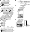
APP internalization is reduced in conditional Mint knock-out (KO) neurons. MTFtg neurons were infected with either lentiviral inactive (Control) or active cre-recombinase to delete all Mint proteins (Mint KO), respectively. A, B, Representative immunoblots and quantification for surface biotinylated proteins that were internalized for 30 min at 37°C and precipitated with Neutravidin agarose beads. Precipitated proteins were blotted for internalized APP (APPI) and GluA1. Input lysates were probed for APPT and Tubulin. Efficient knock-out of Mint proteins were represented in lanes 2, 4, and 6 reflecting Mint1, 2 and 3 protein levels. The amount of internalized APP was taken as the band intensity of APPI relative to APPT present in the lysates and expressed as percentage control. C, D, Representative immunoblots and quantification for control and Mint KO neurons that have been surface biotinylated. Precipitated proteins and total input lysates were immunoblotted for APP and the amount of surface APP (APPS) was taken as the measure of band intensity relative to APPT and expressed as percentage control. E, Live cell endocytosis assay of MTFtg neurons incubated with an extracellular N-terminal APP antibody (22C11) and internalized for 30 min before stripping of excess surface antibody and fixation. F, Internalized APP immunostaining was visualized by confocal microscopy and quantification of APP internalized was taken as a measure of pixel intensity within a defined soma area and expressed as percentage control (n = 3/22 represents number of independent experiments and total number of neurons assessed). G, Live cell recycling assay of MTFtg neurons. After blocking existing cell surface APPs with primary antibody recognizing the N-terminal extracellular domain of APP and cold (non-fluorescence-conjugated) secondary antibody, neurons were incubated at 37°C for 30 min. The newly inserted cell surface APPs were visualized using confocal microscopy. Representative images signify newly inserted cell surface APP from orthogonal projections of confocal z-stacks. H, Newly inserted cell surface APP was visualized by confocal microscopy and quantification of APP reinsertion was taken as a measure of pixel intensity within a defined soma area and expressed as percentage control (n = 2/17 represents number of independent experiments and total number of neurons). *p < 0.05, ***p < 0.005. To avoid bias, the investigator was blind to whether cultured neurons were infected with functional or inactive cre recombinase. Scale bar, 20 μm.
Figure 2.
Figure 2.
Mints are differentially phosphorylated by Src family of kinases in vitro. HEK293T cells were cotransfected with plasmids expressing Src family kinases and putative substrates. A, B, Representative immunoblots of cell lysates expressing constitutively active c-Src, kinase-deficient c-Src (Src inactive), Fyn, Yes, or Lyn kinases with Disabled protein Dab1, Mint1, Mint2 or Mint3 and blotted with a phosphotyrosine-specific antibody 4G10. C, HEK293T cells were transfected with Mint1 or Mint2 in the presence of active or inactive Src kinase or transfected with only a single plasmid. Phosphotyrosine proteins were immunoprecipitated using phosphotyrosine-specific antibody 4G10. Precipitated proteins were analyzed by immunoblotting with Mint1 (lanes 1–5) or Mint2 (lanes 6–10) antibody. D, Phosphotyrosine proteins were immunoprecipitated using phosphotyrosine-specific antibody 4G10 from primary neuronal cultures. Precipitated proteins were analyzed by immunoblotting with Mint1 or Mint2 antibody. E, Representative immunoblots of cell lysates expressing constitutively active c-Src, Fyn, Yes, or Lyn kinases with Mint1, APPswe and wild-type APP and blotted with 4G10. F, Postulated tyrosine phosphorylation sites for Mint2 by Src kinase at amino acid positions 86, 110 and 193 in rat Mint2 was identified. Below are recombinant GST-Mint2 truncated fusion proteins that encompass the Mint2 protein in overlapping fragments: N-terminal region (GST-Mint2-N; residues 1–399), PTB domain (GST-Mint2-PTB; residues 363–546) and two tandem PDZ domains (GST-Mint2-C; residues 546–750). G, In vitro phosphorylation assay combining purified Src with GST-Mint2 fusion proteins described in F. Coomassie Brilliant Blue (CBB) staining indicates equal amount of GST protein. H, Representative immunoblots of lysates from HEK293T cells cotransfected with Src kinase and Mint2 wild-type (Mint2-WT, lanes 1 and 4), or Mint2 phospho-mutants where tyrosines 86, 110 and 193 were mutated to glutamic acid (Mint2-Y3E, lanes 2 and 5) or phenylalanine (Mint2-Y3F, lanes 3 and 6). I, Relative phosphotyrosine immunoreactivity are quantified by normalizing mean pixel intensity of the 4G10 signal to that of Tubulin and expressed as percentage of control. **p < 0.01, ***p < 0.005.
Figure 3.
Figure 3.
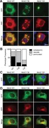
Mint2 phospho-mutants affect APP trafficking. A, COS7 cells were cotransfected with APP and GFP-Mint2-WT, GFP-Mint2-Y3E or GFP-Mint2-Y3F. Representative images of the subcellular localization of GFP-Mint2-WT and phospho-mutants (green), APP immunoreactivity (red) and DAPI nuclear staining (blue) are shown. The top row shows cytoplasmic diffuse localization of both proteins. The middle row represents GFP-Mint2-Y3E colocalized with APP in cytoplasmic vacuoles, whereas the bottom row shows GFP-Mint2-Y3F is sequestered to a distinct perinuclear compartment that differs from that of APP. Phenotypes were assessed in 120 randomly selected cells. B, Quantification representing phenotype frequency as described in A. C, COS7 cells transfected with only GFP-Mint2-WT, GFP-Mint2-Y3E or GFP-Mint2-Y3F did not show any alterations in Mint2 subcellular distribution suggesting that the localization changes observed in A were due to an interaction between Mint2 and APP. D, COS7 cells were cotransfected with a mutant APP plasmid lacking the last 15 aa in the C-terminal region of APP (APPΔC15) and GFP-Mint2-WT, GFP-Mint2-Y3E or GFP-Mint2-Y3F. Scale bars, 20 μm.
Figure 4.
Figure 4.
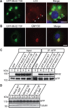
Mint2 phospho-mutants differentially affect APP localization without altering binding affinity to APP. A, COS7 cells were cotransfected with APP and GFP-Mint2-Y3E and stained with various intracellular markers. LC3-positive autophagosome marker colocalized with the GFP-Mint2-Y3E- and APP-containing vacuoles (marked by arrowheads) suggesting that phosphomimetic Mint2-Y3E mutant directs APP to the autophagic vesicles. B, GM-130-positive Golgi marker colocalized with GFP-Mint2-Y3F and APP indicating that phospho-resistant Mint2-Y3F mutant directs APP to the Golgi network pathway. Scale bars, 20 μm. C, HEK293T cells were cotransfected with APP and GFP-Mint2-WT, GFP-Mint2-Y3E, or GFP-Mint2-Y3F and subjected to immunoprecipitation with an APP antibody. Input (lanes 1–4) and precipitated fractions (lanes 5–8) were immunoblotted for Mint2 and APP demonstrating that Mint2 phospho-mutants did not alter the binding affinity to APP. D, Representative immunoblots of cell lysates from HEK293T cells cotransfected with APP and Mint2 phospho-mutants indicating that the phosphorylation state of Mint2 does not alter steady-state levels of APP.
Figure 5.
Figure 5.
Phosphorylation of Mint2 accelerates APP internalization in primary neurons. MTFtg neurons were infected with lentiviral inactive cre-recombinase (Control), active cre-recombinase to delete all Mint proteins (Mint KO), cre-IRES-Mint2-WT, cre-IRES-Mint2-Y3E, or cre-IRES-Mint2-Y3F. A, Representative immunoblot for surface APP (APPS) and total input APP (APPT) levels determined by surface biotinylation assay showed no difference in surface APP expression. B, Representative immunoblot and quantification for internalized APP (APPI) by Mint2-WT and phospho-mutants. Lanes 1–2, 3–4 and 5–6 are duplicates within the same group. C, Biotin-labeled surface proteins were internalized at 37°C for 5 min before lysis and the amount of APPI is expressed as the ratio of the band intensity of biotin-labeled proteins immunoreactive for APP to APPT and expressed as percentage control. D, Live hippocampal neurons were prelabeled with an extracellular APP antibody and after 5 min of internalization, neurons were fixed for immunostaining. Internalized APP (red) and simultaneous staining for early endocytic marker Rab5 (green) revealed enhanced APP internalization and colocalization in neurons expressing phosphomimetic Mint2-Y3E mutant compared with control Mint2-WT and phospho-resistant Mint2-Y3F-expressing neurons. Scale bar, 20 μm. E, Quantification of internalized APP was taken as a measure of pixel intensity within a defined soma area and expressed as percentage control (n = 4/33 represents number of independent experiments and total number of neurons assessed). ***p < 0.005.
Figure 6.
Figure 6.
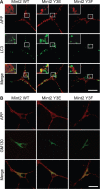
Phosphorylation of Mint2 modulates internalized APP localization. A, B, MTFtg hippocampal neurons were cultured and infected with lentiviral cre-IRES-Mint2-WT, cre-IRES-Mint2-Y3E, or cre-IRES-Mint2-Y3F. Live neurons were prelabeled with an extracellular APP antibody and after 5 min of internalization were fixed for immunostaining. Representative images of internalized APP (red) were visualized following cell permeabilization using fluorophore-conjugated antibody. A, Simultaneous staining for autophagosome LC3 (green) revealed extensive colocalization with internalized APP in phosphomimetic Mint2-Y3E mutant. Boxed inset represents a zoomed view (n = 2/15 represents number of independent experiments and total number of neurons assessed in each experiment). B, Double-labeling with anti-Golgi marker GM130 (green) indicated colocalization with internalized APP in all Mint2 conditions. Scale bars, 20 μm.
Figure 7.
Figure 7.
Phosphorylation of Mint2 increases intracellular Aβ42 accumulation. A, MTFtg hippocampal neurons were cultured and infected with lentiviral cre-IRES-Mint2-WT, cre-IRES-Mint2-Y3E or cre-IRES-Mint2-Y3F and immunostained with Aβ42 or APP antibody. Scale bar represents 10 μm. B, C, Quantitation of pixel intensity over soma area for Aβ42 or APP and expressed as percentage control (B, n = 3/32 represents number of independent experiments and total number of neurons assessed; C, n = 2/17 neurons). D, Conditioned medium from phosphomimetic Mint2-Y3E mutant showed a significant reduction in Aβ42 levels while phospho-resistant Mint2-Y3F mutant increased Aβ42 levels by 24% compared with control neurons infected with Mint2-WT by ELISA. E, Representative immunoblots of soluble APP (sAPP), total APP, and Tubulin showed no significant changes in any of the proteins observed. F, G, Quantification and representative immunoblots for APP recycling assay. Surface biotinylated proteins were internalized for 5 min at 37°C, recycled back to the cell surface for 30 min at 37°C and precipitated with Neutravidin agarose beads. Precipitated proteins were blotted for internalized APP (APPI) and expressed as percentage control. *p < 0.05, ***p < 0.005.
Figure 8.
Figure 8.
Src kinase effects on APP trafficking and processing. MTFtg neurons were cultured and infected with lentivirus encoding GFP (Control) or c-Src. A, Live cell endocytosis assay of MTFtg neurons incubated with an extracellular N-terminal APP antibody (22C11) and internalized for 5 min before stripping of excess surface antibody and fixation. Scale bar, 10 μm. B, Internalized APP immunostaining was visualized by confocal microscopy and quantification of APP internalized was taken as a measure of pixel intensity within a defined soma area and expressed as percentage control (n = 2/40 represents number of independent experiments and total number of neurons assessed). C, Neurons expressing GFP or c-Src were immunostained with LC3, Aβ42, or APP antibody. Scale bar, 10 μm. D–F, Quantifications of pixel intensity over soma area for LC3, Aβ42, and APP and expressed as percentage control (D, n = 2/21 represents number of independent experiments and total number of neurons assessed; E, n = 2/20; F, n = 2/14). G, Secreted Aβ42 levels were examined in neurons expressing GFP (control) or c-Src by ELISA. H, Representative immunoblots of soluble APP, total APP, Src, and Tubulin from neurons infected with control and c-Src (n = 9). *p < 0.05, ***p < 0.005. I, Model for the role of Mint2 phosphorylation in regulating the intracellular fate of internalized APP and Aβ secretion events. Upon activation of clathrin-mediated endocytosis, cell-surface APP and Mint2 is internalized and destined for Rab5-positive early endosomes. APP-Mint2 internalization is accelerated in phosphomimetic Mint2-Y3E mutant. Constitutive phosphorylation of Mint2 sorts internalized APP for autophagosome vesicular trafficking leading to an accumulation of intracellular Aβ42 immunoreactivity and decrease in secreted Aβ42 levels. Alternatively, phospho-resistant Mint2-Y3F mutant showed a significant increase in secreted Aβ42 levels that is due to APP recycling back to the cell surface which has been shown to favor amyloidogenic processing of APP. A population of internalized APP is sorted to the Golgi apparatus.

References

    1. Biederer T, Cao X, Südhof TC, Liu X. Regulation of APP-dependent transcription complexes by Mint/X11s: differential functions of Mint isoforms. J Neurosci. 2002;22:7340–7351. - PMC - PubMed
    1. Blom N, Gammeltoft S, Brunak S. Sequence and structure-based prediction of eukaryotic protein phosphorylation sites. J Mol Biol. 1999;294:1351–1362. - PubMed
    1. Boland B, Kumar A, Lee S, Platt FM, Wegiel J, Yu WH, Nixon RA. Autophagy induction and autophagosome clearance in neurons: relationship to autophagic pathology in Alzheimer's disease. J Neurosci. 2008;28:6926–6937. - PMC - PubMed
    1. Bolte S, Cordelières FP. A guided tour into subcellular colocalization analysis in light microscopy. J Microscopy. 2006;224:213–232. - PubMed
    1. Bonifacino JS. The GGA proteins: adaptors on the move. Nat Rev Mol Cell Biol. 2004;5:23–32. - PubMed

Publication types

MeSH terms

LinkOut - more resources