. 2012 Jul 11;32(28):9639-48.
doi: 10.1523/JNEUROSCI.1064-12.2012.

Wnt-responsive Lgr5-expressing stem cells are hair cell progenitors in the cochlea

Affiliations

Wnt-responsive Lgr5-expressing stem cells are hair cell progenitors in the cochlea

Fuxin Shi et al. J Neurosci. .

Abstract

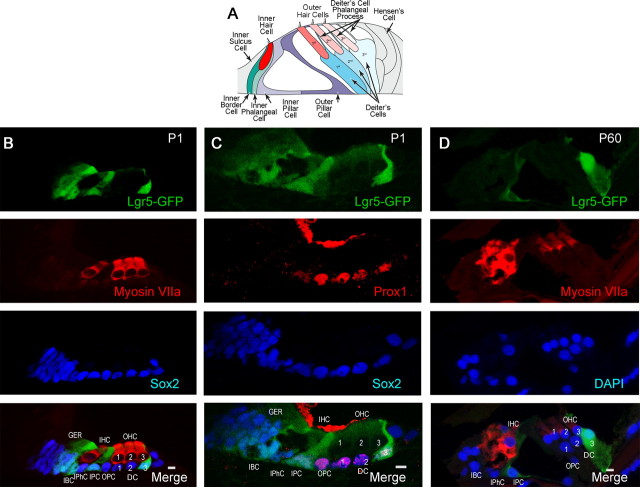
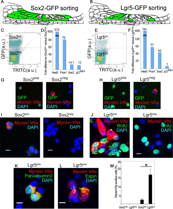
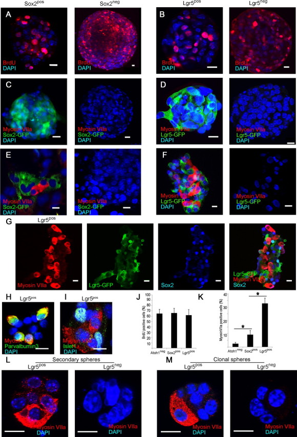
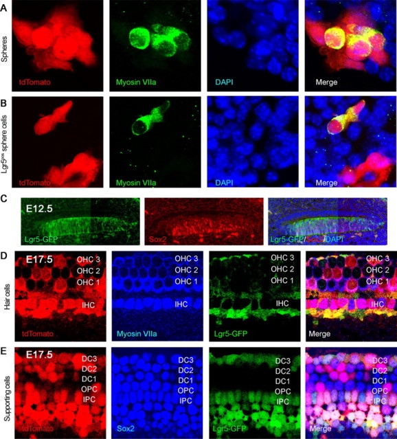
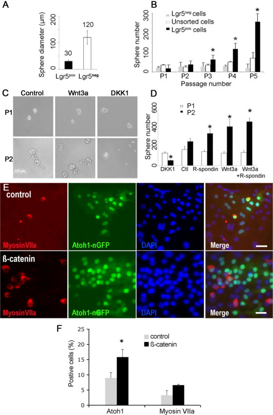
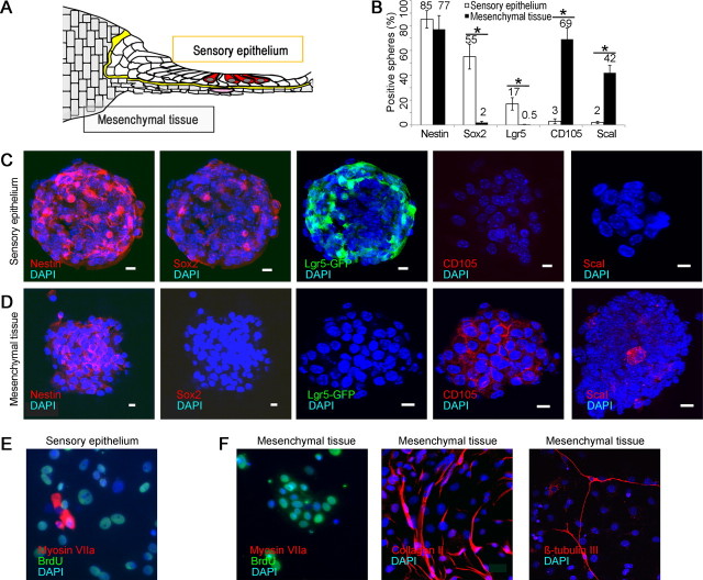
Auditory hair cells are surrounded on their basolateral aspects by supporting cells, and these two cell types together constitute the sensory epithelium of the organ of Corti, which is the hearing apparatus of the ear. We show here that Lgr5, a marker for adult stem cells, was expressed in a subset of supporting cells in the newborn and adult murine cochlea. Lgr5-expressing supporting cells, sorted by flow cytometry and cultured in a single-cell suspension, compared with unsorted cells, displayed an enhanced capacity for self-renewing neurosphere formation in response to Wnt and were converted to hair cells at a higher (>10-fold) rate. The greater differentiation of hair cells in the neurosphere assay showed that Lgr5-positive cells had the capacity to act as cochlear progenitor cells, and lineage tracing confirmed that Lgr5-expressing cells accounted for the cells that formed neurospheres and differentiated to hair cells. The responsiveness to Wnt of cells with a capacity for division and sensory cell formation suggests a potential route to new hair cell generation in the adult cochlea.


Figures

Figure 1.
Figure 1.
Lgr5 expression in a subset of supporting cells. A, Schematic shows arrangement of sensory epithelial cells in the adult organ of Corti (adapted from Thiers et al., 2008). B, At P1, Lgr5 (Lgr5-GFP, first panel) was expressed in the greater epithelial ridge, inner border cells, inner pillar cells, and the third row of Deiter's cells. Hair cells were stained with a myosin VIIa antibody (second panel). All supporting cells and cells in the greater epithelial ridge were stained with an antibody to Sox2 (third panel). The merged image is shown in the bottom panel (Merge). C, Compared with Lgr5 (first panel), Prox1 was expressed in outer pillar cells and Deiter's cells (second panel). Third panel shows the expression of Sox2 for comparison. D, At P60 the third row of Deiter's cells and the inner pillar cells retained expression of Lgr5 (first panel). Hair cells were stained with an antibody to myosin VIIa (second panel). Nuclei were counterstained with DAPI (third panel). The merged image is shown in the bottom panel (Merge). IHC, Inner hair cells; OHC-1, 2, 3, outer hair cells; IPC, inner pillar cells; IPhC, inner phalangeal cells; IBC, inner border cells; DC-1, 2, 3, Deiter's cells; OPC, outer pillar cells; GER, greater epithelial ridge.
Figure 2.
Figure 2.
Lgr5-positive supporting cells acted as hair cell progenitors in vitro. A, All supporting cells and greater epithelial ridge are labeled in the newborn Sox2-GFP mouse. B, The supporting cells and greater epithelial ridge cells shown in Figure 1 are fluorescent in the newborn Lgr5-GFP mouse. C, Sox2-expressing cells from the organ of Corti were separated by flow cytometry (Sox2pos, 23.3%). D, An enrichment in supporting cell markers in the Sox2pos fraction was seen by quantitative RT-PCR (mean, n = 2). E, Lgr5-expressing cells from the organ of Corti were separated by flow cytometry (Lgr5pos, 4.8%). F, An enrichment in supporting cell markers in the Lgr5pos fraction was seen by quantitative RT-PCR (mean, n = 2). G, The Sox2neg but not the Sox2pos fraction contained myosin VIIa-positive cells. Nuclei were stained with DAPI. H, The Lgr5pos fraction was free of myosin VIIa-positive cells which were in the Lgr5neg fraction. I, Myosin VIIa-positive cells were produced after 10 d of culture of Sox2pos but not Sox2neg cells. J, Lgr5pos but not Lgr5neg cells gave rise to myosin VIIa-positive cells after 10 d in culture, but had similar rates of BrdU incorporation. K, Differentiation of Lgr5pos cells resulted in myosin VIIa costaining with parvalbumin 3. L, Espin staining in the myosin VIIa-positive cells. M, The myosin VIIa staining in cultured Lgr5-sorted cells was 6.9 times higher than for Sox2-sorted cells. Means ± SEM are shown, n = 3.
Figure 3.
Figure 3.
Hair cell generation only from Lgr5-expressing neurospheres. A, Neurospheres formed by the Sox2pos and Sox2neg fractions incorporated BrdU. B, Sorted Lgr5pos and Lgr5neg cells formed neurospheres that incorporated BrdU. C, Neurospheres formed by the Sox2pos and Sox2neg fractions did not contain hair cells (Myosin VIIa). D, Neurospheres made from sorted Lgr5pos and Lgr5neg cells showed no labeling for myosin VIIa. E, Myosin VIIa-positive cells were observed upon differentiation of neurospheres made from the Sox2pos cells but not the Sox2neg cells. F, After differentiation, neurospheres made from Lgr5pos cells generated myosin VIIa-positive cells. No hair cells were observed upon differentiation of neurospheres made from the Lgr5neg cells. G, Differentiated Lgr5pos neurospheres retained Lgr5 and Sox2 surrounding the myosin VIIa-positive cells that lost expression of Lgr5. H, Myosin VIIa-staining cells from Lgr5pos neurospheres costained for parvalbumin 3. I, Cells surrounding the myosin VIIa-positive cells expressed islet1. J, Neurospheres made from Atoh1 (negatively sorted), or Sox2 or Lgr5 (positively sorted) cells had similar rates of cell division. K, Increasing capacity for hair cell differentiation was found in neurospheres made from Atoh1, Sox2 or Lgr5-sorted cells. Means ± SEM are shown, n = 3. Scale bars, 10 μm. L, Secondary neurospheres made by sorting Lgr5pos cells differentiated into myosin VIIa-positive hair cells. No hair cells were observed after differentiation of secondary neurospheres from Lgr5neg cells. M, Clonal neurospheres from single Lgr5pos but not from single Lgr5neg cells from cochlear sensory epithelium gave rise to hair cells upon differentiation.
Figure 4.
Figure 4.
Lgr5-positive cells were progenitors for hair cells in vitro and in vivo. A, Neurospheres were obtained from Lgr5-CreER mice crossed to tdTomato reporter mice that had been treated with tamoxifen at P1 to label Lgr5-expressing cells. The label for tdTomato in hair cells that were positive for myosin VIIa indicated a derivation from Lgr5-expressing supporting cells. B, When tamoxifen was added to third generation neurospheres from Lgr5-CreER reporter mice, labeling by tdTomato was observed in cells positive for hair cell markers, indicating derivation of hair cells from Lgr5-positive cells in the spheres. C, At E12.5, Lgr5 (Lgr5-GFP, left) colocalized with Sox2-positive progenitor cells in the cochlear duct (middle). D, Hair cell generation was traced back to Lgr5-positive progenitor cells using a tdTomato lineage tag (E12–E17). E, Supporting cells beyond the restricted Lgr5-GFP-positive subset were tdTomato-positive.
Figure 5.
Figure 5.
Cochlear neurospheres respond to Wnt/β-catenin signaling. A, Neurospheres from Lgr5pos compared with Lgr5neg neurospheres cells were smaller and more homogenous. B, Neurospheres from Lgr5pos compared with Lgr5neg cells had a significantly higher rate of expansion (indicated by asterisk) upon multiple passages (p < 0.05, n = 3). C, Neurospheres are shown at passaging from the first to the second generation in the absence of other factors (Control) or in the presence of either Wnt3a-conditioned medium (Wnt3a) or Dickkopf1 (DKK1). D, Cells treated with R-spondin1, Wnt3a, or Wnt3a and R-spondin1 generated significantly more neurospheres, and treatment with DKK1 significantly reduced the number of neurospheres (p < 0.05, n = 3). E, Transduction of cochlear neurospheres with adenovirus containing the β-catenin gene (β-catenin) gave rise to more Atoh1 (green)- and myosin VIIa (red)-positive cells than an empty adenoviral vector (Control). Nuclei were stained with DAPI (blue). F, Quantification of Atoh1 and myosin VIIa-positive cells (n = 3, 5000 cells counted). Scale bar, 20 μm.
Figure 6.
Figure 6.
Lack of progenitors for hair cells in mesenchymal tissue. A, Separation of sensory epithelium from mesenchymal tissue after treatment with thermolysin is depicted. B, Spheres from sensory epithelium stained for neural stem cell markers and spheres from mesenchyme did not (50 spheres counted; n = 3; asterisk indicates p < 0.05). C, Neurospheres produced from sensory epithelial spheres after separation with thermolysin, expressing nestin, prosensory markers, Sox2, and Lgr5, but not mesenchymal markers, CD105 and Sca1. D, Mesenchymal spheres (positive for nestin, CD105 and Sca1) did not express Sox2 or Lgr5. E, Cells from sensory epithelium formed neurospheres that were labeled for BrdU and gave rise to myosin VIIa-positive hair cells under differentiating conditions. F, When cultured under differentiating conditions, the mesenchymal spheres were labeled for BrdU but did not give rise to cells expressing hair cell markers (Myosin VIIa). The mesenchymal cells differentiated into collagen II-positive chondrocytes and βIII-tubulin-positive neurons.

References

    1. Adachi K, Mirzadeh Z, Sakaguchi M, Yamashita T, Nikolcheva T, Gotoh Y, Peltz G, Gong L, Kawase T, Alvarez-Buylla A, Okano H, Sawamoto K. Beta-catenin signaling promotes proliferation of progenitor cells in the adult mouse subventricular zone. Stem Cells. 2007;25:2827–2836. - PubMed
    1. Barker N, Clevers H. Leucine-rich repeat-containing G-protein-coupled receptors as markers of adult stem cells. Gastroenterology. 2010;138:1681–1696. - PubMed
    1. Barker N, van Es JH, Kuipers J, Kujala P, van den Born M, Cozijnsen M, Haegebarth A, Korving J, Begthel H, Peters PJ, Clevers H. Identification of stem cells in small intestine and colon by marker gene Lgr5. Nature. 2007;449:1003–1007. - PubMed
    1. Barker N, Ridgway RA, van Es JH, van de Wetering M, Begthel H, van den Born M, Danenberg E, Clarke AR, Sansom OJ, Clevers H. Crypt stem cells as the cells-of-origin of intestinal cancer. Nature. 2009;457:608–611. - PubMed
    1. Beattie MS, Harrington AW, Lee R, Kim JY, Boyce SL, Longo FM, Bresnahan JC, Hempstead BL, Yoon SO. ProNGF induces p75-mediated death of oligodendrocytes following spinal cord injury. Neuron. 2002;36:375–386. - PMC - PubMed

Publication types

MeSH terms

Substances