Clinical Trial
. 2012 Jul 11;32(28):9677-89.
doi: 10.1523/JNEUROSCI.4742-11.2012.

An effector-reduced anti-β-amyloid (Aβ) antibody with unique aβ binding properties promotes neuroprotection and glial engulfment of Aβ

Affiliations
Clinical Trial

An effector-reduced anti-β-amyloid (Aβ) antibody with unique aβ binding properties promotes neuroprotection and glial engulfment of Aβ

Oskar Adolfsson et al. J Neurosci. .

Abstract

Passive immunization against β-amyloid (Aβ) has become an increasingly desirable strategy as a therapeutic treatment for Alzheimer's disease (AD). However, traditional passive immunization approaches carry the risk of Fcγ receptor-mediated overactivation of microglial cells, which may contribute to an inappropriate proinflammatory response leading to vasogenic edema and cerebral microhemorrhage. Here, we describe the generation of a humanized anti-Aβ monoclonal antibody of an IgG4 isotype, known as MABT5102A (MABT). An IgG4 subclass was selected to reduce the risk of Fcγ receptor-mediated overactivation of microglia. MABT bound with high affinity to multiple forms of Aβ, protected against Aβ1-42 oligomer-induced cytotoxicity, and increased uptake of neurotoxic Aβ oligomers by microglia. Furthermore, MABT-mediated amyloid plaque removal was demonstrated using in vivo live imaging in hAPP((V717I))/PS1 transgenic mice. When compared with a human IgG1 wild-type subclass, containing the same antigen-binding variable domains and with equal binding to Aβ, MABT showed reduced activation of stress-activated p38MAPK (p38 mitogen-activated protein kinase) in microglia and induced less release of the proinflammatory cytokine TNFα. We propose that a humanized IgG4 anti-Aβ antibody that takes advantage of a unique Aβ binding profile, while also possessing reduced effector function, may provide a safer therapeutic alternative for passive immunotherapy for AD. Data from a phase I clinical trial testing MABT is consistent with this hypothesis, showing no signs of vasogenic edema, even in ApoE4 carriers.


Figures
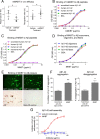
Figure 1.
Figure 1.
The anti-Aβ MABT mAb binds with high affinity to different Aβ peptides and has antiaggregation properties. A, An in vivo efficacy study administering mMABT to hAPP(V717I) mice showed a reduction in Aβ plaque load (left panel) and improved memory performance (right panel). Results show means (±95% CI). B, C, An Aβ ELISA was used to compare the binding to human and murine Aβ1–42 and human Aβ1–40, for mMABT (B) and MABT (C). D, MABT was also tested for binding to different Aβ1–42 assembly states, showing equal binding for oligomers, monomers, and fibers. E, MABT binds Aβ plaques present in brain sections from transgenic hAPP(V717I) mice (top panels) and human AD temporal neocortical sections (bottom panels). F, In vitro functionality was shown by the ability of MABT to impede Aβ1–42 aggregation (left panel), and to disassemble preformed Aβ1–42 aggregates (right panel) in a ThT-based assay, with an Aβ1–42 to MABT molar ratio of 10:1. An anti-Aβ IgG mAb with a N-terminal epitope was used as control. Results show the mean (±SD) of three independent experiments. *p < 0.05; **p < 0.01. G, MABT was also tested in an Aβ1–42 self-assembly assay that does not rely on ThT fluorescence upon binding to multimeric Aβ assemblies, as described in Materials and Methods. The means (±SEM) of two assays are shown.
Figure 2.
Figure 2.
The MABT inhibits cytotoxicity of Aβ1–42 oligomers on primary mixed cortical cultures. A, Mixed cortical cells from P1 rats were treated with 2.5 or 5 μm Aβ1–42 oligomers with or without 100 μg/ml of MABT or an IgG control mAb. An MTT assay was used to determine cell viability as described in Materials and Methods. The means (±SEM) of five independent experiments are shown. *p < 0.05; **p < 0.01. B, In a comparable assay, but measuring ATP production as a marker of metabolic activity, cells were treated with 10 μm Aβ1–42 oligomers with or without 200 μg/ml MABT. Results show the means (±SEM) of two independent assays. C, Neurotoxicity following extended Aβ1–42 oligomer treatment was tested by morphological analyses. Cells as above were treated for 4 d with 10 μm Aβ1–42 oligomers, with or without 50 μg/ml of MABT, and then stained for TuJ1 (green) and DAPI (blue). D, Silver-stained SDS-PAGE showing the highly cytotoxic Aβ oligomer preparations consisting of a mixture of Aβ1–42 oligomer assemblies, ranging in size from dimers and trimers to higher-molecular-weight multimers.
Figure 3.
Figure 3.
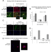
Aβ1–42 oligomer binding to neurites is reduced by the anti-Aβ IgG4 mAb. A, Mixed cortical cells from P1 rats were treated with 2 μm Aβ1–42 oligomers, with or without 100 μg/ml of the MABT or an IgG control for 30 min. The panels from left to right show the following: treatment with buffer control, Aβ1–42 oligomers, and Aβ1–42 oligomers with MABT. Green is TuJ1, red is anti-Aβ (clone 6E10), and blue is DAPI. The bottom row shows all three markers, whereas the top row shows only Aβ and DAPI. The two insets illustrate the binding of Aβ1–42 oligomers to neurites (left) and the inhibition of this binding by the MABT mAb (right). B, Quantitative measures of fluorescence are shown for 30 min and 18 h treatments. Mean results (±SEM) of two experiments are shown. C, Aβ1–42 tagged with HyLite Fluor-488 verifies that MABT inhibits binding of Aβ1–42 to neurites. Cortical cultures from P1 rats were treated as described for A, except that Aβ1–42 tagged with HyLite Fluor 488 was used. Green is Aβ1–42 and blue is DAPI, with one representative experiment shown. D, Green fluorescence was quantified, with the mean (±SD) of the two independent experiments shown. E, Upon treatment with MABT, Aβ1–42 oligomers appeared to be taken up by cells resembling microglia. Green is Aβ1–42 and blue is DAPI.
Figure 4.
Figure 4.
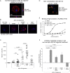
Aβ1–42 oligomers are taken up via an FcR-mediated mechanism into microglia upon MABT treatment. A, Confocal imaging was used to show that Aβ1–42 oligomers complexed to MABT are taken up into microglia. Aβ1–42 oligomers are shown in red, and blue is DAPI. An apical-to-distal slice was obtained from a three-dimensional image rendered from Z-stacks. Confocal imaging was done on mixed cortical cells labeled for Aβ (red) and DAPI (blue) as described in Materials and Methods. B, C, Microglia were identified and scanned for the maximum fluorescence intensity through a series of confocal stacks representing intracellular locations (B), and the total area of fluorescent signal above a minimum threshold was quantified (C). Each mark on the graph represents the total area of Aβ staining within a single cell. A minimum of 20 cells was analyzed for each treatment condition. Data were compared using one-way ANOVA followed by Tukey's post hoc multiple comparison. Means (±SEM) are shown. D, Microglia (Iba1+) was verified as the cell type taking up Aβ1–42 complexed to MABT. Red is Iba1, green is HyLite Fluor 488-labeled Aβ1–42, and blue is DAPI. E, Differential binding to FcγRIIIa-V158 was verified in a binding assay. F, An anti-Aβ mAb requires effector function via FcγR binding for full protective effect against Aβ1–42 oligomer toxicity. Mixed cortical cells from P1 rats were treated with Aβ1–42 oligomers, with or without MABT, MABT-IgG1-D265A, MABT-IgG1, or an IgG1 control mAb. The graph shows the mean (±SEM) percentage increase in cell survival compared with Aβ1–42 oligomer treated cells, from five independent experiments. Statistical analysis was done using one-way ANOVA followed by Tukey's post hoc multiple comparison.
Figure 5.
Figure 5.
Systemic dosing of MABT modulates individual amyloid plaques in vivo. A, Amyloid plaques in hAPP(V717I)/PS1 animals were labeled with methoxy-X04 injected intraperitoneally, visualized by in vivo two-photon microscopy, and tracked over multiple weeks. B, The relative change in plaque volume over time plotted as fold increase from the initial imaging session. On average, the individual plaque size decreased in volume after systemic dosing with MABT (14 plaques, 2 animals).
Figure 6.
Figure 6.
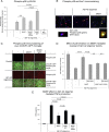
MABT reduces microglial activation as measured by p38MAPK activity and TNFα secretion. A, When complexed to Aβ1–42 oligomers, the IgG1 wild-type backbone significantly increases p38MAPK activation over that shown for Aβ1–42 oligomer-treated microglia. Mixed cortical cells from P1 rats were treated with 10 μm Aβ1–42 oligomers for 30 min with or without 100 μg/ml MABT, MABT-IgG1-D265A, or MABT-IgG1 wild type. An IgG1 mAb not binding to Aβ was used as control. The activity of p38MAPK was measured by a phosphospecific ELISA as described in Materials and Methods. The mean (±SEM) of four independent experiments is shown. Statistical analysis was done using one-way ANOVA followed by Tukey's post hoc multiple comparison. B, Activation of p38MAPK with Aβ1–42 oligomers complexed with mAb is specific to microglia. Cells were treated as described above and then stained for phospho-p38MAPK (green), Iba1 (red), and DAPI (blue). The panels from left to right show the following: treatment with buffer control, Aβ1–42 oligomers, and Aβ1–42 oligomers with MABT. C, Pure microglia from CX3CR1-GFP mice were used to verify microglia-specific p38MAPK activity. The top panels show phospho-p38MAPK immunofluorescence (red), whereas the bottom panels are a merge of phospho-p38 and GFP (green) on a phase contrast image. D, p38MAPK activity is required for the full protective effect of MABT against Aβ1–42 oligomer toxicity. Mixed cortical cells from P1 rats were treated with 10 μm Aβ1–42 oligomers alone or together with 100 μg/ml MABT or MABT-IgG1-D265A, in the presence or absence of 1 μm SB239063, a p38MAPK-specific inhibitor. A cytotoxicity assay using the MTT readout was performed after 24 h. The mean (±SEM) of four experiments is shown. Statistical analysis was done using one-way ANOVA followed by Tukey's post hoc multiple comparison. E, IgG1 is less effective in reducing Aβ1–42-mediated proinflammatory release by microglia as measured by TNFα levels from conditioned media of enriched microglia following 24 h of treatment. The mean (±SEM) of three independent experiments is shown. Statistical analysis was done using one-way ANOVA followed by Tukey's post hoc multiple comparison.
Figure 7.
Figure 7.
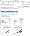
MABT phase I clinical study results. A, Phase I clinical study design. B, Actual study population. C, The serum MABT concentration–time profiles following both single (SD phase) or weekly (MD phase) intravenous administration increased in proportion to dose and were characterized by slow clearance (∼3 ml · d−1 · kg−1) and a long half-life (18–23 d). D, Increases in plasma total Aβ1–40 and total Aβ1–42 correlated well with serum MABT concentration following single or weekly administration in the SD phase; serum MABT and plasma total Aβ samples were collected just before MABT administration through study day 112. In the MD phase, samples were collected just before MABT administration on day 0, at 1 h and 7 d after each dose and to 119 d after last dose through study day 140. Serum MABT concentrations were determined using a quantitative ELISA with a limit of detection (LOD) of 0.0225 μg/ml. Total plasma levels of Aβ1–40 and Aβ1–42 were determined using an electrochemiluminescence assay with a LOD of 0.01 and 0.125 nm, respectively. Aβ1–42 was not detectable in patients given placebo (or at baseline in patients given MABT) so is not shown.

References

    1. Bandyopadhyay S, Hartley DM, Cahill CM, Lahiri DK, Chattopadhyay N, Rogers JT. Interleukin-1alpha stimulates non-amyloidogenic pathway by alpha-secretase (ADAM-10 and ADAM-17) cleavage of APP in human astrocytic cells involving p38 MAP kinase. J Neurosci Res. 2006;84:106–118. - PubMed
    1. Bateman DA, McLaurin J, Chakrabartty A. Requirement of aggregation propensity of Alzheimer amyloid peptides for neuronal cell surface binding. BMC Neurosci. 2007;8:29. - PMC - PubMed
    1. Bitan G, Kirkitadze MD, Lomakin A, Vollers SS, Benedek GB, Teplow DB. Amyloid beta-protein (Abeta) assembly: Abeta 40 and Abeta 42 oligomerize through distinct pathways. Proc Natl Acad Sci U S A. 2003;100:330–335. - PMC - PubMed
    1. Carlson C, Estergard W, Oh J, Suhy J, Jack CR, Jr, Siemers E, Barakos J. Prevalence of asymptomatic vasogenic edema in pretreatment Alzheimer's disease study cohorts from phase 3 trials of semagacestat and solanezumab. Alzheimers Dement. 2011;7:396–401. - PubMed
    1. Carty NC, Wilcock DM, Rosenthal A, Grimm J, Pons J, Ronan V, Gottschall PE, Gordon MN, Morgan D. Intracranial administration of deglycosylated C-terminal-specific anti-Abeta antibody efficiently clears amyloid plaques without activating microglia in amyloid-depositing transgenic mice. J Neuroinflamm. 2006;3:11. - PMC - PubMed

Publication types

MeSH terms