. 2012 Aug 21;126(8):963-74.
doi: 10.1161/CIRCULATIONAHA.112.094722. Epub 2012 Jul 11.

Aldosterone inactivates the endothelin-B receptor via a cysteinyl thiol redox switch to decrease pulmonary endothelial nitric oxide levels and modulate pulmonary arterial hypertension

Affiliations

Aldosterone inactivates the endothelin-B receptor via a cysteinyl thiol redox switch to decrease pulmonary endothelial nitric oxide levels and modulate pulmonary arterial hypertension

Bradley A Maron et al. Circulation. .

Abstract

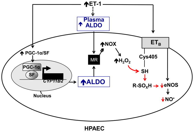
Background: Pulmonary arterial hypertension (PAH) is characterized, in part, by decreased endothelial nitric oxide (NO(·)) production and elevated levels of endothelin-1. Endothelin-1 is known to stimulate endothelial nitric oxide synthase (eNOS) via the endothelin-B receptor (ET(B)), suggesting that this signaling pathway is perturbed in PAH. Endothelin-1 also stimulates adrenal aldosterone synthesis; in systemic blood vessels, hyperaldosteronism induces vascular dysfunction by increasing endothelial reactive oxygen species generation and decreasing NO(·) levels. We hypothesized that aldosterone modulates PAH by disrupting ET(B)-eNOS signaling through a mechanism involving increased pulmonary endothelial oxidant stress.

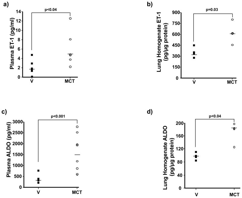
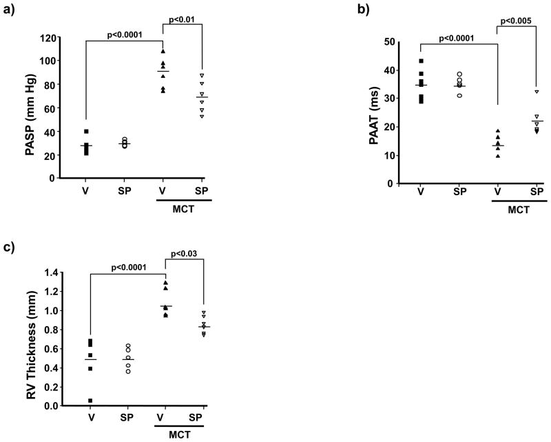
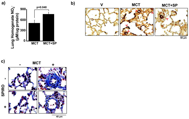
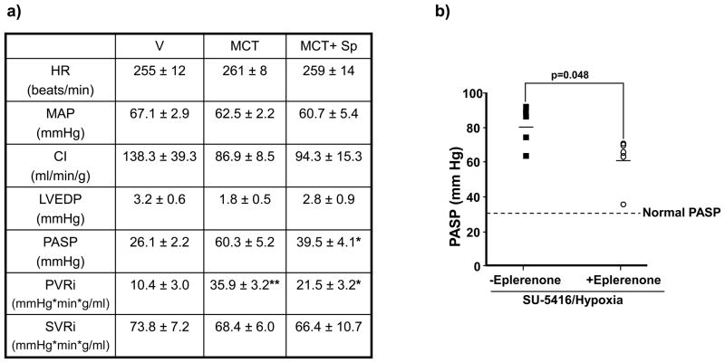
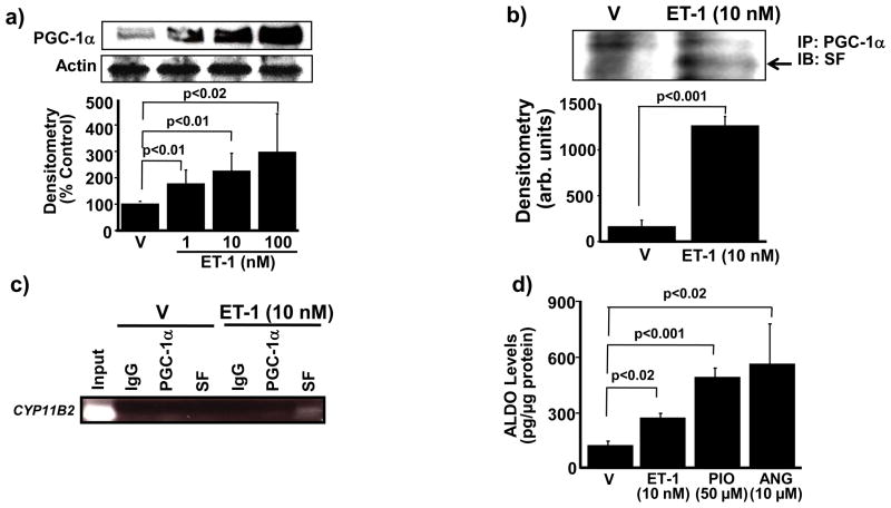
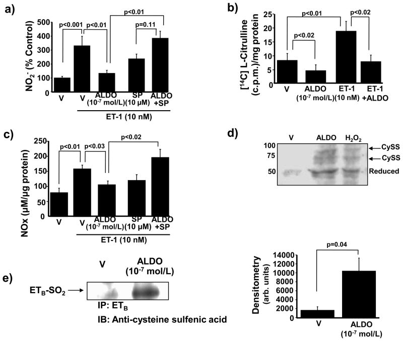
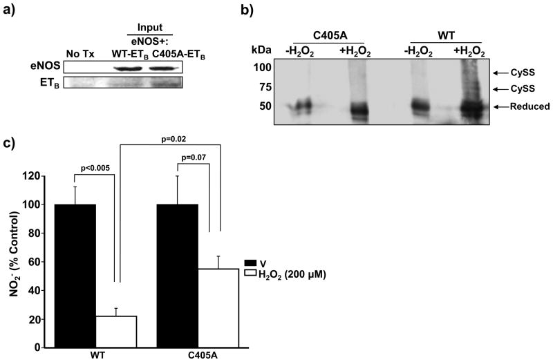
Methods and results: In rats with PAH, elevated endothelin-1 levels were associated with elevated aldosterone levels in plasma and lung tissue and decreased lung NO(·) metabolites in the absence of left-sided heart failure. In human pulmonary artery endothelial cells, endothelin-1 increased aldosterone levels via peroxisome proliferator-activated receptor gamma coactivator-1α/steroidogenesis factor-1-dependent upregulation of aldosterone synthase. Aldosterone also increased reactive oxygen species production, which oxidatively modified cysteinyl thiols in the eNOS-activating region of ET(B) to decrease endothelin-1-stimulated eNOS activity. Substitution of ET(B)-Cys405 with alanine improved ET(B)-dependent NO(·) synthesis under conditions of oxidant stress, confirming that Cys405 is a redox-sensitive thiol that is necessary for ET(B)-eNOS signaling. In human pulmonary artery endothelial cells, mineralocorticoid receptor antagonism with spironolactone decreased aldosterone-mediated reactive oxygen species generation and restored ET(B)-dependent NO(·) production. Spironolactone or eplerenone prevented or reversed pulmonary vascular remodeling and improved cardiopulmonary hemodynamics in 2 animal models of PAH in vivo.

Conclusions: Our findings demonstrate that aldosterone modulates an ET(B) cysteinyl thiol redox switch to decrease pulmonary endothelium-derived NO(·) and promote PAH.


Conflict of interest statement

Conflict of Interest Disclosures:

None.

Figures

Figure 1
Figure 1
Elevated levels of ET-1 are associated with hyperaldosteronism in PAH. (a,b) Levels of ET-1 (n=4–6) and (c,d) aldosterone (ALDO) (n=4–8) were measured in plasma and lung tissue homogenates of Sprague-Dawley rats 25 days following treatment with vehicle control (V) or monocrotaline (MCT) (50 mg/kg). Horizontal line represents the median for each condition.
Figure 2
Figure 2
Aldosterone promotes PAH in vivo. Sprague-Dawley rats were treated with vehicle control (V) or monocrotaline (MCT) (50 mg/kg) and randomized immediately to V or spironolactone (SP) (25 mg/kg/d) for 25 days (n=6 rats per condition). The contribution of aldosterone to PAH was assessed by (a) right heart catheterization to measure pulmonary artery (assumed to be equivalent to right ventricular) systolic pressure (PASP); echocardiography to assess changes in (b) pulmonary artery acceleration time (PAAT); and, (c) right ventricular (RV) free-wall thickness. Horizontal line represents the mean for each condition.
Figure 3
Figure 3
Spironolactone increases pulmonary vascular NO levels and attenuates pulmonary vascular remodeling in PAH. (a) The effect of spironolactone (SP)(25 mg/kg/d) on pulmonary vascular NO levels in PAH was assessed by measuring nitrite (NO2) in lung tissue homogenates from Sprague-Dawley rats treated with vehicle control (V) or monocrotaline (MCT) (50 mg/kg) (n=4). (b) Tissue sections were stained with anti-smooth muscle cell α-actin antibody and the number of muscularized distal pulmonary arterioles (red arrows) was counted in 20 consecutive fields per section (100x magnification). Compared to V-treated rats with PAH, spironolactone decreased significantly the number of α-actin-stained muscularized distal pulmonary arterioles (76.0 [64–95] vs. 59.5 [59–61] muscularized pulmonary arterioles/20 high powered fields, p<0.005, n=5). (c) Gomori’s trichrome stain was performed on paraffin-embedded lung sections and perivascular collagen deposition in pulmonary arterioles measuring 20–50 μm located distal to terminal bronchioles (400x magnification) was measured. Compared to V-treated rats with PAH, spironolactone decreased perivascular collagen deposition by 77% (p<0.001, n=4–5 rats per condition). Representative photomicrographs are shown.
Figure 4
Figure 4
The effect of mineralocorticoid receptor antagonism on reversal or prevention of adverse cardiopulmonary hemodynamics in two models of experimental PAH. (a) In a reversal study, Sprague-Dawley rats were randomized to receive vehicle control (V) or spironolactone (SP) (25 mg/kg/d) 14 days following the administration of V or monocrotaline (MCT) (50 mg/kg), and cardiopulmonary hemodynamics were assessed by cardiac catheterization 10 days later. *p<0.02 vs. MCT, n=6 rats per condition; **p<0.04 vs. V, n=4 rats per condition. Data are presented as mean ± S.E. (b) In a prevention study, Sprague-Dawley rats were injected with SU-5416 and exposed to chronic hypoxia for 21 days. Immediately following exposure to hypoxia, rats were randomized to receive standard chow or eplerenone (0.6 gm/1 gm chow) until completion of the study. The effect of eplerenone on pulmonary artery systolic pressure (PASP) was assessed by cardiac catheterization (n=5 rats per condition). HR, heart rate; CI, cardiac index; LVEDP, left ventricular end-diastolic pressure; PVRi, pulmonary vascular resistance index; SVRi, systemic vascular resistance index.
Figure 5
Figure 5
ET-1 stimulates PGC-1α-dependent association of SF with CYP11B2 to increase aldosterone levels. (a) The effect of ET-1 on PGC-1α expression was assessed by Western analysis (n=4). (b) Co-immunoprecipitation experiments demonstrated that incubation of HPAECs with ET-1 (10 nM) for 24 h induced the association of PGC-1α with steroidogenesis factor-1 (SF) (n=3). (c) Chromatin immunoprecipitation (n=3) of cell lysates using antibodies to PGC-1α, SF, and immunoglobulin-G (IgG) as a negative control was followed by PCR amplification of the proximal region of the CYP11B2 promoter region containing the gonadotrope-specific element. (d) The functional effect of PGC-1α stimulation on aldosterone production was assessed in cells treated with the selective PGC-1α agonist pioglitazone (50 μM) for 24 h (n=4), or with ET-1 (10 nM) or angiotensin II (ANG)(10 μM) for 24 h as positive controls. PGC-1α, PPAR-γ co-activator-1α; arb. units, arbitrary units; IP, immunoprecipitation, IB; immunoblot. Data are presented as mean ± S.E.M. Representative blots are shown.
Figure 6
Figure 6
Aldosterone decreases ETB-dependent synthesis of NO. (a) HPAECs were exposed to vehicle (V) or aldosterone (ALDO) (10−7 mol/L) for 24 h in the presence or absence of spironolactone (SP) (10 μM) and NO2 formation was assessed. Prior to analysis, cells were exposed to ET-1 (10 nM) for 10 min to stimulate ETB signaling (n=4). (b) The effect of ALDO on ETB-dependent activation of eNOS was determined (n=4). c.p.m., counts per minute. (c) The effect of ALDO on ETB-dependent NO generation was assessed by measuring total NO metabolite levels (NOx: NO2 + NO3) (n=3). (d) HPAECs were exposed to V, hydrogen peroxide (H2O2) (200 μM) for 20 min, or ALDO (10−7 mol/L) for 24 h to assess changes to the redox status and de novo disulfide bond formation by ETB cysteinyl thiols. For each disulfide formed, a 20-kDa shift in band location of the reduced ETB protein occurs on the Western blot using an antibody specific to the region of ETB containing Cys405 (n=4). Cyss, disulfide bond. A representative blot is shown. (e) The region of ETB containing Cys405 was immunoprecipitated from cells treated with V or ALDO (10−7 mol/L) for 24 h and immunoblotting was performed to detect differences in protein sulfenic acid levels (R-SOH) (n=3). IP, immunoprecipitation, IB; immunoblot. Data are presented as mean ± S.E.M. Representative blots are shown.
Figure 7
Figure 7
Oxidation of Cys405 impairs ETB-dependent NO generation. (a) COS-7 cells were transiently transected with wild type (WT)-eNOS and WT-ETB or mutant ETB DNA containing a substitution of alanine for cysteine at position 405 (C405A-ETB) and protein expression was confirmed. No Tx, untransfected. (b) Disulfide bond formation was assessed by Western immunoblotting of PEG-conjugated maleimide-labeled cell extracts exposed to H2O2 (200 μmol/L for 20 min). Compared to WT-ETB-transfected cells, in which H2O2 (200 μmol/L for 20 min) induced the formation of 1 or 2 disulfide bonds, C405A-ETB was resistant to disulfide bond formation (n=4) Cyss, disulfide bond. (c) COS-7 cells expressing WT-eNOS and WT-ETB or C405A-ETB were exposed to vehicle (V) control or hydrogen peroxide (H2O2) (200 μmol/L) for 60 min. After that time, the cell culture medium was replaced and cells were treated with ET-1 (10 nM) for 10 min. and nitrite (NO2) levels were measured (n=4). Data are presented as mean ± S.E.M. Representative blots are shown.
Figure 8
Figure 8
A proposed mechanism by which hyperaldosteronism decreases pulmonary endothelial eNOS activation and NO generation in PAH. Hyperaldosteronism (ALDO) in pulmonary arterial hypertension (PAH) may occur via i) endothelin-1 (ET-1)-mediated activation of PPARγ coactivator-1α (PGC-1α)/steroidogenesis factor-1 (SF) to increase CYP11B2 (aldosterone synthase) gene transcription in HPAECs, and/or ii) upregulation of adrenal ALDO synthesis via ET-1 and/or overactivation of the renin-angiotensin pathway. Stimulation of the mineralocorticoid receptor (MR) in HPAECs by ALDO activates NADPH oxidase type 4 (NOX4) to increase levels of hydrogen peroxide (H2O2), which, in turn, oxidatively modifies redox sensitive, functional cysteinyl thiol(s) in the ETB receptor (Cys405) to impair ETB-dependent activation of eNOS and decrease synthesis of nitric oxide (NO). eNOS, endothelial nitric oxide synthase; R-SOXH, higher oxidative intermediaries of cysteine.

References

    1. Archer SL, Weir EK, Wilkins MR. Basic science of pulmonary arterial hypertension for clinicians: new concepts and experimental therapies. Circulation. 2010;121:2045–2066. - PMC - PubMed
    1. Farber HW, Loscalzo J. Pulmonary arterial hypertension. N Engl J Med. 2004;351:1655–1665. - PubMed
    1. Michelakis ED. The role of the NO axis and its therapeutic implications in pulmonary arterial hypertension. Heart Fail Rev. 2003;8:5–21. - PubMed
    1. Leopold JA, Dam A, Maron BA, Scribner AW, Liao R, Handy DE, Stanton RC, Pitt B, Loscalzo J. Aldosterone impairs vascular reactivity by decreasing glucose-6-phosphate dehydrogenase activity. Nat Med. 2007;13:189–197. - PMC - PubMed
    1. Weber KT. Aldosterone in congestive heart failure. N Engl J Med. 2001;345:1689–1697. - PubMed

Publication types

MeSH terms