. 2012 Sep;86(18):10093-102.
doi: 10.1128/JVI.00930-12. Epub 2012 Jul 11.

Herpes simplex virus is equipped with RNA- and protein-based mechanisms to repress expression of ATRX, an effector of intrinsic immunity

Affiliations

Herpes simplex virus is equipped with RNA- and protein-based mechanisms to repress expression of ATRX, an effector of intrinsic immunity

Igor Jurak et al. J Virol. 2012 Sep.

Abstract

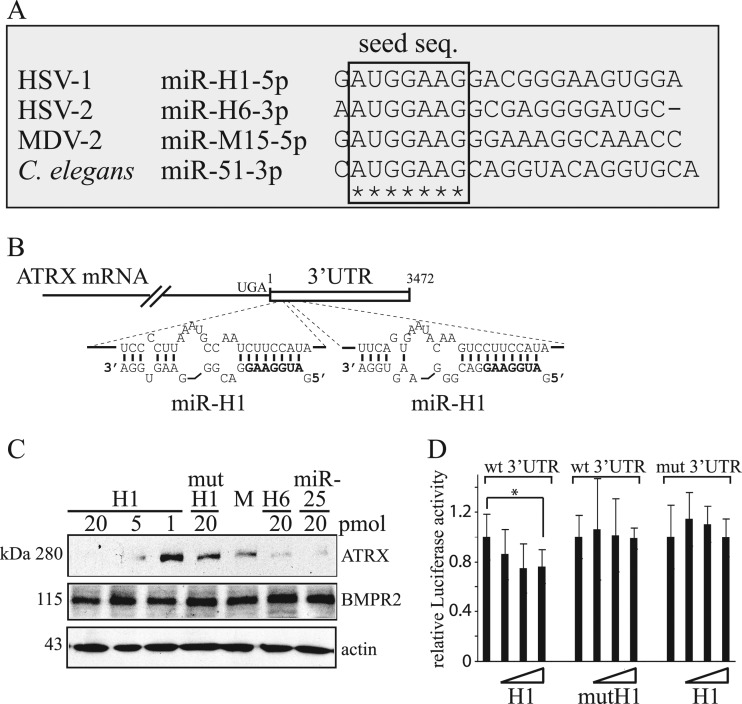
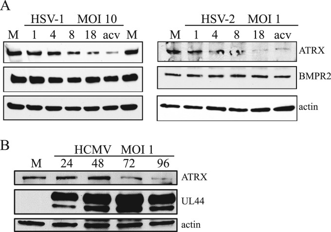
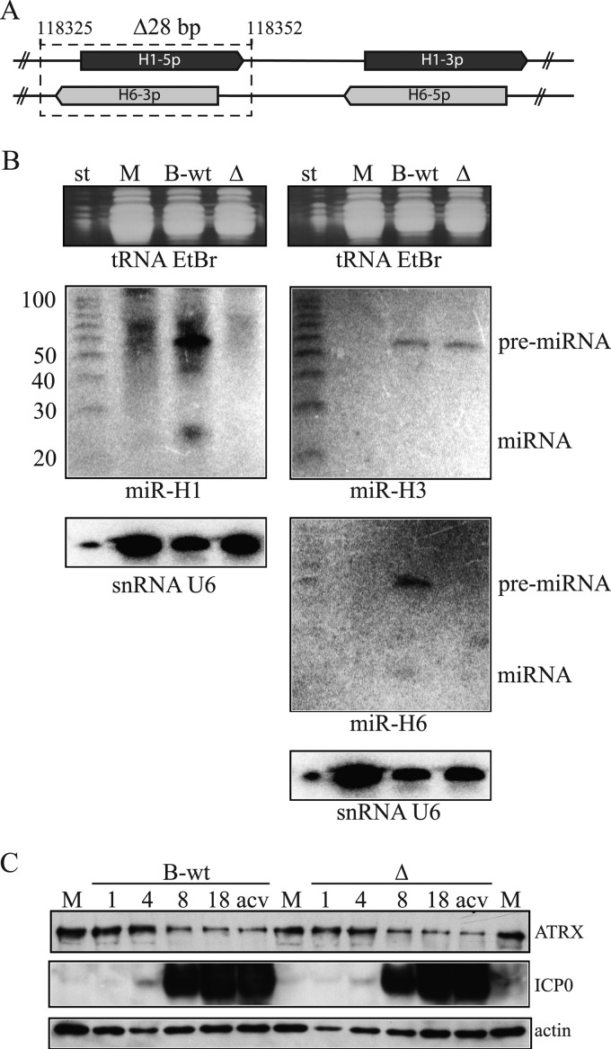
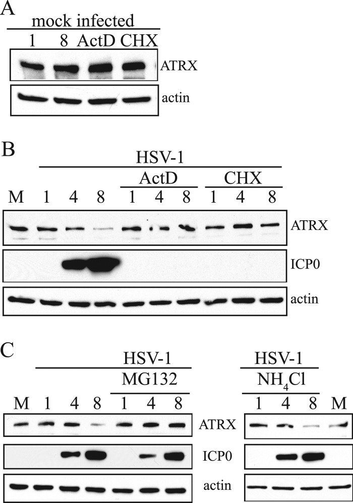
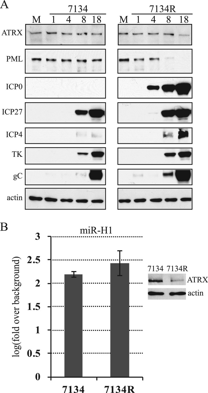
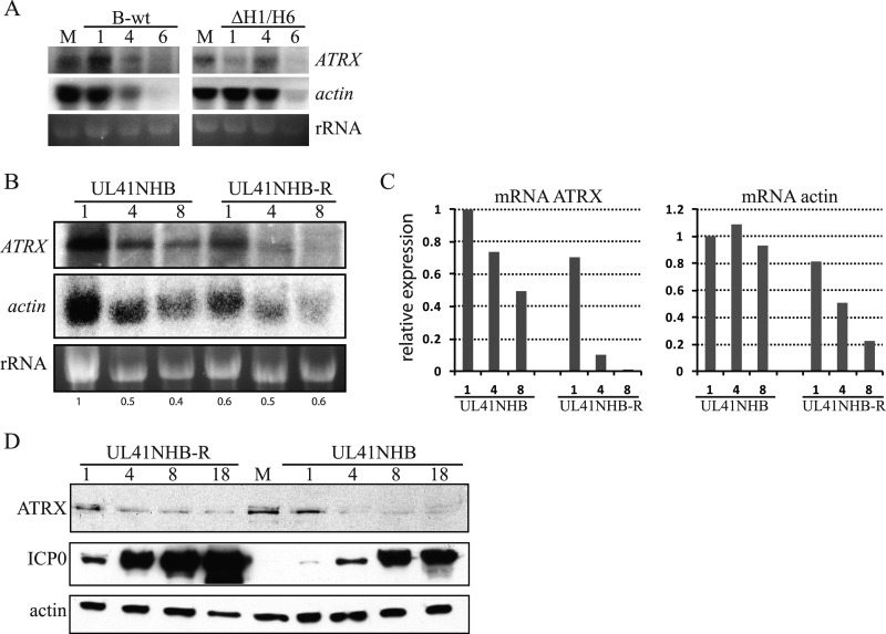
Intrinsic immunity is a first-line intracellular defense against virus infection, and viruses have evolved mechanisms to counteract it. During herpes simplex virus (HSV) infection, nuclear domain 10 (ND10) components localize adjacent to incoming viral genomes and generate a repressive environment for viral gene expression. Here, we found that the ND10 component, alpha-thalassemia/mental retardation syndrome X-linked (ATRX) protein, is predicted to be a target of HSV-1 miR-H1 and HSV-2 miR-H6. These microRNAs (miRNAs) share a seed sequence and are abundant during lytic infection. Mimics of both miRNAs could deplete endogenous ATRX, and an miR-H1 mimic could repress the expression of a reporter linked to the 3' untranslated region of ATRX mRNA, identifying a cellular mRNA targeted by an HSV miRNA. Interestingly, ATRX protein and its mRNA were depleted in cells lytically infected with HSV, and ATRX protein was also depleted in cells infected with human cytomegalovirus. However, infection with an HSV-1 mutant lacking miR-H1 still resulted in ATRX depletion. This depletion was sensitive to a proteasome inhibitor and was largely ablated by a deletion of the gene encoding the immediate-early ICP0 protein. Additionally, a deletion of the gene encoding the tegument protein Vhs ablated most of the depletion of ATRX mRNA. Thus, HSV is equipped with multiple mechanisms to limit the expression of ATRX. As ATRX is implicated in repression of lytic viral gene expression, our results suggest roles for these different mechanisms during various phases of HSV infection.


Figures

Fig 1
Fig 1
HSV-1 miR-H1 and HSV-2 miR-H6 seed sequence conservation and targeting of ATRX mRNA. (A) Sequence alignment of miR-H1, miR-H6, miR-M15, and miR-51 expressed by HSV-1, HSV-2, Marek's disease virus type 2 (MDV-2), and Caenorhabditis elegans, respectively. Identical nucleotides are indicated with stars, and the seed sequence (nucleotides 2 to 8) is indicated within a frame. (B) Schematic representation of ATRX mRNA and its potential targeting by miR-H1. The mRNA coding sequence and 3′UTR are shown as a solid line and an unfilled box, respectively. UGA indicates the stop codon for the coding region, and the numbers indicate the first and the last nucleotide of the 3′UTR sequence. Below are shown expanded views of miR-H1 (lower sequence) binding to sequences within the 3′UTR predicted by RNAhybrid (67). The seed sequence of miR-H1 is shown in bold. (C) Western blot analysis of HEK-293 cells sequentially transfected three times with the indicated amounts of miRNA mimics and analyzed 72 h after transfection. H1, HSV-1 miR-H1; mutH1, mutated miR-H1; M, mock transfected; H6, HSV-2 miR-H6; miR-25, human miR-25. The proteins detected in the analysis and their sizes are indicated to the right and to the left of the panels, respectively. (D) HEK-293 cells were cotransfected with luciferase-expressing and β-galactosidase-expressing constructs and mimics of miRNAs, and luciferase activity was measured 24 h after transfection. The luciferase reporter gene fused to either the 3′UTR of ATRX mRNA or a mutant 3′UTR is indicated as wt 3′UTR or mut 3′UTR, respectively. Wild-type or mutant mimics are indicated as H1 and mutH1, respectively, and the increasing amounts of transfected mimics (0.1, 0.5, and 2 pmol) are shown as unfilled triangles. The experiment was performed in triplicate, and luciferase activity was normalized to the activity of β-galactosidase. The error bars represent standard deviations, and a statistically significant difference is indicated with an asterisk (P = 0.026; t test).
Fig 2
Fig 2
ATRX protein levels are downregulated during lytic HSV and HCMV infection. (A) Vero cells were mock infected (M) or infected with HSV-1 or HSV-2 at an MOI of 10 or 1, respectively. Samples for the analysis were collected at different hours after infection, indicated above the top panels, and the proteins were analyzed by Western blotting using antibodies against the proteins indicated to the right. M, mock-infected cells; acv, infection in the presence of acyclovir. (B) Similar to panel A, except that HFF cells were mock-infected (M) or infected with HCMV at an MOI of 1.
Fig 3
Fig 3
miR-H1 is not required for the depletion of ATRX protein. (A) Schematic of the genomic locus spanning miR-H1 and miR-H6. The double-stranded DNA HSV-1 genome is depicted with solid lines, and the location of the 5p and 3p strands of miR-H1 (dark gray) and miR-H6 (light gray) are shown as arrows. The 28-bp deletion is shown as a dotted line surrounding the miR-H1-5p and miR-H6-3p loci. The numbers indicate the first and the last nucleotides of the deletion. (B) Small enriched RNAs were extracted from mock-infected (M) or B-wt- and ΔH1/H6 (Δ)-infected HEK-293 cells (MOI of 10) and analyzed on two separate polyacrylamide gels (left and right), stained with ethidium bromide (top), and blotted to a membrane for hybridization with probes for the RNAs indicated below each of the panels. After each hybridization and exposure to a phosphorimager, membranes were stripped and sequentially hybridized with each of the remaining probes. The nucleotide sizes of the RNA markers (st) are indicated to the left of the middle panels. The positions of detected pre-miRNAs and miRNAs are indicated to the right of the middle panels. (C) HEK-293 cells were mock infected (M) or infected with wt or ΔH1/H6 (Δ) at an MOI of 10. Samples for the analysis were collected at different times after infection, indicated above the top panels, and the proteins were analyzed by Western blotting using antibodies against the proteins indicated to the right. acv, infection in the presence of acyclovir.
Fig 4
Fig 4
ATRX protein depletion is sensitive to a proteasome inhibitor. (A) Proteins extracted from uninfected HEK-293 cells that were either mock treated or treated with actinomycin D (ActD) or cycloheximide (CHX) for 8 h were analyzed by Western blot with antibodies against the proteins indicated to the right of the panels. (B) HEK-293 cells were mock infected, HSV-1 infected (MOI of 10), or HSV-1 infected and treated with ActD or CHX, and samples for the analysis were collected at different hours postinfection, indicated above the top panels, and the proteins were analyzed by Western blotting using antibodies against the proteins indicated to the right. (C) The experiment was performed as described in the legend to panel B, except that cells were treated with MG132 or NH4Cl, as indicated above the panels, and the proteins analyzed are indicated between the panels.
Fig 5
Fig 5
ICP0 is required for depletion of ATRX protein. (A) HEK-293 cells were mock infected (M) or infected with ICP0-null virus (7134) and its marker-rescued derivative (7134R) at an MOI of 1. Samples for the analysis were collected at different hours after infection, indicated above the top panels, and the proteins were analyzed by Western blotting using antibodies against the proteins indicated to the left. (B) Similar to panel A, except that total RNA and proteins were extracted from mock-, 7134-, and 7134R-infected cells at 18 hpi. The expression of HSV-1 miR-H1 and cellular let-7a were measured by qRT-PCR. Each bar represents log10 mean changes in expression relative to that of a mock-infected control sample and normalized to values of let-7a. The experiment was performed in triplicate, and error bars indicate standard deviations. Differences were not statistically significant (P = 0.275; t test). The panel to the right represents detection of ATRX and actin proteins by Western blotting from the same experiment.
Fig 6
Fig 6
Vhs is required for depletion of ATRX mRNA. (A) Total RNAs were extracted from mock (M)-, B-wt-, or ΔH1/H6-infected HEK-293 cells (MOI of 5) and separated by agarose gel electrophoresis, stained for RNA with ethidium bromide (bottom panels), and blotted to a membrane for hybridization with probes for the RNAs indicated to the right of the panels. (B) Similar to panel A, except that cells were infected with UL41NHB-R or UL41NHB (MOI of 5). (C) Signal densities from panel B were quantified using ImageJ (NIH) software. Each bar represents the relative expression of the mRNA indicated above the diagrams normalized to rRNA and setting the value from UL41NHB-infected cells at 1 hpi as 1. (D) Proteins were extracted from mock-infected cells (M) or cells infected with UL41NHB-R or UL41NHB at an MOI of 5. Samples for the analysis were collected at the hours after infection indicated above the top panels, and the proteins indicated to the left were analyzed by Western blotting.

References

    1. Adler M, Tavalai N, Muller R, Stamminger T. 2011. Human cytomegalovirus immediate-early gene expression is restricted by the nuclear domain 10 component Sp100. J. Gen. Virol. 92:1532–1538 - PubMed
    1. Ahn JH, Brignole EJ, III, Hayward GS. 1998. Disruption of PML subnuclear domains by the acidic IE1 protein of human cytomegalovirus is mediated through interaction with PML and may modulate a RING finger-dependent cryptic transactivator function of PML. Mol. Cell. Biol. 18:4899–4913 - PMC - PubMed
    1. Baek D, et al. 2008. The impact of microRNAs on protein output. Nature 455:64–71 - PMC - PubMed
    1. Barbato C, et al. 2009. Computational challenges in miRNA target predictions: to be or not to be a true target? J. Biomed. Biotechnol. 2009:803069. - PMC - PubMed
    1. Becker Y, Tavor E, Asher Y, Berkowitz C, Moyal M. 1993. Effect of herpes simplex virus type-1 UL41 gene on the stability of mRNA from the cellular genes: beta-actin, fibronectin, glucose transporter-1, and docking protein, and on virus intraperitoneal pathogenicity to newborn mice. Virus Genes 7:133–143 - PubMed

Publication types

MeSH terms