. 2012 Oct;86(19):10408-17.
doi: 10.1128/JVI.01155-12. Epub 2012 Jul 11.

Glycan binding avidity determines the systemic fate of adeno-associated virus type 9

Affiliations

Glycan binding avidity determines the systemic fate of adeno-associated virus type 9

Shen Shen et al. J Virol. 2012 Oct.

Abstract

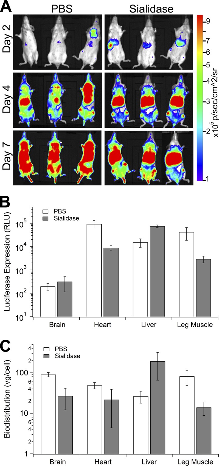
Glycans are key determinants of host range and transmissibility in several pathogens. In the case of adeno-associated viruses (AAV), different carbohydrates serve as cellular receptors in vitro; however, their contributions in vivo are less clear. A particularly interesting example is adeno-associated virus serotype 9 (AAV9), which displays systemic tropism in mice despite low endogenous levels of its primary receptor (galactose) in murine tissues. To understand this further, we studied the effect of modulating glycan binding avidity on the systemic fate of AAV9 in mice. Intravenous administration of recombinant sialidase increased tissue levels of terminally galactosylated glycans in several murine tissues. These conditions altered the systemic tropism of AAV9 into a hepatotropic phenotype, characterized by markedly increased sequestration within the liver sinusoidal endothelium and Kupffer cells. In contrast, an AAV9 mutant with decreased glycan binding avidity displayed a liver-detargeted phenotype. Altering glycan binding avidity also profoundly affected AAV9 persistence in blood circulation. Our results support the notion that high glycan receptor binding avidity appears to impart increased liver tropism, while decreased avidity favors systemic spread of AAV vectors. These findings may not only help predict species-specific differences in tropism for AAV9 on the basis of tissue glycosylation profiles, but also provide a general approach to tailor AAV vectors for systemic or hepatic gene transfer by reengineering capsid-glycan interactions.


Figures

Fig 1
Fig 1
Intravenous sialidase alters endogenous tissue glycosylation patterns in vivo. (A) Fluorescent lectin staining of heart, liver, brain, and skeletal muscle tissues harvested from BALB/c mice. FITC-ECL was used to detect tissue glycans containing terminal β1,4-galactose and FITC-MALI to detect α2,3-sialylated glycans. (B) Fluorescent lectin staining of heart, liver, brain, and skeletal muscle harvested from BALB/c mice administered intravenous sialidase to enzymatically remove α2,3-sialic acid residues in vivo. Confocal micrographs were obtained using a Zeiss 710 confocal laser scanning microscope with a 40× objective at zoom 0.6. Scale bar = 50 μm.
Fig 2
Fig 2
High glycan binding avidity redirects systemic AAV9 to the liver. (A) Live-animal bioluminescence images of AAV9-mediated luciferase transgene expression in BALB/c mice. The animals were injected with PBS (control; left) or recombinant sialidase (right) 3 h prior to AAV9-CBA-luciferase administration via the tail vein. The images were obtained using a Xenogen IVIS Lumina system at 2 days, 4 days, and 7 days postadministration (n = 3). The rainbow scale represents RLU. (B) Luciferase transgene expression in brain, heart, liver, and skeletal muscle tissue lysates. Mice were intravenously injected with PBS or sialidase 3 h prior to injection of AAV9. Tissues were harvested at 2 weeks postadministration. Luciferase transgene expression (RLU) was normalized to total cellular protein in tissue lysates (n = 3). The data are represented as means ± SEM. (C) AAV9 vector genome biodistribution in brain, heart, liver, and skeletal muscle tissues harvested from BALB/c mice pretreated with PBS or sialidase at 3 days postadministration (n = 3). Viral genome copy numbers in different tissues/animals were obtained using quantitative PCR and normalized to the total copy number of the constitutive mouse lamin gene in host genomic DNA. The primer sequences utilized for qPCR are provided in Materials and Methods. The data are represented as means ± SEM.
Fig 3
Fig 3
High glycan avidity potentiates AAV9 sequestration by liver sinusoidal endothelium and Kupffer cells. (A) Liver sections were obtained 15 min after injection of AAV9 in BALB/c mice pretreated with intravenous PBS (middle row) or sialidase (bottom row). Tissue sections were stained with fluorescent lectin to detect β1,4-galactose, FITC-ECL (green); anti-AAV9 capsid antibody, ADK9 (red); and anti-CD16/CD32 endothelial cell marker antibody (magenta). The color images were merged, along with nuclear DAPI staining (blue), to generate the overlay panel. Untreated mouse liver is shown as a control (top row). The large arrows indicate liver sinusoidal endothelial cells, and the small arrows indicate nonendothelial cells. (B) Liver sections obtained from BALB/c mice pretreated with intravenous PBS or sialidase and immunostained with FITC-ECL (green), ADK9 (red), anti-F4/80 Kupffer cell marker antibody (magenta), and nuclear DAPI staining (blue). The large arrows indicate liver sinusoidal endothelial cells, and the small arrows indicate Kupffer cells. Fluorescent micrographs were obtained using a Zeiss 710 confocal laser scanning microscope with a 63× objective. Scale bar = 50 μm.
Fig 4
Fig 4
Low glycan binding avidity detargets AAV9-W503R from the liver. (A) The AAV9-W503R mutant displays decreased binding affinity for terminal galactose residues on the surfaces of CHO Lec2 cells lacking sialic acid (n = 4). Curve fitting based on a single-binding-site model was utilized to obtain binding potential values (Bmax/Kd′) for the mutant and parental AAV9. The data are represented as means ± SEM. (B) Transduction efficiencies of AAV9 and AAV9-W503R on CHO Pro5 (constitutive glycan composition) and Lec2 (sialic acid-deficient) cells at different multiplicities of infection (n = 4). Luciferase transgene expression in cell lysates was analyzed 24 h postinfection; **, P < 0.01; ***, P < 0.005. The data are represented as means ± SEM. (C) AAV9 and AAV9-W503R accumulation in the livers of BALB/c mice at 15 min after intravenous administration. Immunostaining was carried out using anti-AAV9 capsid antibody, ADK9 (red); anti-CD16/CD32 endothelial cell marker (magenta); and nuclear DAPI staining (blue). Fluorescent images were collected as described above. The large arrows indicate liver sinusoidal endothelial cells, and the small arrows indicate nonendothelial cells. Scale bar = 20 μm. (D) Transgene expression (tdTomato reporter) mediated by AAV9 and AAV9-W503R in liver and heart in BALB/c mice (n = 3). Tissues were harvested and imaged 2 weeks postinfection with a 10× objective.
Fig 5
Fig 5
Vascular endothelial cells limit cardiac uptake of AAV9 capsids. Confocal micrographs of cardiac tissue sections harvested from BALB/c mice pretreated with PBS (second row from top) or sialidase (third row) obtained at 15 min after intravenous AAV9 injection. Immunostaining was carried out using FITC-ECL (green), ADK9 (red), anti-CD16/CD32 (magenta), and nuclear DAPI (blue) staining. Colocalization of mutant AAV9-W503R with vascular endothelium in the heart (bottom row) and cardiac tissue sections (top row) from untreated BALB/c mice is also shown. The arrows indicate cardiac endothelium. Scale bar = 50 μm.
Fig 6
Fig 6
Glycan binding avidity affects the blood circulation profile of AAV9. (A) Blood circulation kinetics of the AAV9-W503R mutant or AAV9 in BALB/c mice pretreated with PBS or sialidase. Viral genome copy numbers in blood obtained at 15 min, 30 min, 1 h, 3 h, 6 h, and 24 h postadministration were obtained by quantitative PCR (n = 3). Curve fitting based on a biexponential, two-compartment model was utilized to obtain the pharmacokinetic parameters listed in Table 1. The data are represented as means ± SEM. (B) Proposed mathematical model for predicting the impact of high glycan binding avidity on viral blood clearance and liver sequestration. Rates of viral elimination from the blood (ke) and viral dissemination from the blood to the liver (k12) and the liver to the blood (k21) are shown. The large arrows indicate higher rates of dissemination and clearance from the blood. This model specifically explains the pharmacokinetics of wild-type AAV9 following enzymatic desialylation in mice. (C) Proposed mathematical model for predicting the impact of low glycan binding avidity on viral blood clearance and liver sequestration. Different rates (k values) are shown in Table 3. The dashed arrows indicate lower rates of dissemination and clearance from the blood. This model specifically explains the pharmacokinetics of mutant AAV9-W503R in mice.

Similar articles

Cited by

References

    1. Agbandje-McKenna M, Kleinschmidt J. 2011. AAV capsid structure and cell interactions. Methods Mol. Biol. 807:47–92 - PubMed
    1. Altheide TK, et al. 2006. System-wide genomic and biochemical comparisons of sialic acid biology among primates and rodents: evidence for two modes of rapid evolution. J. Biol. Chem. 281:25689–25702 - PubMed
    1. Asokan A, et al. 2010. Reengineering a receptor footprint of adeno-associated virus enables selective and systemic gene transfer to muscle. Nat. Biotechnol. 28:79–82 - PMC - PubMed
    1. Asokan A, Schaffer DV, Jude Samulski R. 2012. The AAV vector toolkit: poised at the clinical crossroads. Mol. Ther. 20:699–708 - PMC - PubMed
    1. Bell CL, Gurda BL, Van Vliet K, Agbandje-McKenna M, Wilson JM. 2012. Identification of the galactose binding domain of the AAV9 capsid. J. Virol. 86:7326–7333 - PMC - PubMed

Publication types

MeSH terms

LinkOut - more resources