. 2012 Oct;86(19):10384-98.
doi: 10.1128/JVI.00801-12. Epub 2012 Jul 11.

A multivalent adsorption apparatus explains the broad host range of phage phi92: a comprehensive genomic and structural analysis

Affiliations

A multivalent adsorption apparatus explains the broad host range of phage phi92: a comprehensive genomic and structural analysis

David Schwarzer et al. J Virol. 2012 Oct.

Abstract

Bacteriophage phi92 is a large, lytic myovirus isolated in 1983 from pathogenic Escherichia coli strains that carry a polysialic acid capsule. Here we report the genome organization of phi92, the cryoelectron microscopy reconstruction of its virion, and the reinvestigation of its host specificity. The genome consists of a linear, double-stranded 148,612-bp DNA sequence containing 248 potential open reading frames and 11 putative tRNA genes. Orthologs were found for 130 of the predicted proteins. Most of the virion proteins showed significant sequence similarities to proteins of myoviruses rv5 and PVP-SE1, indicating that phi92 is a new member of the novel genus of rv5-like phages. Reinvestigation of phi92 host specificity showed that the host range is not limited to polysialic acid-encapsulated Escherichia coli but includes most laboratory strains of Escherichia coli and many Salmonella strains. Structure analysis of the phi92 virion demonstrated the presence of four different types of tail fibers and/or tailspikes, which enable the phage to use attachment sites on encapsulated and nonencapsulated bacteria. With this report, we provide the first detailed description of a multivalent, multispecies phage armed with a host cell adsorption apparatus resembling a nanosized Swiss army knife. The genome, structure, and, in particular, the organization of the baseplate of phi92 demonstrate how a bacteriophage can evolve into a multi-pathogen-killing agent.


Figures
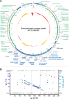
Fig 1
Fig 1
Schematic overview of the phi92 genome. (A) Genome map of phi92. The circular map represents the linear phi92 genome, including both DTRs (drawn as brown boxes). Predicted proteins encoded on the direct or complementary strand are shown in blue or green, respectively. Hypothetical proteins without homologs in other organisms are depicted in pale blue and pale green. The predicted function of selected phi92 proteins is indicated outside and the gene numbers are inside the circle, each in the corresponding color. Underlined names indicate proteins identified by MS (cf. Fig. 3). The cluster of predicted tRNA is shown in black characters. The GC content is depicted in the center of the map, whereas values above or below the average GC content value (37.4%) are shown in orange or red, respectively (see also Datasets S1 and S2 in the supplemental material). (B) Proteome plot of phi92 [gp (phi92)] (x axis) versus protein orthologs from rv5 [magenta, left axis; gp (rv5)] and PVP-SE1 [turquoise, right axis; gp (PVP-SE1)] with more than 35% sequence similarity over the whole protein sequence (see Tables S1 and S2 in the supplemental material) or from other organisms (“other”) or when no homologs were found in the databases (“none”). The three PVP-SE1 proteins (gp030, gp104, and gp204) that are homologous to phi92 gp74 are connected by a turquoise line. The dashed box borders the virion-associated proteins encoded in the structural operon.
Fig 2
Fig 2
Comparison of structural operons of rv5-like phages. (A) Comparison of the structural operons of phi92, rv5, and PVP-SE1 drawn at the same scale (bar, 1 kb). Genes encoding a protein are represented by boxes filled with the respective colors according to the predicted function. Gene numbers are given below the boxes. The PVP-SE1 and rv5 gp numbers are according to a recent publication (63). In contrast, in the genome Genbank file (NC_016071.1) of PVP-SE1, gp015 to gp144 are shifted by one (i.e., gene 16 encodes gp015, etc.). tRNA genes are given as black arrows. Note that gene 66 of phage PVP-SE1 is encoded on the complementary strand. The genes can be grouped according to the locations and functions of their products in the phage virion (bars above) as represented in different colors. Functions assigned to phi92 proteins are given above the phi92 genome data. Gray solid lines and dashed lines between two organisms represent homologous proteins and protein domains, correspondingly, based on BLASTP search analysis (phi92 versus rv5 and PVP-SE1; E-value < 10−5 [cf. Tables S1 and S2 in the supplemental material]) or on recently published data (rv5 versus PVP-SE1) (63), respectively. Proteins conserved in all three phages (underscored gene numbers) are connected by thick gray lines. Terminase gene 65 of phage rv5 contains two introns (red I and II). (B) Identity/similarity matrix based on a CLUSTAL W alignment of 21 homolog proteins of rv5-like phages (gp119, gp120, gp121, gp124, gp126, gp127, gp128, gp130, gp131, gp132, gp133, gp135, gp136, gp137, gp138, gp139, gp140, gp141, gp145, gp146, and gp148 of phi92 and the respective orthologs of rv5 and PVP-SE1 [underscored numbers in panel A]) concatenated into one polypeptide sequence for each phage. (C) Phylogenetic tree based on the CLUSTAL W alignment calculated as described for panel B.
Fig 3
Fig 3
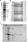
Identification of phi92 virion proteins. (A) 1-DE and MS analysis. Approximately 4 μg of phi92 protein was separated using 14% 1-DE. The marker bands (Precision Plus) on the left indicate the molecular masses. The bar represents 5 mm in the original scale of the gel. (B) The table summarizes the phi92 proteins described for panel A identified by MS. For proteins highlighted by a gray background, the calculated (Calc.) molecular mass (MM) values deviate less than 20% from the apparent (App.) molecular mass value determined using the 1-DE gel as described for panel A. §, proteins identified by single peptide matches. (C) 2-DE and MS analysis. Approximately 800 μg of phi92 protein was separated by isoelectric focusing (pI 3 to 11) followed by 10% SDS-polyacrylamide gel electrophoresis (SDS-PAGE) using 2-DE analysis. The original size of the gel was ∼20 by 30 cm2. The bar represents 2 cm in the original scale. Spots containing E. coli proteins are underscored and italicized. See also Table 2 and Dataset S2 in the supplemental material.
Fig 4
Fig 4
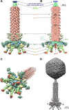
CryoEM structure of phi92. (A) A raw cryoEM image of phi92. Three separate reconstructions were calculated for areas highlighted with boxes colored in yellow, blue, and red. (B) The structure of phi92 capsid with the imposed icosahedral symmetry. On one face of the icosahedron, the HK97 capsid protein structure is fitted into the cryoEM density. (C) The location of the decoration protein gp123 (gold) on the capsid shell formed by gp124 (moss green). The magenta triangle highlights one face of the icosahedron. (D) Interpretation of the capsid cryoEM density with the help of the crystal structure of the HK97 capsid protein. In panels B, C, and D, the cryoEM map is contoured at 1.5 σ from the mean.
Fig 5
Fig 5
Structure of phi92 tail and baseplate. (A and B) Side and cutaway views, respectively, of the phi92 tail structure. A composite map, which includes two overlapping maps (tail sheath and the baseplate) produced separately, is shown. The map is interpreted in terms of the component proteins that were known. (C) A tilted view of the tail colored identically to the views in panels A and B. (D) The structure of phage phi92 produced by combining three reconstructions (the 5-fold-averaged capsid, the 6-fold-averaged tail, and the 6-fold-averaged baseplate). In panels A, B, and C, the cryoEM maps are contoured at 1.5 σ from the mean.

References

    1. Altschul SF, et al. 1997. Gapped BLAST and PSI-BLAST: a new generation of protein database search programs. Nucleic Acids Res. 25:3389–3402 - PMC - PubMed
    1. Altschul SF, et al. 2005. Protein database searches using compositionally adjusted substitution matrices. FEBS J. 272:5101–5109 - PMC - PubMed
    1. Baker TS, Henderson R. 2006. Electron cryomicroscopy, p 451–463 In International tables for crystallography, vol. F Springer, New York, NY
    1. Barry GT, Goebel WF. 1957. Colominic acid, a substance of bacterial origin related to sialic acid. Nature 179:206. - PubMed
    1. Bateman A, Eddy SR, Mesyanzhinov VV. 1997. A member of the immunoglobulin superfamily in bacteriophage T4. Virus Genes 14:163–165 - PubMed

Publication types

MeSH terms

Associated data

LinkOut - more resources