. 2012 Sep;23(17):3473-84.
doi: 10.1091/mbc.E12-02-0132. Epub 2012 Jul 11.

Collagen/β1 integrin signaling up-regulates the ABCC1/MRP-1 transporter in an ERK/MAPK-dependent manner

Affiliations

Collagen/β1 integrin signaling up-regulates the ABCC1/MRP-1 transporter in an ERK/MAPK-dependent manner

Mohammed-Amine El Azreq et al. Mol Biol Cell. 2012 Sep.

Abstract

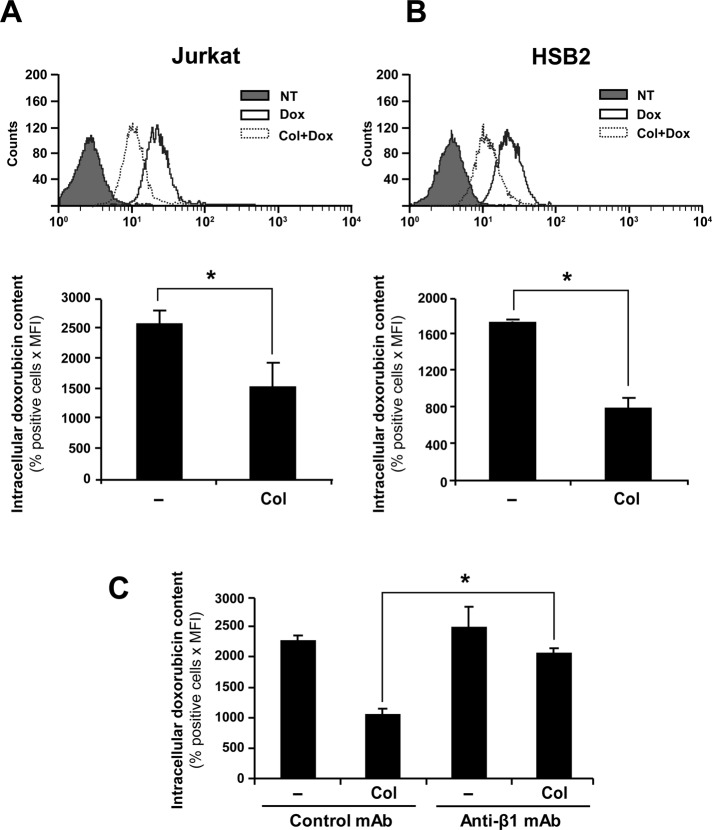
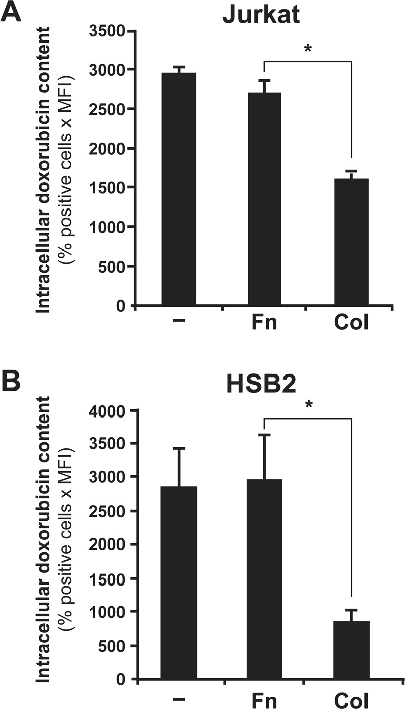
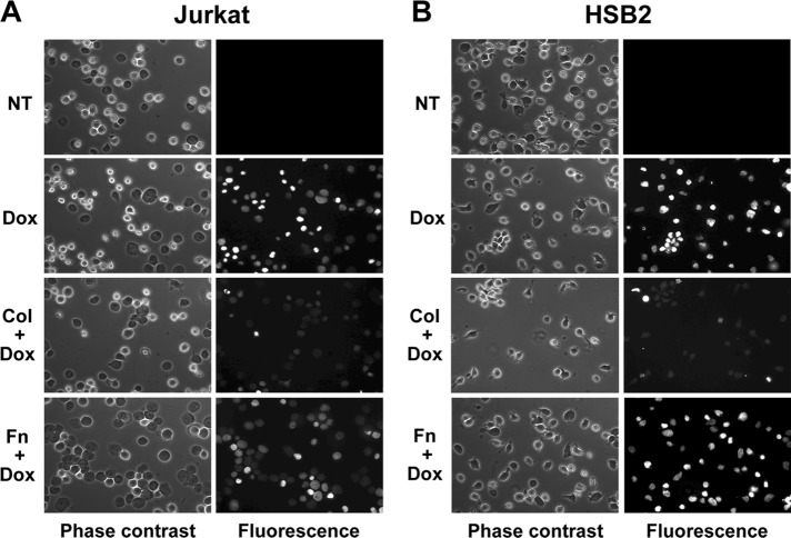
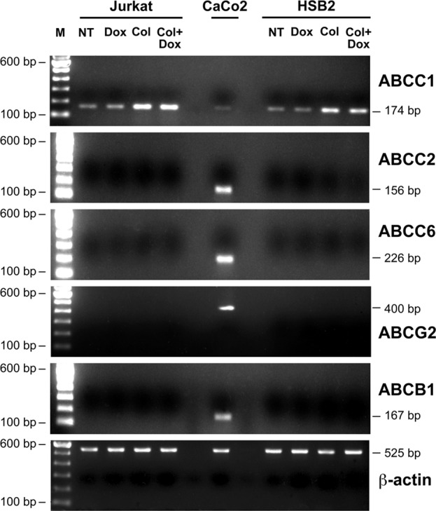
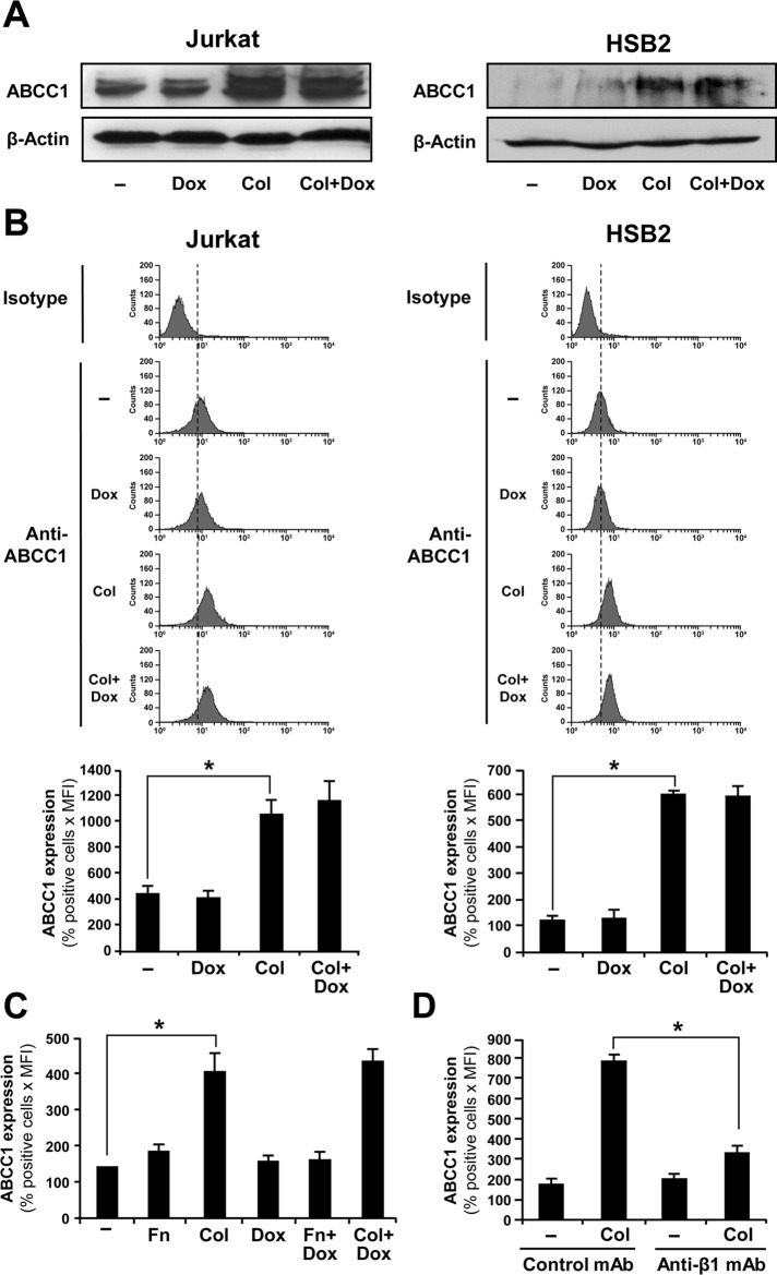
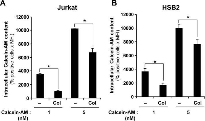
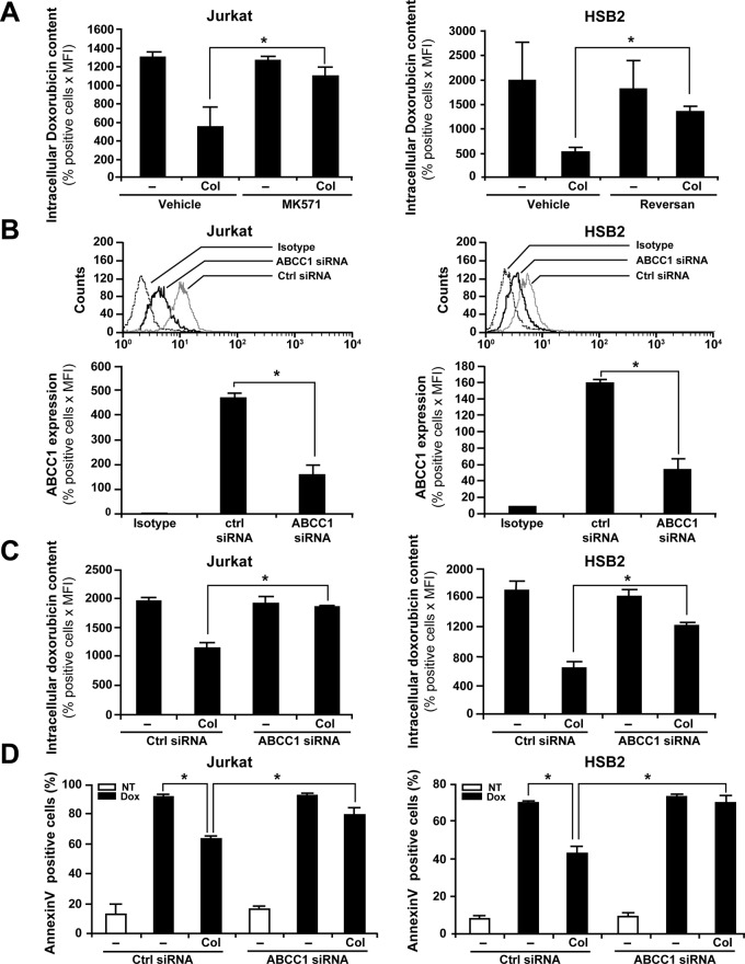
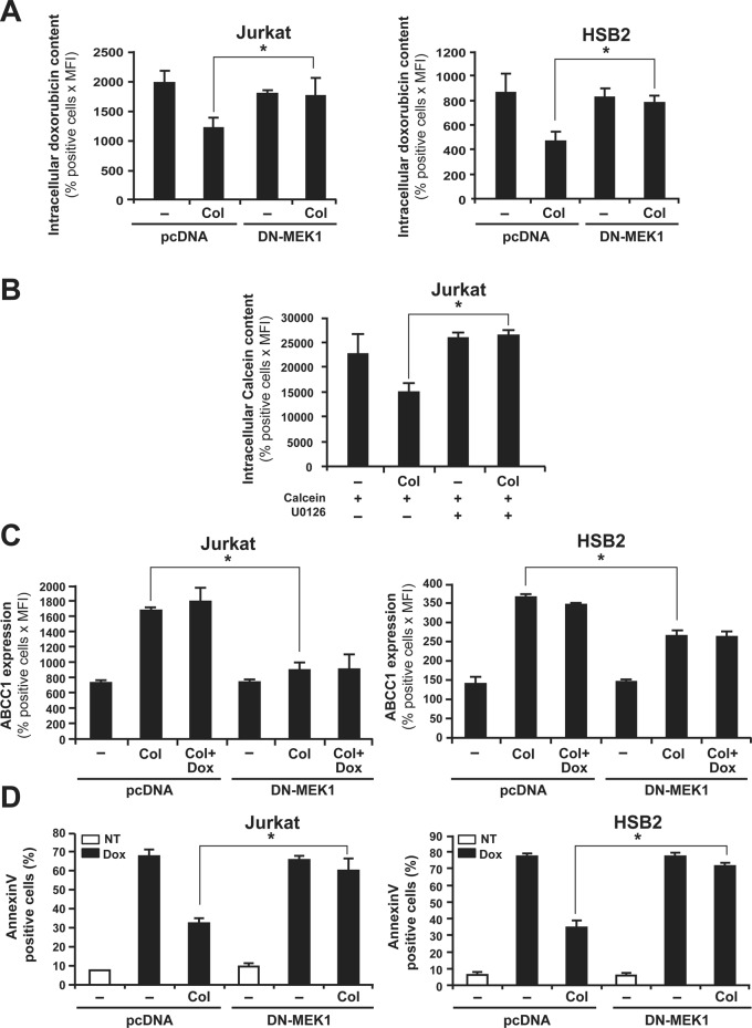
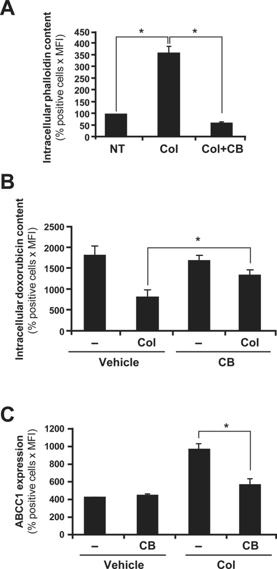
The mechanisms by which β1 integrins regulate chemoresistance of cancer cells are still poorly understood. In this study, we report that collagen/β1 integrin signaling inhibits doxorubicin-induced apoptosis of Jurkat and HSB2 leukemic T-cells by up-regulating the expression and function of the ATP-binding cassette C 1 (ABCC1) transporter, also known as multidrug resistance-associated protein 1. We find that collagen but not fibronectin reduces intracellular doxorubicin content and up-regulates the expression levels of ABCC1. Inhibition and knockdown studies show that up-regulation of ABCC1 is necessary for collagen-mediated reduction of intracellular doxorubicin content and collagen-mediated inhibition of doxorubicin-induced apoptosis. We also demonstrate that activation of the extracellular signal-regulated kinase (ERK)/mitogen-activated protein kinase signaling pathway is involved in collagen-induced reduction of intracellular doxorubicin accumulation, collagen-induced up-regulation of ABCC1 expression levels, and collagen-mediated cell survival. Finally, collagen-mediated up-regulation of ABCC1 expression and function also requires actin polymerization. Taken together, our results indicate for the first time that collagen/β1 integrin/ERK signaling up-regulates the expression and function of ABCC1 and suggest that its activation could represent an important pathway in cancer chemoresistance. Thus simultaneous targeting of collagen/β1 integrin and ABCC1 may be more efficient in preventing drug resistance than targeting each pathway alone.


Figures

FIGURE 1:
FIGURE 1:
Collagen/β1 integrin reduces intracellular amounts of doxorubicin. Jurkat (A) and HSB2 (B) cells were preactivated or not with collagen (Col) for 4 h and then treated with 250 ng/ml doxorubicin (Dox) for an additional 2 h at 37°C in the dark. The cells were then washed with PBS, and the intracellular doxorubicin content was analyzed by flow cytometry using the FL-2 settings. Top, flow cytometric profile of intracellular doxorubicin content (NT, nontreated; cells that had not been treated with doxorubicin). Intracellular doxorubicin content (bottom) was quantified according to the following formula: positive cells (%) times mean fluorescence intensity (MFI). (C) Jurkat cells were pretreated with 10 μg/ml anti-β1 (anti-β1 mAb) or IgG isotypic control antibody (control mAb) for 1 h before being activated with collagen. The cells were then treated with doxorubicin, and intracellular doxorubicin amount was determined as described. The results presented in A and B (bottom) and in C represent mean values ± SD from three independent experiments. *p < 0.05 where indicated.
FIGURE 2:
FIGURE 2:
Fibronectin had no effect on intracellular doxorubicin content. Jurkat (A) and HSB2 (B) cells were preactivated or not with collagen (Col) or fibronectin (Fn) before the addition of doxorubicin. The cells were then washed with PBS, and the intracellular doxorubicin content was quantified by flow cytometry. The results represent mean values ± SD from three independent experiments. *p < 0.05 where indicated.
FIGURE 3:
FIGURE 3:
Collagen but not fibronectin also reduces intracellular doxorubicin content as measured by fluorescence microscopy. Jurkat (A) and HSB2 (B) cells were preactivated with collagen (Col), fibronectin (Fn), or left unstimulated (NT) for 4 h and then were treated or not with doxorubicin (Dox) for 2 h. The cells were washed, fixed, and mounted between slides and cover slips and were observed by phase contrast and by using a red excitation fluorescence filter. The results are representative of three independent experiments.
FIGURE 4:
FIGURE 4:
ABCC1 is the major ABC transporter expressed in leukemic T-cell lines. Jurkat and HSB2 cells were preactivated with collagen (Col) or left unstimulated (NT) for 4 h and were then treated with doxorubicin (Dox) for 2 h. mRNA levels for the different ABC transporters and for β-actin were determined by reverse transcriptase-PCR using specific primers (Table 1). The CaCo2 cell line was used as a positive control for ABCC2, ABCC6, ABCB1, and ABCG2 mRNA detection. M, the 100–base pair DNA ladder marker. The results are representative of three independent experiments.
FIGURE 5:
FIGURE 5:
Collagen but not fibronectin up-regulates ABCC1 protein levels through β1 integrin. (A) Immunoblot analysis of ABCC1 expression. Jurkat (left) and HSB2 (right) cells were preactivated with collagen (Col) for 4 h and then treated with 250 ng/ml doxorubicin (Dox) for 2 h. The cells were lysed and the cell lysates analyzed by immunoblot using the anti-ABCC1 mAb. The blots were stripped and reprobed with control anti–β-actin antibody to ensure equal loading. The results are representative of two independent experiments. (B) Flow cytometry analysis of ABCC1 expression in Jurkat and HSB2 cells. After collagen activation and doxorubicin treatment, the cells were washed with PBS, and ABCC1 expression was assessed by flow cytometry by staining the cells with FITC-coupled anti-ABCC1 antibody or appropriate FITC-conjugated isotypic antibody. Top, representative flow cytometric profiles of ABCC-1 expression. Bottom, quantification of ABCC-1 expression. (C) Fibronectin had no effect on ABCC1 levels. Jurkat cells were preactivated with collagen (Col) or fibronectin (Fn) and then treated or not with doxorubicin (Dox). The cells were then washed with PBS, and ABCC1 expression was assessed by flow cytometry. (D) Collagen-mediated up-regulation of ABCC1 levels is dependent on β1 integrin. Jurkat cells were pretreated with the control or anti–β1 integrin blocking antibodies for 1 h and then activated or not with collagen (Col), and ABCC1 expression was assessed by flow cytometry. The data in B (bottom), C, and D represent mean values ± SD of positive cells (%) times the mean fluorescence intensity from three independent experiments. *p < 0.05 where indicated.
FIGURE 6:
FIGURE 6:
Collagen down-regulates the intracellular accumulation of calcein-AM. Jurkat (A) and HSB2 (B) cells were preactivated or not with collagen (Col) for 4 h and then treated with increasing concentrations of calcein-AM for 30 min at 37°C in the dark. The cells were washed with PBS, and the intracellular calcein-AM content was analyzed by flow cytometry using the FL-1 settings. The results represent mean values ± SD of positive cells (%) times the mean fluorescence intensity from three independent experiments. *p < 0.05 where indicated.
FIGURE 7:
FIGURE 7:
ABCC1 inhibition abrogates collagen-induced drug efflux and protection against doxorubicin-mediated apoptosis. (A) ABCC1 inhibitors reduce collagen-mediated doxorubicin efflux. Jurkat (left) and HSB2 (right) cells were pretreated for 1 h with 20 μM of MK571 or Reversan, respectively, or with the vehicle. The cells were then activated or not with collagen (Col) and treated with doxorubicin. The cells were washed with PBS, and intracellular doxorubicin content was analyzed by flow cytometry using the FL-2 settings. (B) ABCC1 siRNA reduces ABCC1 levels. Jurkat and HSB2 cells were transfected with ABCC1 or control (Ctrl) siRNAs as described in Materials and Methods. Efficiency of ABCC1 silencing in Jurkat and HSB2 cells was monitored by flow cytometry analysis of ABCC1 expression levels using FITC-coupled anti-ABCC1 mAb as described in Materials and Methods. Control isotypic staining of the cells is shown. Bottom, quantification of ABCC1 expression in control and in ABCC1 siRNA-transfected cells. (C) ABCC1 siRNA reduces collagen-mediated doxorubicin efflux. Transfected Jurkat and HSB2 cells were activated or not with collagen (Col) before their treatment with doxorubicin. The cells were washed with PBS, and intracellular doxorubicin content was assessed by flow cytometry. (D) ABCC1 siRNA reverses the protective effect of collagen on doxorubicin-induced apoptosis. Transfected Jurkat and HSB2 cells were left untreated (NT) or activated with collagen (Col) for 4 h and then treated with doxorubicin (Dox). After 16 h of drug treatment, apoptosis was determined by annexin V staining and flow cytometry. The results in the different panels represent mean values ± SD from three independent experiments. *p < 0.05 where indicated.
FIGURE 8:
FIGURE 8:
ERK inhibition abrogates collagen-induced drug efflux, ABCC1 expression, and resistance against doxorubicin-mediated apoptosis. (A) DN-MEK-1 inhibits the ability of collagen to reduce intracellular doxorubicin content. Cells were transfected with DN-MEK1 or pcDNA (empty plasmid). After transfection, the cells were activated or not with collagen (Col) and then treated with doxorubicin. The intracellular doxorubicin content was measured by flow cytometry. (B) The MEK-1 inhibitor abrogates collagen-mediated calcein efflux. Jurkat cells were preincubated or not for 1 h with 10 μM of the MEK/ERK inhibitor (U0126) before their activation or not with collagen (Col) and treatment with calcein-AM. Intracellular calcein content was then assessed by flow cytometry. (C) DN-MEK-1 abolishes the up-regulation of ABCC1 expression levels by collagen. Expression of ABCC1 in transfected cells was determined by ABCC1 staining and flow cytometry analysis as described in Materials and Methods. (D) DN-MEK-1 abolishes the protective effect of collagen on doxorubicin-induced apoptosis. Transfected cells were activated or not with collagen and then treated or not with doxorubicin (Dox) for 16 h (NT, nontreated). Apoptosis was determined by annexin V staining and flow cytometry analysis. The results represent mean values ± SD from three independent experiments. *p < 0.05 where indicated.
FIGURE 9:
FIGURE 9:
Actin polymerization is required for collagen-induced drug efflux and ABCC1 expression. (A) Cytochalasin B (CB) abrogates collagen-induced F-actin formation. Jurkat cells were left untreated (NT) or activated with collagen (Col) in the presence or absence of CB (10 μM). The F-actin content was assessed by staining with Alexa Fluor 594–conjugated phalloidin (1 μM) and flow cytometry analysis as described in Materials and Methods. (B) Actin cytoskeleton disruption abrogates collagen-induced drug efflux. Jurkat cells were activated or not with collagen (Col) in the presence or absence of CB and then treated with doxorubicin. Intracellular doxorubicin content was then assessed by flow cytometry analysis. (C) Actin cytoskeleton disruption abrogates collagen-induced ABCC1 expression. Jurkat cells were activated or not with collagen (Col) in the presence or absence of CB. ABCC1 expression was then assessed by intracellular staining and flow cytometry as described in Materials and Methods. The results represent mean values ± SD of positive cells (%) times the mean fluorescence from three independent experiments. *p < 0.05 where indicated.

Similar articles

Cited by

References

    1. Abdul-Ghani R, Serra V, Gyorffy B, Jurchott K, Solf A, Dietel M, Schafer R. The PI3K inhibitor LY294002 blocks drug export from resistant colon carcinoma cells overexpressing MRP1. Oncogene. 2006;25:1743–1752. - PubMed
    1. An Y, Ongkeko WM. ABCG2: the key to chemoresistance in cancer stem cells? Expert Opin Drug Metab Toxicol. 2009;5:1529–1542. - PubMed
    1. Angelini A, Ciofani G, Baccante G, Di Febbo C, Carmine DI, Cuccurullo F, Porreca E. Modulatory effects of heparin on cellular accumulation and cytotoxicity of doxorubicin in MRP1-overexpressing HL60/doxo cells. Anticancer Res. 2007;27:351–355. - PubMed
    1. Aoudjit F, Guo W, Gagnon-Houde JV, Castaigne JG, Alcaide-Loridan C, Charron D, Al-Daccak R. HLA-DR signaling inhibits Fas-mediated apoptosis in A375 melanoma cells. Exp Cell Res. 2004;299:79–90. - PubMed
    1. Aoudjit F, Vuori K. Engagement of the alpha2beta1 integrin inhibits Fas ligand expression and activation-induced cell death in T cells in a focal adhesion kinase-dependent manner. Blood. 2000;95:2044–2051. - PubMed

Publication types

MeSH terms

Substances