

Sensitization of the trigeminovascular pathway: perspective and implications to migraine pathophysiology
- PMID: 22787491
- PMCID: PMC3391624
- DOI: 10.3988/jcn.2012.8.2.89
Sensitization of the trigeminovascular pathway: perspective and implications to migraine pathophysiology
Abstract
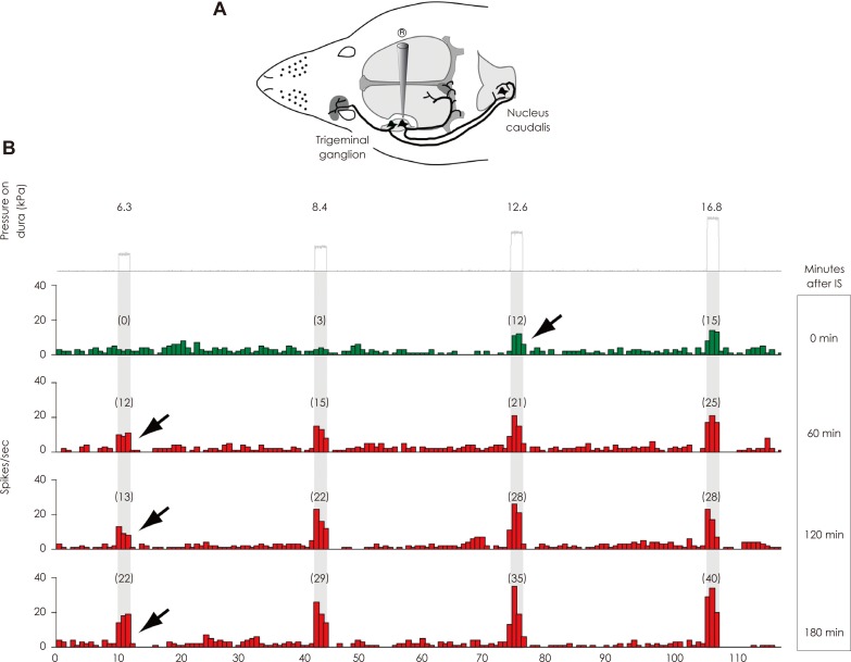
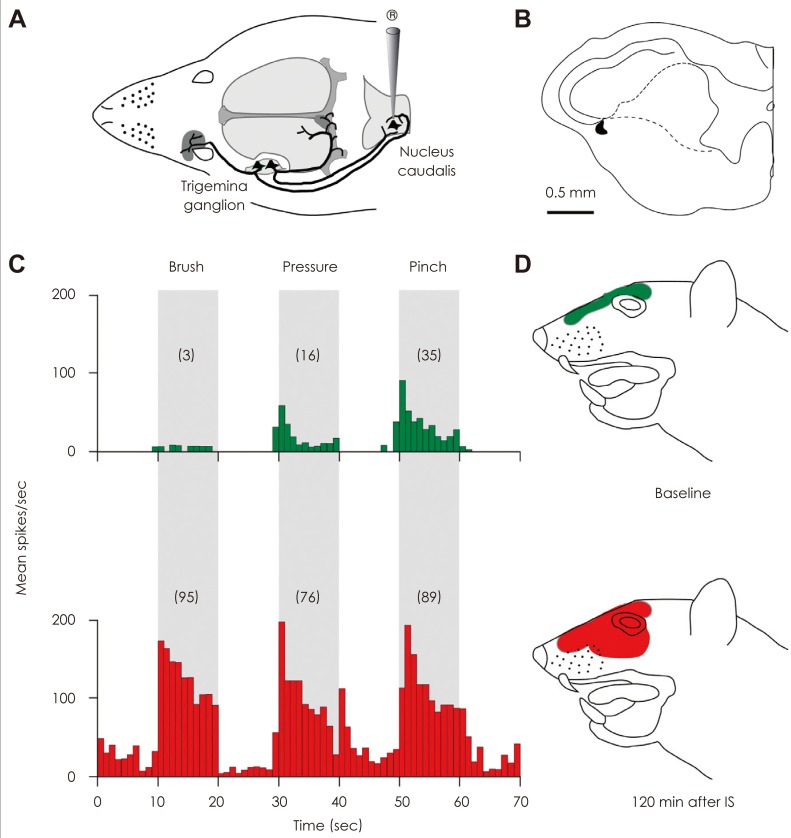
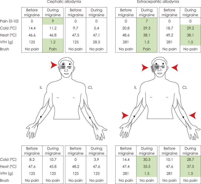
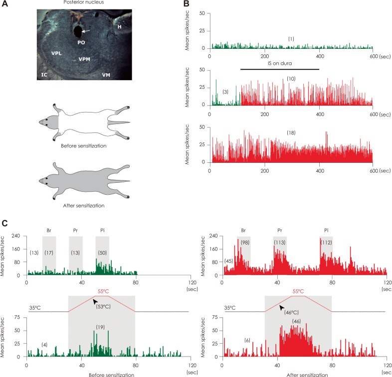
Migraine headache is commonly associated with signs of exaggerated intracranial and extracranial mechanical sensitivities. Patients exhibiting signs of intracranial hypersensitivity testify that their headache throbs and that mundane physical activities that increase intracranial pressure (such as bending over or coughing) intensify the pain. Patients exhibiting signs of extracranial hypersensitivity testify that during migraine their facial skin hurts in response to otherwise innocuous activities such as combing, shaving, letting water run over their face in the shower, or wearing glasses or earrings (termed here cephalic cutaneous allodynia). Such patients often testify that during migraine their bodily skin is hypersensitive and that wearing tight cloth, bracelets, rings, necklaces and socks or using a heavy blanket can be uncomfortable and/or painful (termed her extracephalic cutaneous allodynia). This review summarizes the evidence that support the view that activation of the trigeminovascular pathway contribute to the headache phase of a migraine attack, that the development of throbbing in the initial phase of migraine is mediated by sensitization of peripheral trigeminovascular neurons that innervate the meninges, that the development of cephalic allodynia is propelled by sensitization of second-order trigeminovascular neurons in the spinal trigeminal nucleus which receive converging sensory input from the meninges as well as from the scalp and facial skin, and that the development of extracephalic allodynia is mediated by sensitization of third-order trigeminovascular neurons in the posterior thalamic nuclei which receive converging sensory input from the meninges, facial and body skin.
Keywords: headache; noiciceptrion; pain; thalamus; trigeminal; triptans.
Conflict of interest statement
The authors have no financial conflicts of interest.
Figures
References
-
- Anthony M. The treatment of migraine--old methods, new ideas. Aust Fam Physician. 1993;22:1434–1435. 1438–1439, 1442–1443. - PubMed
-
- Blau JN, Dexter SL. The site of pain origin during migraine attacks. Cephalalgia. 1981;1:143–147. - PubMed
-
- Rasmussen BK, Jensen R, Schroll M, Olesen J. Epidemiology of headache in a general population--a prevalence study. J Clin Epidemiol. 1991;44:1147–1157. - PubMed
-
- Wolff HG. Headache and Other Head Pain. 2nd ed. New York: Oxford University Press; 1963.
-
- Wolff HG, Tunis MM, Goodell H. Studies on headache; evidence of damage and changes in pain sensitivity in subjects with vascular headaches of the migraine type. AMA Arch Intern Med. 1953;92:478–484. - PubMed
Grants and funding
LinkOut - more resources
Full Text Sources
Other Literature Sources
