

A case of an anomalous hypertrophied muscle band in the left ventricle
- PMID: 22787527
- PMCID: PMC3391635
- DOI: 10.4250/jcu.2012.20.2.97
A case of an anomalous hypertrophied muscle band in the left ventricle
Abstract
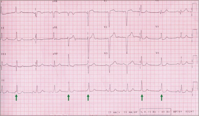
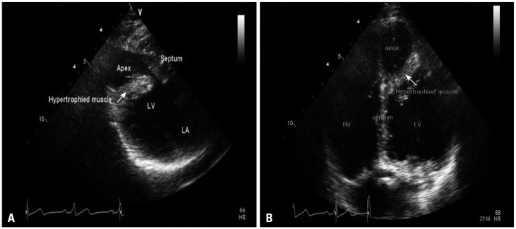
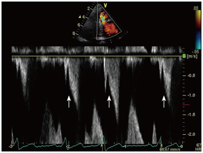
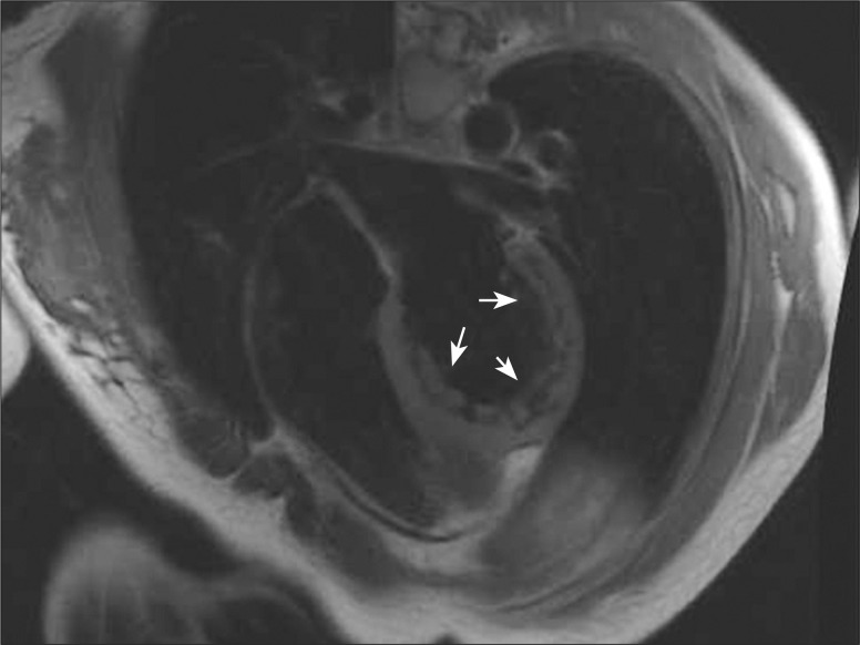
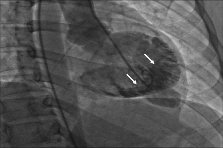
A hypertrophied muscle band (HMB) in the left ventricle (LV), which can be misinterpreted as apical hypertrophic cardiomyopathy, is a rare echocardiographic finding in a patient with normal LV wall thickness. Not only are symptoms produced, but changes in the electrocardiogram (ECG) are limited to the repolarization phase and show no progression even in a large HMB. Hence, we report a case of a 25-year-old woman who visited a local medical clinic due to epigastric discomfort in January 2007. The 24-hour Holter ECG showed multiple premature ventricular complexes. An HMB (3.23 × 10.8 cm) was observed on two-dimensional echocardiography that ran toward the interventricular septum (IVS) across the LV and divided the LV into apical and basal cavities at the apical one-third of the LV. Although LV wall thickness showed normal range, flow acceleration was observed between the HMB and IVS and revealed dagger-shaped with a high pressure gradient up to 30 mmHg in continuous wave Doppler examination. Circumferential band-like myocardial hypertrophy was observed at the LV apex on cardiac magnetic resonance imaging. Myocardial thinning and prominent trabeculae were present from the proximal to distal HMB. However, contractility was normal at the myocardial thinning site, regional wall motion abnormality was not observed in cine images. Focal fatty accumulation was evident at the base of the HMB. Coronary angiography revealed no significant stenosis, whereas left ventriculography showed septation at the apical one-third of the LV. The patient was discharged without any medication.
Keywords: Echocardiography; Left ventricle; Muscles.
Figures
References
-
- Salazar J. Left ventricular anomalous muscle band and electrocardiographic repolarization changes. Pediatr Cardiol. 1997;18:434–436. - PubMed
-
- Maron BJ. Apical hypertrophic cardiomyopathy: the continuing saga. J Am Coll Cardiol. 1990;15:91–93. - PubMed
-
- Sakamoto T, Tei C, Murayama M, Ichiyasu H, Hada Y. Giant T wave inversion as a manifestation of asymmetrical apical hypertrophy (AAH) of the left ventricle. Echocardiographic and ultrasono-cardiotomographic study. Jpn Heart J. 1976;17:611–629. - PubMed
-
- Yamaguchi H, Ishimura T, Nishiyama S, Nagasaki F, Nakanishi S, Takatsu F, Nishijo T, Umeda T, Machii K. Hypertrophic nonobstructive cardiomyopathy with giant negative T waves (apical hypertrophy): ventriculographic and echocardiographic features in 30 patients. Am J Cardiol. 1979;44:401–412. - PubMed
Publication types
LinkOut - more resources
Full Text Sources
Miscellaneous
