

Detection and segmentation of cell nuclei in virtual microscopy images: a minimum-model approach
- PMID: 22787560
- PMCID: PMC3394088
- DOI: 10.1038/srep00503
Detection and segmentation of cell nuclei in virtual microscopy images: a minimum-model approach
Abstract
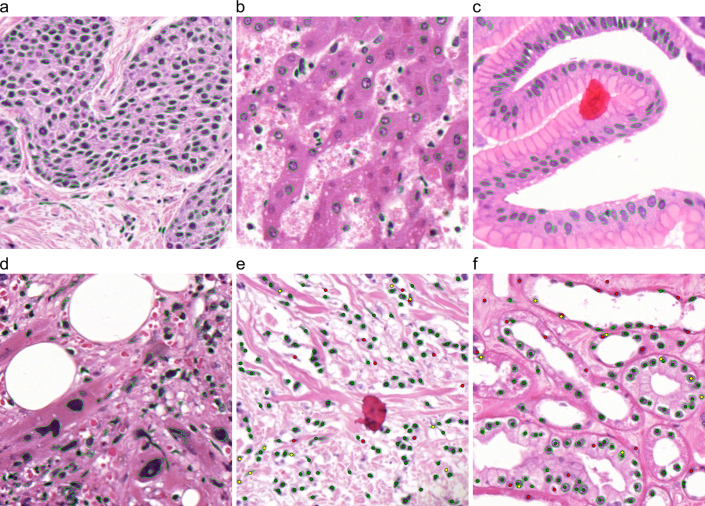
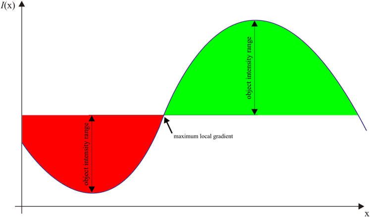
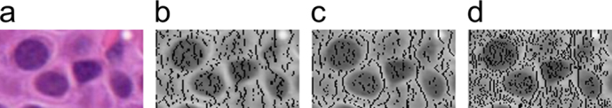
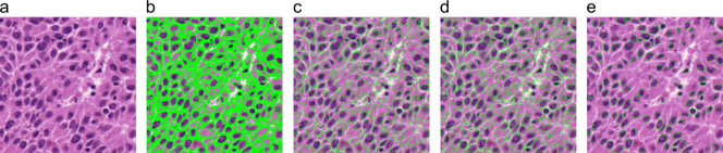
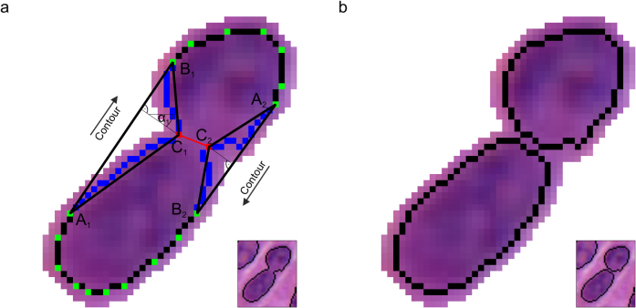
Automated image analysis of cells and tissues has been an active research field in medical informatics for decades but has recently attracted increased attention due to developments in computer and microscopy hardware and the awareness that scientific and diagnostic pathology require novel approaches to perform objective quantitative analyses of cellular and tissue specimens. Model-based approaches use a priori information on cell shape features to obtain the segmentation, which may introduce a bias favouring the detection of cell nuclei only with certain properties. In this study we present a novel contour-based "minimum-model" cell detection and segmentation approach that uses minimal a priori information and detects contours independent of their shape. This approach avoids a segmentation bias with respect to shape features and allows for an accurate segmentation (precision = 0.908; recall = 0.859; validation based on ∼8000 manually-labeled cells) of a broad spectrum of normal and disease-related morphological features without the requirement of prior training.
Figures
References
-
- Bibbo M., Bartels P. H., Dytch H. E. & Wied G. L. Computed cell image information. Monogr Clin Cytol 9, 62–100 (1984). - PubMed
-
- Bengtsson E. The measuring of cell features. Anal. Quant. Cytol. Histol 9, 212–217 (1987). - PubMed
-
- Bamford P. Unsupervised cell nucleus segmentation with active contours. Signal Processing 71, 203–213 (1998).
-
- Bartels P. H., Gahm T. & Thompson D. Automated microscopy in diagnostic histopathology: From image processing to automated reasoning. Int. J. Imaging Syst. Technol 8, 214–223 (1997).
-
- Jiang & Yang An evolutionary tabu search for cell image segmentation. IEEE Trans. Syst. Man, Cybern. B 32, 675–678 (2002). - PubMed
Publication types
MeSH terms
LinkOut - more resources
Full Text Sources
Other Literature Sources
Molecular Biology Databases
