. 2012 Dec;167(7):1431-47.
doi: 10.1111/j.1476-5381.2012.02103.x.

The association of statins plus LDL receptor-targeted liposome-encapsulated doxorubicin increases in vitro drug delivery across blood-brain barrier cells

Affiliations

The association of statins plus LDL receptor-targeted liposome-encapsulated doxorubicin increases in vitro drug delivery across blood-brain barrier cells

Ml Pinzón-Daza et al. Br J Pharmacol. 2012 Dec.

Abstract

Background and purpose: The passage of drugs across the blood-brain barrier (BBB) limits the efficacy of chemotherapy in brain tumours. For instance, the anticancer drug doxorubicin, which is effective against glioblastoma in vitro, has poor efficacy in vivo, because it is extruded by P-glycoprotein (Pgp/ABCB1), multidrug resistance-related proteins and breast cancer resistance protein (BCRP/ABCG2) in BBB cells. The aim of this study was to convert poorly permeant drugs like doxorubicin into drugs able to cross the BBB.

Experimental approach: Experiments were performed on primary human cerebral microvascular endothelial hCMEC/D3 cells, alone and co-cultured with human brain and epithelial tumour cells.

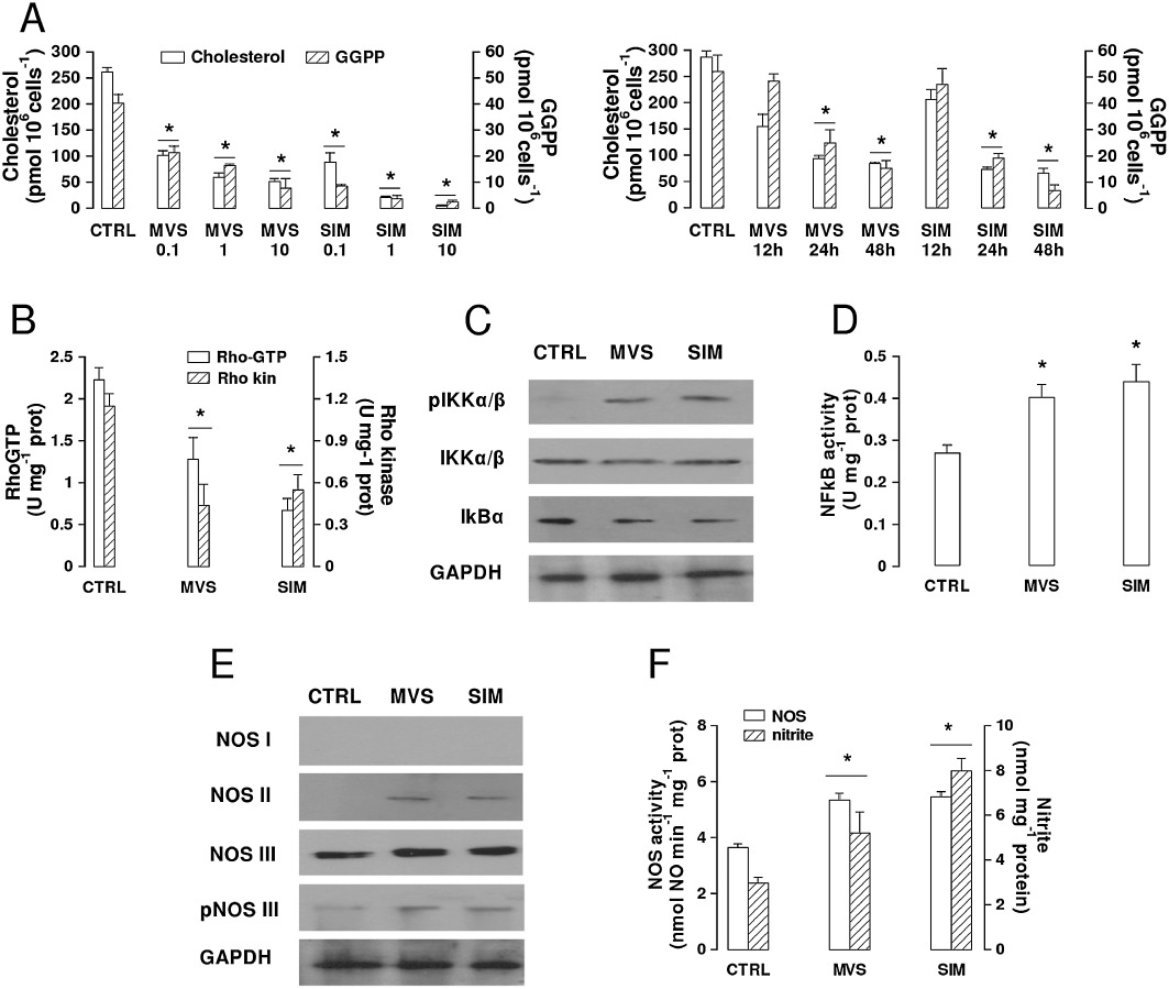
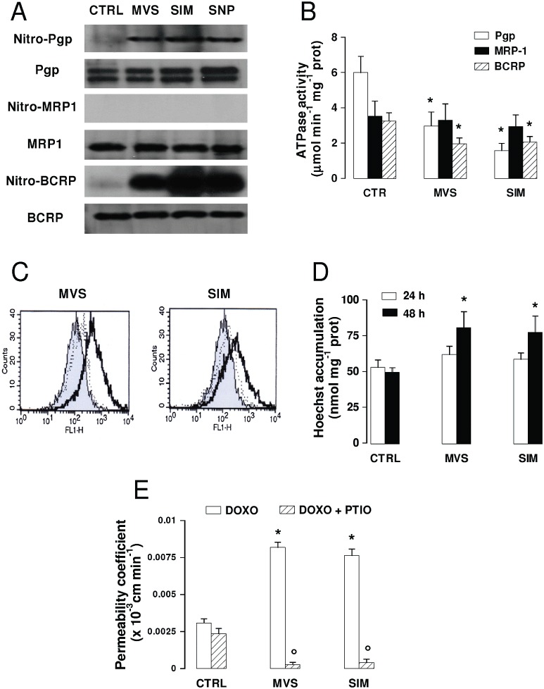
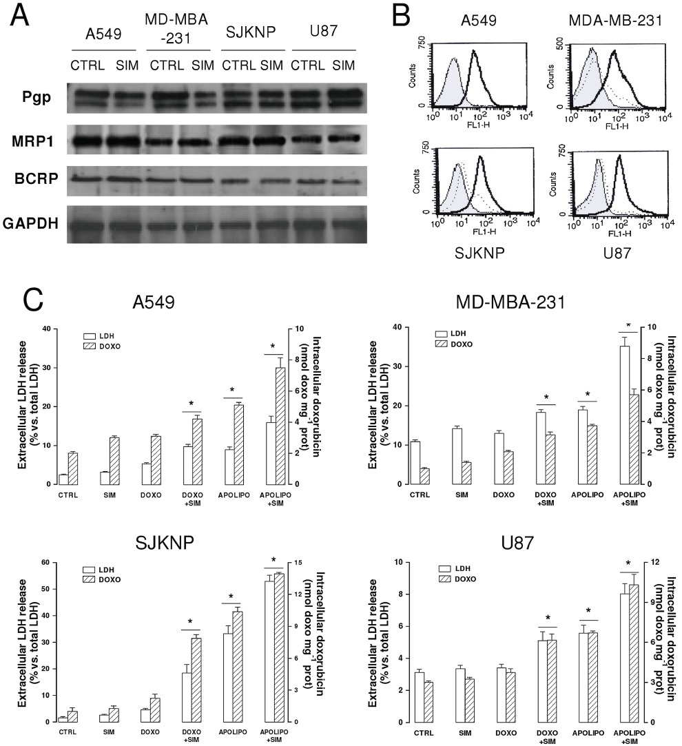
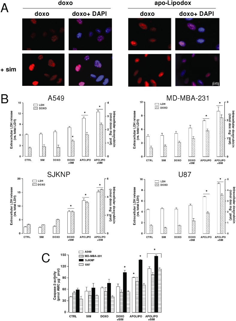
Key results: Statins reduced the efflux activity of Pgp/ABCB1 and BCRP/ABCG2 in hCMEC/D3 cells by increasing the synthesis of NO, which elicits the nitration of critical tyrosine residues on these transporters. Statins also increased the number of low-density lipoprotein (LDL) receptors exposed on the surface of BBB cells, as well as on tumour cells like human glioblastoma. We showed that the association of statins plus drug-loaded nanoparticles engineered as LDLs was effective as a vehicle for non-permeant drugs like doxorubicin to cross the BBB, allowing its delivery into primary and metastatic brain tumour cells and to achieve significant anti-tumour cytotoxicity.

Conclusions and implications: We suggest that our 'Trojan horse' approach, based on the administration of statins plus a LDL receptor-targeted liposomal drug, might have potential applications in the pharmacological therapy of different brain diseases for which the BBB represents an obstacle.


Figures

Figure 1
Figure 1
Effects of statins on cholesterol and GGPP synthesis, RhoA/RhoA kinase activity, NF-kB pathway and NO synthesis in BBB cells. (A) Cholesterol and GGPP synthesis. hCMEC/D3 cells were incubated in the absence (CTRL) or presence of different concentrations of 0.1, 1, 10 µmol·L−1 mevastatin (MVS) and simvastatin (SIM) for 24 h (left panel), or in the presence of 0.1 µmol·L−1statin for 12, 24 or 48 h (right panel). In the subsequent 24 h, the cells were labelled with [3H]-acetate, then cholesterol (open columns) and GGPP (hatched columns) synthesis was measured as reported under Methods. Data are presented as means ± SD (n= 3). Versus CTRL: *P < 0.05. For the subsequent experiments, 0.1 µmol·L−1MVS or SIM for 24 h was used. (B) RhoA/RhoA kinase activity. Samples were subjected to ELISA assays to measure the amount of RhoA-GTP (open bars) and the activity of RhoA kinase (hatched bars). The experiments were performed in duplicate, as described in the Methods section. Data are presented as means ± SD (n= 3). Versus CTRL: *P < 0.05. (C) Western blot detection of phospho-Ser(176/180)-IKK α/β, IKK α/β and IkB-α protein in extracts from hCMEC/D3 cells. The expression of GAPDH was used to check the equal protein loading. The figure is representative of three experiments with superimposable results. (D) NF-kB activity. The activity of NF-kB was detected in the nuclear extracts measuring the DNA-binding capacity of NF-kB on its target sequence (see Methods). Measurements were performed in duplicate and data are presented as means ± SD (n= 3). Versus CTRL: *P < 0.05. (E) Western blot detection of NOS isoforms (nNOS/NOS I; iNOS/NOS II; eNOS/NOS III) and of phospho-(Ser 1177)eNOS/NOS III protein in extracts from hCMEC/D3 cells. The expression of GAPDH was used to check the equal protein loading. The figure is representative of three experiments with superimposable results. (F) NO synthesis. NOS activity in cell lysates and nitrite accumulation in the extracellular medium were measured by use of spectrophotometric assays, as reported in Methods. Data are presented as means ± SD (n= 3). Versus CTRL: *P < 0.05.
Figure 2
Figure 2
Effects of statins on ABC transporters activity and doxorubicin permeability across BBB cells. The hCMEC/D3 cells were incubated in the absence (CTRL) or presence of 0.1 µmol·L−1mevastatin (MVS) or simvastatin (SIM) for 24 h or 48 h, then subjected to the following investigations. (A) Nitration of ABC transporters. After a 24 h incubation, cells were lysed and the whole cell extracts were immunoprecipitated with an anti-nitrotyrosine polyclonal antibody. The immunoprecipitated proteins were subjected to Western blotting, using an anti-Pgp, an anti-MRP1 or an anti-BCRP antibody (see Methods). The NO donor sodium nitroprusside (100 µmol·L−1 for 24 h, SNP) was used as a positive control of nitration. The figure is representative of three experiments with similar results. (B) ATPase activity was measured spectrophotometrically after immunoprecipitation of Pgp, MRP1, BCRP from membrane fractions, as described in the Methods. Measurements were performed in duplicate and data are presented as means ± SD (n= 3). Versus CTRL: *P < 0.05. (C) Rhodamine assay. Cells were incubated for 20 min at 37°C with the fluorescent Pgp substrate rhodamine 123. The intracellular fluorescence was assessed by flow cytometry analysis in untreated cells (grey peak) and in cells treated with statins for 24 h (dotted line) or 48 h (continuous line). The figures shown here are representative of three similar experiments performed in duplicate. (D) Hoechst 33342 assay. Cells were incubated for 15 min at 37°C with Hoechst 33342, lysed and analysed fluorimetrically for the intracellular content of the dye. Measurements were performed in duplicate and data are presented as mean ± SD (n= 3). Versus CTRL: *P < 0.05. (E) Transport of doxorubicin across BBB monolayer. hCMEC/D3 cells were grown up to the confluence in Transwell insert, in fresh medium or in the presence of statins for 48 h, alone or co-incubated with the NO scavenger PTIO (100 µmol·L−1). 5 µmol·L−1doxorubicin was then added in the upper chamber. After 3 h, the medium was recovered by the lower chamber and the amount of doxorubicin was measured fluorimetrically. Measurements were performed in duplicate and data are presented as means ± SD (n= 4). Versus CTRL: *P < 0.05; versus condition without PTIO: °P < 0.001.
Figure 3
Figure 3
Effects of statins on the expression of LDL receptors and on the permeability of LDL receptor-targeted liposomal doxorubicin across the BBB. The hCMEC/D3 cells were incubated in the absence (CTRL) or presence of 0.1 µmol·L−1mevastatin (MVS) or simvastatin (SIM) for 24 h (A, B), 48 h (B, D and E), then subjected to the following investigations. (A) RT-PCR of LDL receptors. Total RNA was extracted, reverse transcribed and amplified by RT-PCR, as indicated in Methods. Measurements were performed in triplicate and data are presented as means ± SD (n= 3). Versus CTRL: *P < 0.005. (B) Flow cytometry analysis of surface LDL receptors in untreated cells (grey peak) or treated with MVS (continuous line) or SIM (dotted line) for 48 h. The figures shown here are representative of 3 similar experiments, performed in triplicate. (C) Apo-Lipodox imaging by TEM. The micrographs (28 500× magnification) are representative of three similar experiments. (D) Transport of apo-Lipodox across the BBB monolayer. The hCMEC/D3 cells were grown to the confluence in Transwell insert, in fresh medium or in the presence of statins or of the NO scavenger PTIO (100 µmol·L−1 for 48 h), then 5 µmol·L−1 apo-Lipodox was added in the upper chamber. After 3 h, the amount of the drug recovered by the lower chamber medium was measured fluorimetrically. Measurements were performed in duplicate and data are presented as means ± SD (n= 4). Versus CTRL: *P < 0.005; versus condition without PTIO: °P < 0.001. (E) Transport of Lipodox and apo-Lipodox across the BBB monolayer. The hCMEC/D3 cells were grown up to the confluence in Transwell insert, in fresh medium or in the presence of SIM, then 5 µmol·L−1 Lipodox or apo-Lipodox was added in the upper chamber. When indicated, the free LDL receptor-targeted apoB100 peptide (the same that was conjugated with apo-Lipodox), was co-incubated with the liposomes (100 µmol·L−1; B100). After 3 h, the amount of doxorubicin recovered by the lower chamber medium was measured fluorimetrically. Measurements were performed in duplicate and data are presented as means ± SD (n= 3). Versus CTRL: *P < 0.005; versus SIM: °P < 0.05.
Figure 4
Figure 4
Characterization of different CNS- and non-CNS-derived tumour cells for ABC transporters expression, surface LDL receptors and resistance to doxorubicin. Human glioblastoma U87-MG cells, human neuroblastoma SJKNP cells, human breast cancer MDA-MB-231, human lung cancer A549 cells were grown in the absence (CTRL) or presence of simvastatin (0.1 µmol·L−1for 48 h; SIM). When indicated, 5 µmol·L−1doxorubicin (DOXO) or apo-Lipodox (APOLIPO) were added in the last 24 h. (A) Western blot analysis of ABC transporters. The expression of Pgp, MRP1 and BCRP on whole cells extracts was detected by Western blotting. The expression of GAPDH was used to check the equal protein loading. The figure is representative of three experiments with superimposable results. (B) Flow cytometry analysis of surface LDL receptors in untreated cells (dotted line) or SIM-treated cells (continuous line). Negative controls, with non-immune isotypic antibodies, are represented by the grey peak. The figures shown here are representative of two similar experiments, each performed in duplicate. (C) Intracellular doxorubicin accumulation and toxicity. Culture supernatant was checked for the extracellular activity of LDH; cells were detached and lysed to quantify the intracellular amount of doxorubicin, as described in Methods. The hatched columns in the conditions ‘CTRL’ and ‘SIM’ refer to the cell autofluorescence, measured in the absence of doxorubicin or apo-Lipodox administration. Measurements were performed in duplicate and data are presented as means ± SD (n= 3). Versus CTRL: *P < 0.05.
Figure 5
Figure 5
Drug delivery and antitumour efficacy of statins plus LDL receptor-targeted liposomal doxorubicin in co-culture models. The hCMEC/D3 cells were grown for 7 days up to confluence in Transwell inserts, whereas U87-MG, SJKNP, A549 or MDA-MB-231 cells were seeded at day 4 in the lower chamber. At day 0, supernatant in the upper chamber was replaced with fresh medium without (CTRL) or with simvastatin (0.1 µmol·L−1for 48 h; SIM). 5 µmol·L−1doxorubicin (DOXO) or apo-Lipodox (APOLIPO) were added in the upper chamber of Transwell in the last 24 h, then the following investigations were performed. (A) Microscope analysis of doxorubicin accumulation. U87-MG cells were seeded on sterile glass coverslips, treated as reported above, then analysed by fluorescence microscopy to detect the intracellular accumulation of doxorubicin. The cells were also counterstained with the nuclear fluorescent probe DAPI. The micrographs are representative of three experiments with similar results. (B) Culture supernatant of tumour cells was checked for the extracellular activity of LDH, cells were detached and lysed to quantify the intracellular amount of doxorubicin, as described in Methods. The hatched columns of the conditions ‘CTRL’ and ‘SIM’ refer to the cell autofluorescence, measured in the absence of doxorubicin or apo-Lipodox administration. Measurements were performed in duplicate and data are presented as means ± SD (n= 4). Versus CTRL: *P < 0.05. (C) Apoptosis induction. The activation of caspase-3 in tumour cells lysates was measured fluorimetrically as described in the Methods section. Measurements were performed in duplicate and data are presented as means ± SD (n= 3). Versus CTRL: *P < 0.05.

Similar articles

Cited by

References

    1. Ahn KS, Sethi G, Chaturvedi MM, Aggarwal BB. Simvastatin, 3-hydroxy-3-methylglutaryl coenzyme A reductase inhibitor, suppresses osteoclastogenesis induced by receptor activator of nuclear factor-kB ligand through modulation of NF-kB pathway. Int J Cancer. 2008;123:1733–1740. - PubMed
    1. Ambruosi A, Khalansky AS, Yamamoto H, Gelperina SE, Begley DJ, Kreuter J. Biodistribution of polysorbate 80-coated doxorubicin-loaded [14C]-poly(butyl cyanoacrylate) nanoparticles after intravenous administration to glioblastoma-bearing rats. J Drug Target. 2006;14:97–105. - PubMed
    1. Ananda S, Nowak AK, Cher L, Dowling A, Brown C, Simes J, et al. Phase 2 trial of temozolomide and pegylated liposomal doxorubicin in the treatment of patients with glioblastoma multiforme following concurrent radiotherapy and chemotherapy. J Clin Neurosci. 2011;18:1444–1448. - PubMed
    1. Bababeygy SR, Polevaya NV, Youssef S, Sun A, Xiong A, Prugpichailers T, et al. HMG-CoA reductase inhibition causes increased necrosis and apoptosis in an in vivo mouse glioblastoma multiforme model. Anticancer Res. 2009;29:4901–4908. - PubMed
    1. Bauer B, Hartz AMS, Miller DS. Tumor necrosis factor α and endothelin-1 increase P-glycoprotein expression and transport activity at the blood-brain barrier. Mol Pharmacol. 2007;71:667–675. - PubMed

Publication types

MeSH terms