. 2012 Sep 13;55(17):7346-59.
doi: 10.1021/jm300346w. Epub 2012 Jul 12.

Druggability analysis and structural classification of bromodomain acetyl-lysine binding sites

Affiliations
Free PMC article

Druggability analysis and structural classification of bromodomain acetyl-lysine binding sites

Lewis R Vidler et al. J Med Chem. .
Free PMC article

Abstract

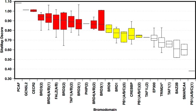
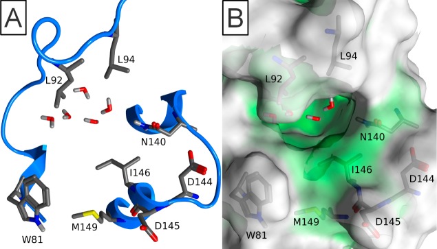
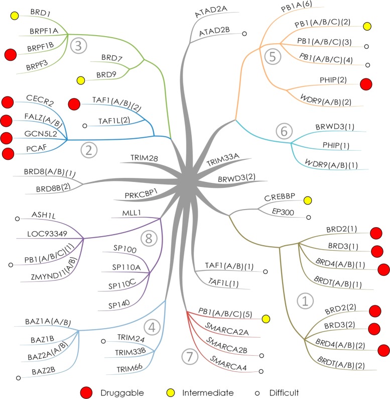
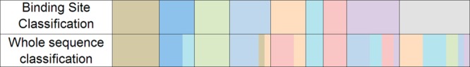
Bromodomains are readers of the epigenetic code that specifically bind acetyl-lysine containing recognition sites on proteins. Recently the BET family of bromodomains has been demonstrated to be druggable through the discovery of potent inhibitors, sparking an interest in protein-protein interaction inhibitors that directly target gene transcription. Here, we assess the druggability of diverse members of the bromodomain family using SiteMap and show that there are significant differences in predicted druggability. Furthermore, we trace these differences in druggability back to unique amino acid signatures in the bromodomain acetyl-lysine binding sites. These signatures were then used to generate a new classification of the bromodomain family, visualized as a classification tree. This represents the first analysis of this type for the bromodomain family and can prove useful in the discovery of inhibitors, particularly for anticipating screening hit rates, identifying inhibitors that can be explored for lead hopping approaches, and selecting proteins for selectivity screening.


Figures

Figure 1
Figure 1
(A) Conserved protein fold of bromodomains comprising the four canonical helices αZ, αA, αB, and αC. (B) Surface representation of a typical KAc binding site. (C) Typical binding of KAc to bromodomain. All illustrated by FALZ (PDB 3QZS).
Figure 2
Figure 2
Selected published bromodomain inhibitors.
Figure 3
Figure 3
Conserved water molecules in the binding site of BRD4(A/B)(1) (PDB 3MXF).
Figure 4
Figure 4
(A) Plot of Dscores obtained when conserved water molecules were included in analysis against the same structures with all water molecules removed. Linear line of best fit added to plot. (B) Histogram of same data showing distribution of scores with normal distribution fitted to this data. Colors indicate druggability classification: red, druggable; yellow, intermediate; white, difficult.
Figure 5
Figure 5
Box-plots showing range and distribution of druggability for each bromodomain across available structures passing imposed filters (including presence of binding site water molecules). Ranked by median Dscore. Colors indicate druggability classification: red, druggable; yellow, intermediate; white, difficult. (a) Four outliers removed from druggability assessment (see group 4 text).
Figure 6
Figure 6
Eight residues around the binding site used in analysis (BRD4(A/B)(1) used as reference, PDB 3MXF). (A) Binding site residues shown as ribbon representation. (B) Binding site residues shown with transparent surface representation.
Figure 7
Figure 7
Bromodomain classification tree generated on the basis of eight binding site amino acid signatures showing bromodomain druggability. Numbering and branch colors consistent with groupings from Table 2. Druggability classification colors consistent with Figure 4 and Figure 5. Druggabilities of ASH1L, ATAD2B, BRPF1B, PB1(A/B/C)(3), PB1(A/B/C)(4), and SMARCA2B assessed using structures failing imposed filters included (see group text).
Figure 8
Figure 8
Comparison between composition of groups from binding site and whole sequence classifications. Groups from left to right in same number order as Table 2 with the same coloring. Whole sequence classification colors generated from binding site classification group which shares highest percentage similarity.
Figure 9
Figure 9
Bromodomain binding site similarity groups exemplified by the surfaces of representative example. Bromodomains aligned with BRD4(A/B)(1) PDB 3MXF and colors generated using MOE Pocket coloring: green = enclosed and white = exposed. Pocket colors are used to highlight binding sites and does not represent pockets identified by SiteMap used for druggability assessment. Images captured from the same viewpoint except image G at the same orientation as Figure 6. (A) BRD4(A/B)(1) PDB 3MXF structure representative of group 1. (B) PCAF PDB 3GG3_B structure representative of group 2. (C) BRD1 PDB 3RCW_A structure representative of Group 3. (D) BAZ2B PDB 3G0L structure representative of group 4. (E) PHIP(2) PDB 3MB3 structure representative of group 5. (F) SMARCA4 PDB 2GRC structure representative of group 7. (G) PB1(A/B/C)(1) PDB 3IU5 structure representative of group 8. (H) CREBBP PDB 3P1C_B structure.
Figure 10
Figure 10
Comparison of BRD2(2) structures. Surface colors consistent with Figure 9. (A) BRD2(2) PDB 3E3K_C structure showing typical BET family conformation. (B) BRD2(2) PDB 2E3K_B structure showing atypical conformation. (C) CREBBP PDB 3DWY_A structure showing similarity to BRD2(2) atypical conformation.
Figure 11
Figure 11
(A) PB1(A/B/C)(5) structure representative of Group 7. (B) PB1(A/B/C)(5) structure demonstrating unusual conformation. (C) Overlaid backbones of usual conformation (PDB 3G0J_A) in green and unusual (PDB 3G0J_B) in blue. Orientation and coloring consistent with Figure 9.

Comment in

References

    1. Kouzarides T. Chromatin Modifications and Their Function. Cell 2007, 128, 693–705. - PubMed
    1. Jenuwein T.; Allis C. D. Translating the Histone Code. Science 2001, 293, 1074–1080. - PubMed
    1. Zeng L.; Zhou M. M. Bromodomain: An acetyl-lysine binding domain. FEBS Lett. 2002, 513, 124–128. - PubMed
    1. Filippakopoulos P.; Picaud S.; Mangos M.; Keates T.; Lambert J.-P.; Barsyte-Lovejoy D.; Felletar I.; Volkmer R.; Müller S.; Gingras A.-C.; Pawson T.; Arrowsmith C.; Knapp S. Histone Recognition and Large-Scale Structural Analysis of the Human Bromodomain Family. Cell 2012, 149, 214–231. - PMC - PubMed
    1. Mutjaba S.; Zeng L.; Zhou M. M. Structure and acetyl-lysine recognition of the bromodomain. Oncogene 2007, 26, 5521–5527. - PubMed

Publication types