

High bone turnover assessed by 18F-fluoride PET/CT in the spine and sacroiliac joints of patients with ankylosing spondylitis: comparison with inflammatory lesions detected by whole body MRI
- PMID: 22788874
- PMCID: PMC3472173
- DOI: 10.1186/2191-219X-2-38
High bone turnover assessed by 18F-fluoride PET/CT in the spine and sacroiliac joints of patients with ankylosing spondylitis: comparison with inflammatory lesions detected by whole body MRI
Abstract
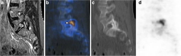
Background: This study compares the frequency and distribution of increased activity on 18 F-fluoride PET/CT with the presence of bone marrow edema on whole-body MR imaging in the spine and sacroiliac joints (SIJ) of patients with active ankylosing spondylitis (AS).
Methods: Ten patients (6 men and 4 women), between 30 and 58 years old (median 44) with active AS, were prospectively examined with both whole-body MRI and 18 F-fluoride PET/CT. Patients fulfilled modified NY criteria and had a Bath Ankylosing Spondylitis Disease Activity Index (BASDAI) of at least 4. Increased radiotracer uptake in PET/CT and bone marrow edema in whole-body MRI of spine and SIJ was evaluated independently by two blinded observers for each modality. Kappa statistics were used to compare interobserver agreement as well as scores of consensus reading of the two imaging modalities.
Results: Analysis of interobserver agreement for PET/CT yielded a kappa value of 0.68 for spinal lesions and of 0.88 for SIJ lesions. The corresponding kappa values for the MRI modality were 0.64 and 0.93, respectively. More spinal lesions were detected by MRI in comparison to PET/CT (68 vs. 38), whereas a similar number of SIJ quadrants scored positive in both modalities (19 vs. 17). Analysis of agreement of lesion detection between both imaging modalities yielded a kappa value of only 0.25 for spinal lesions and of 0.64 for SIJ lesions.
Conclusion: Increased 18 F-fluoride uptake in PET/CT is only modestly associated with bone marrow edema on MRI in the spine and SIJ of patients with AS, suggesting different aspects of bone involvement in AS.
Figures


References
LinkOut - more resources
Full Text Sources
Research Materials
