

Neonatal desensitization supports long-term survival and functional integration of human embryonic stem cell-derived mesenchymal stem cells in rat joint cartilage without immunosuppression
- PMID: 22788986
- PMCID: PMC3528094
- DOI: 10.1089/scd.2012.0116
Neonatal desensitization supports long-term survival and functional integration of human embryonic stem cell-derived mesenchymal stem cells in rat joint cartilage without immunosuppression
Abstract
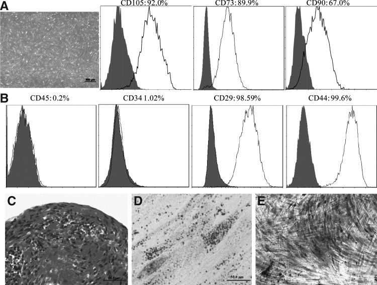
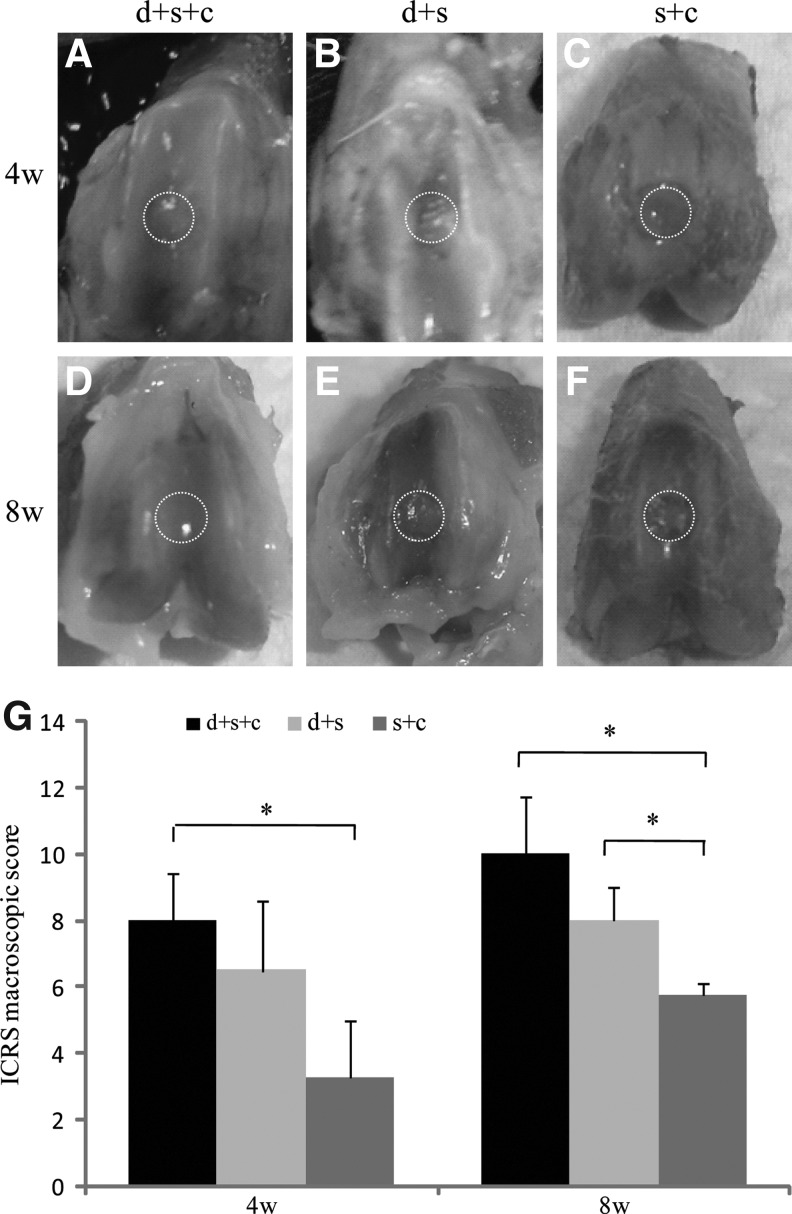
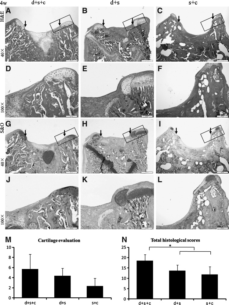
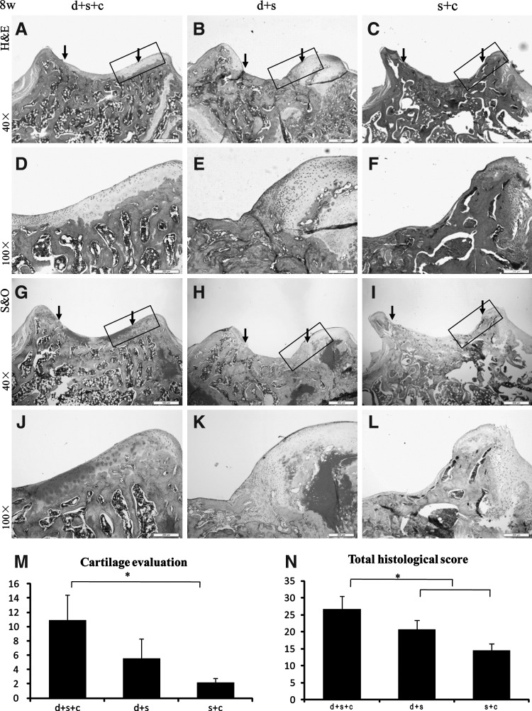
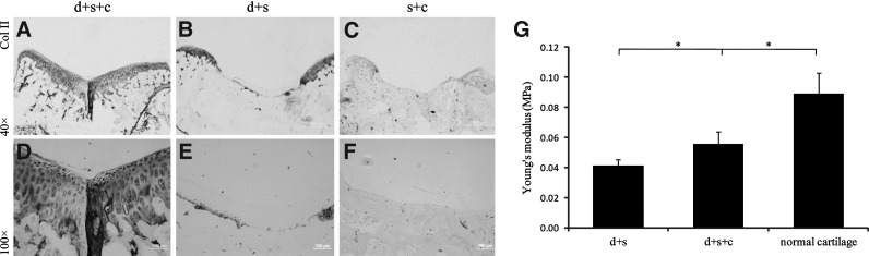
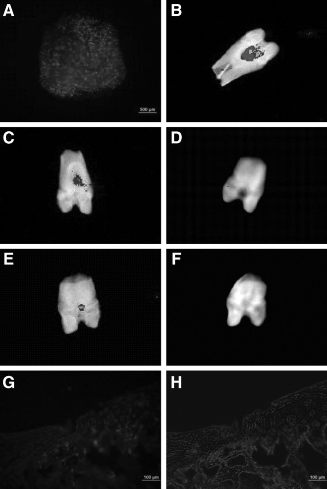
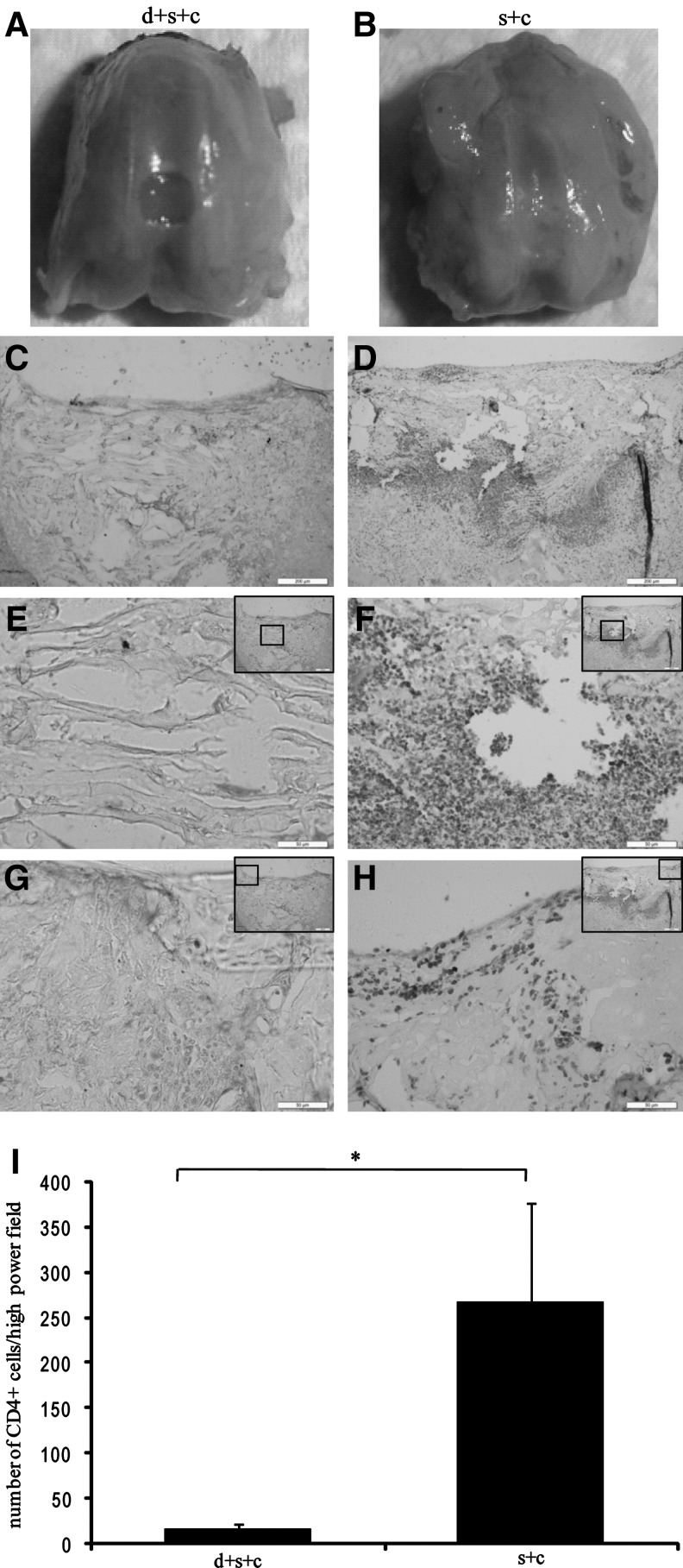
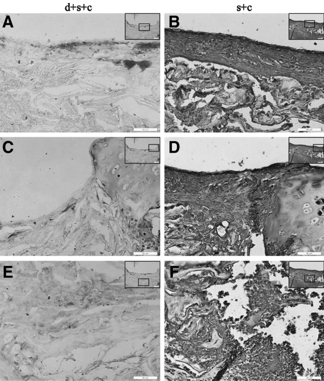
Immunological response hampers the investigation of human embryonic stem cells (hESCs) or their derivates for tissue regeneration in vivo. Immunosuppression is often used after surgery, but exhibits side effects of significant weight loss and allows only short-term observation. The purpose of this study was to investigate whether neonatal desensitization supports relative long-term survival of hESC-derived mesenchymal stem cells (hESC-MSCs) and promotes cartilage regeneration. hESC-MSCs were injected on the day of birth in rats. Six weeks after neonatal injection, a full-thickness cylindrical cartilage defect was created and transplanted with a hESC-MSC-seeded collagen bilayer scaffold (group d+s+c) or a collagen bilayer scaffold (group d+s). Rats without neonatal injection were transplanted with the hESC-MSC-seeded collagen bilayer scaffold to serve as controls (group s+c). Cartilage regeneration was evaluated by histological analysis, immunohistochemical staining, and biomechanical test. The role of hESC-MSCs in cartilage regeneration was analyzed by CD4 immunostaining, cell death detection, and visualization of human cells in regenerated tissues. hESC-MSCs expressed CD105, CD73, CD90, CD29, and CD44, but not CD45 and CD34, and possessed trilineage differentiation potential. Group d+s+c exhibited greater International Cartilage Repair Society (ICRS) scores than group d+s or group s+c. Abundant collagen type II and improved mechanical properties were detected in group d+s+c. There were less CD4+ inflammatory cell infiltration and cell death at week 1, and hESC-MSCs were found to survive as long as 8 weeks after transplantation in group d+s+c. Our study suggests that neonatal desensitization before transplantation may be an efficient way to develop a powerful tool for preclinical study of human cell-based therapies in animal models.
Figures
Similar articles
-
Comparative analysis of the therapeutic effects of mesenchymal stem cells and exosomes on cartilage regeneration: exploring their synergistic potential with hyaluronic acid for treating articular cartilage defects.Eur J Orthop Surg Traumatol. 2025 Apr 10;35(1):154. doi: 10.1007/s00590-025-04284-7. Eur J Orthop Surg Traumatol. 2025. PMID: 40210743
-
Scleraxis-overexpressed human embryonic stem cell-derived mesenchymal stem cells for tendon tissue engineering with knitted silk-collagen scaffold.Tissue Eng Part A. 2014 Jun;20(11-12):1583-92. doi: 10.1089/ten.TEA.2012.0656. Epub 2014 Feb 6. Tissue Eng Part A. 2014. PMID: 24328506
-
Enrichment of human ESC-derived multipotent mesenchymal stem cells with immunosuppressive and anti-inflammatory properties capable to protect against experimental inflammatory bowel disease.Stem Cells. 2011 Feb;29(2):251-62. doi: 10.1002/stem.569. Stem Cells. 2011. PMID: 21732483
-
Mesenchymal stem cells in cartilage regeneration.Curr Stem Cell Res Ther. 2014;9(6):469-88. doi: 10.2174/1574888x09666140709111444. Curr Stem Cell Res Ther. 2014. PMID: 25005451 Review.
-
The potential role of genetically-modified pig mesenchymal stromal cells in xenotransplantation.Stem Cell Rev Rep. 2014 Feb;10(1):79-85. doi: 10.1007/s12015-013-9478-8. Stem Cell Rev Rep. 2014. PMID: 24142483 Free PMC article. Review.
Cited by
-
Chondrogenesis of human amniotic fluid stem cells in Chitosan-Xanthan scaffold for cartilage tissue engineering.Sci Rep. 2021 Feb 4;11(1):3063. doi: 10.1038/s41598-021-82341-x. Sci Rep. 2021. PMID: 33542256 Free PMC article.
-
BMSCs reduce rat granulosa cell apoptosis induced by cisplatin and perimenopause.BMC Cell Biol. 2013 Mar 19;14:18. doi: 10.1186/1471-2121-14-18. BMC Cell Biol. 2013. PMID: 23510080 Free PMC article.
-
Intrauterine desensitization enables long term survival of human oligodendrocyte progenitor cells without immunosuppression.iScience. 2023 Apr 11;26(5):106647. doi: 10.1016/j.isci.2023.106647. eCollection 2023 May 19. iScience. 2023. PMID: 37168574 Free PMC article.
-
Regeneration of Articular Cartilage by Human ESC-Derived Mesenchymal Progenitors Treated Sequentially with BMP-2 and Wnt5a.Stem Cells Transl Med. 2017 Jan;6(1):40-50. doi: 10.5966/sctm.2016-0020. Epub 2016 Aug 5. Stem Cells Transl Med. 2017. PMID: 28170184 Free PMC article.
-
The use of mesenchymal stem cells for cartilage repair and regeneration: a systematic review.J Orthop Surg Res. 2017 Mar 9;12(1):39. doi: 10.1186/s13018-017-0534-y. J Orthop Surg Res. 2017. PMID: 28279182 Free PMC article.
References
-
- Brittberg M. Lindahl A. Nilsson A. Ohlsson C. Isaksson O. Peterson L. Treatment of deep cartilage defects in the knee with autologous chondrocyte transplantation. N Engl J Med. 1994;331:889–895. - PubMed
-
- Kino-Oka M. Maeda Y. Sato Y. Maruyama N. Takezawa Y. Khoshfetrat AB. Sugawara K. Taya M. Morphological evaluation of chondrogenic potency in passaged cell populations. J Biosci Bioeng. 2009;107:544–551. - PubMed
-
- McNickle AG. L'Heureux DR. Yanke AB. Cole BJ. Outcomes of autologous chondrocyte implantation in a diverse patient population. Am J Sports Med. 2009;37:1344–1350. - PubMed
-
- Wakitani S. Aoki H. Harada Y. Sonobe M. Morita Y. Mu Y. Tomita N. Nakamura Y. Takeda S. Watanabe TK. Tanigami A. Embryonic stem cells form articular cartilage, not teratomas, in osteochondral defects of rat joints. Cell Transplant. 2004;13:331–336. - PubMed
-
- Dattena M. Pilichi S. Rocca S. Mara L. Casu S. Masala G. Manunta L. Manunta A. Passino ES. Pool RR. Cappai P. Sheep embryonic stem-like cells transplanted in full-thickness cartilage defects. J Tissue Eng Regen Med. 2009;3:175–187. - PubMed
Publication types
MeSH terms
Substances
LinkOut - more resources
Full Text Sources
Other Literature Sources
Research Materials
Miscellaneous
