. 2012 Aug;36(4):2547-55.
doi: 10.1111/j.1460-9568.2012.08161.x. Epub 2012 Jul 12.

Identification of chronic stress-activated regions reveals a potential recruited circuit in rat brain

Affiliations

Identification of chronic stress-activated regions reveals a potential recruited circuit in rat brain

Jonathan N Flak et al. Eur J Neurosci. 2012 Aug.

Abstract

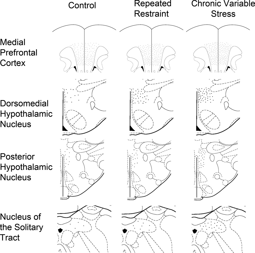
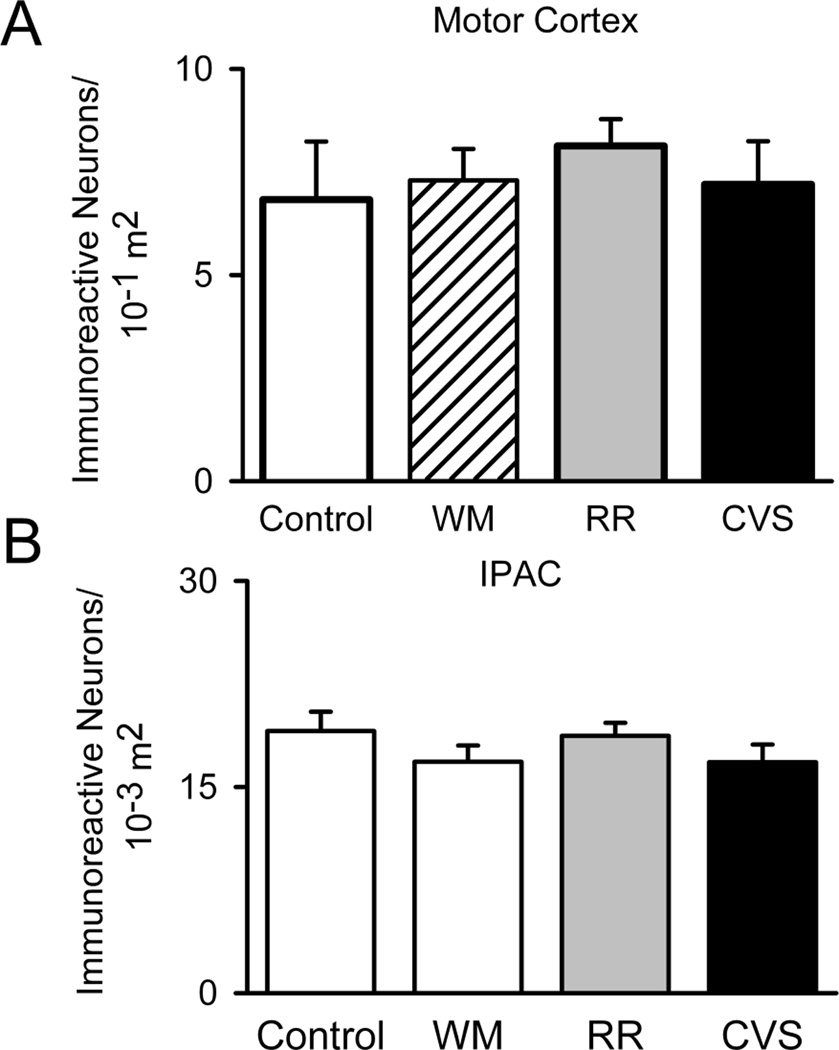
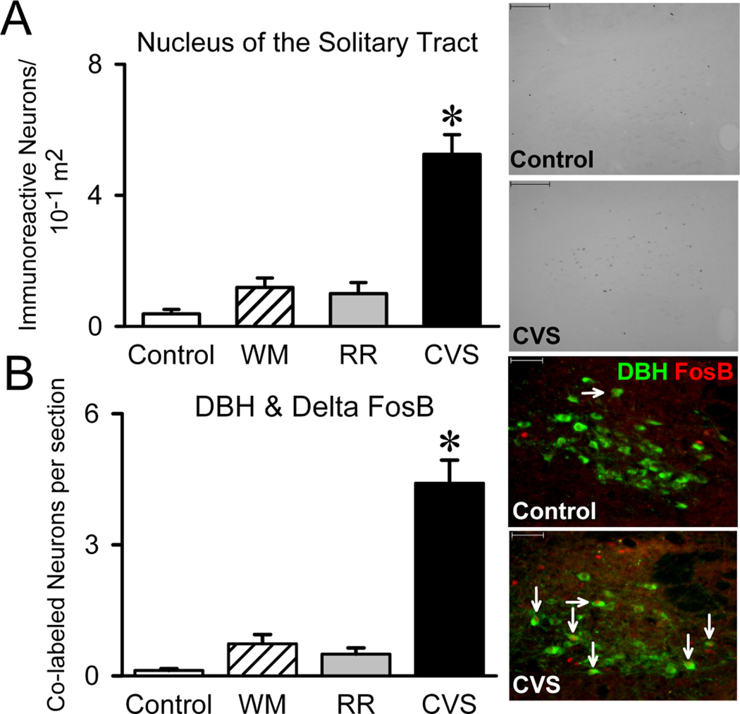
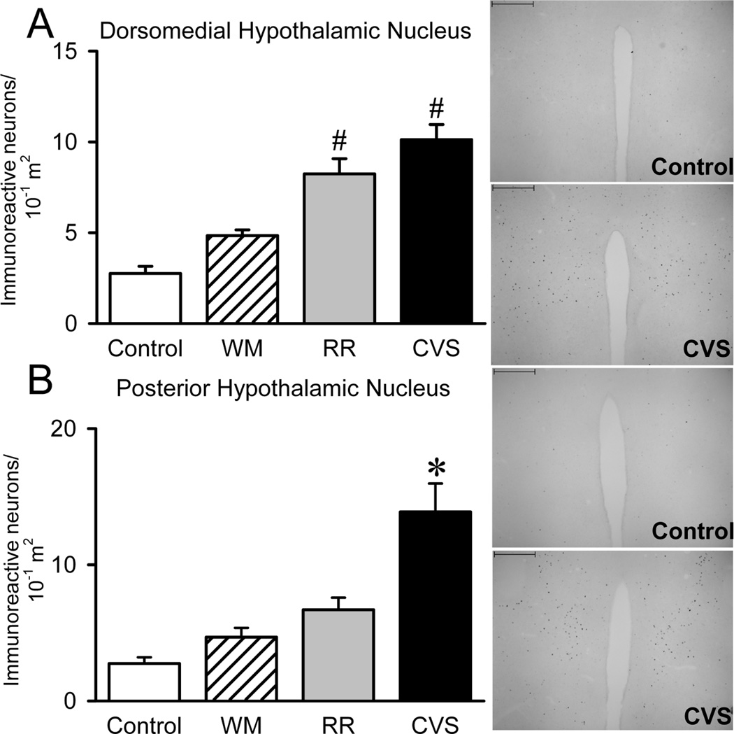
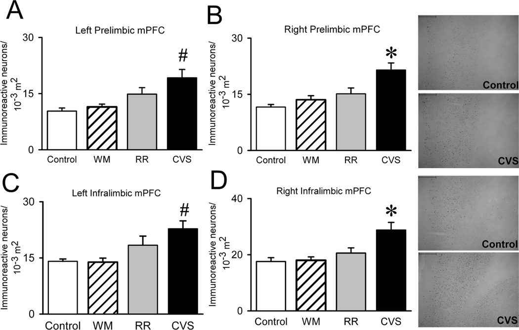
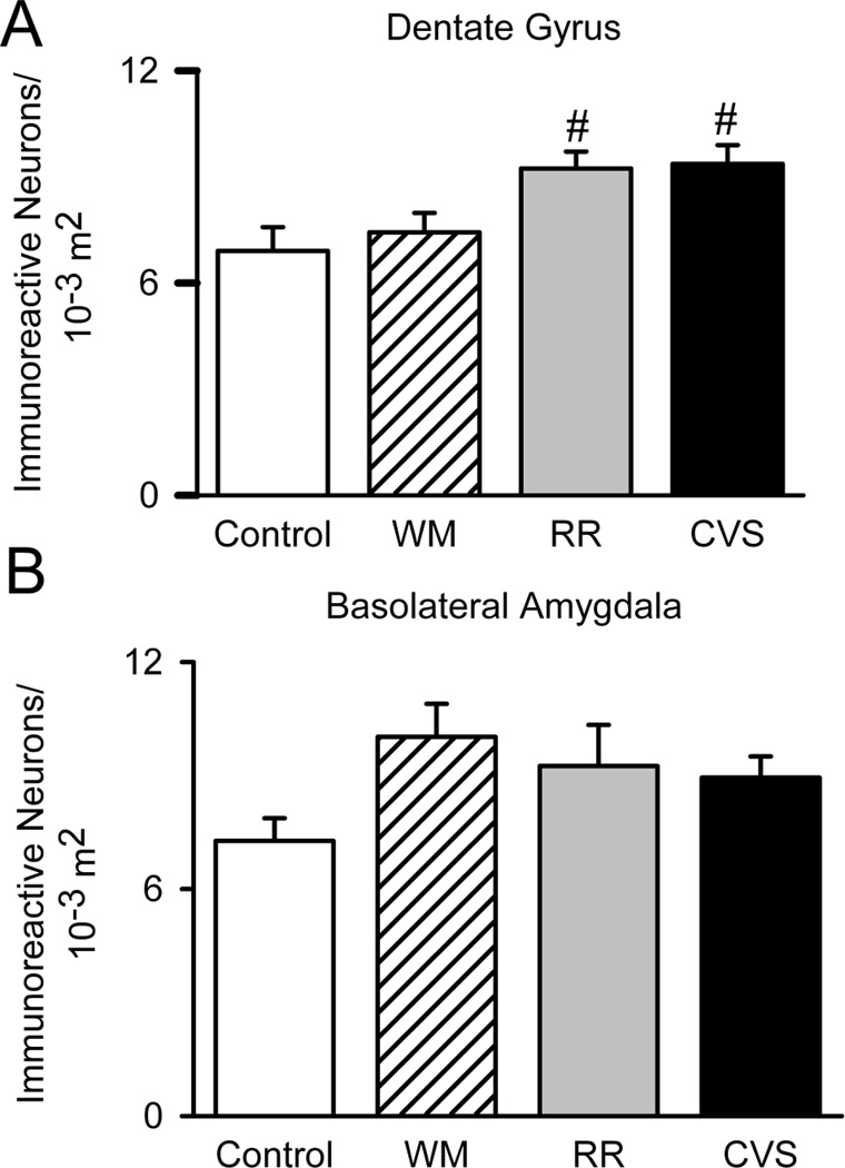
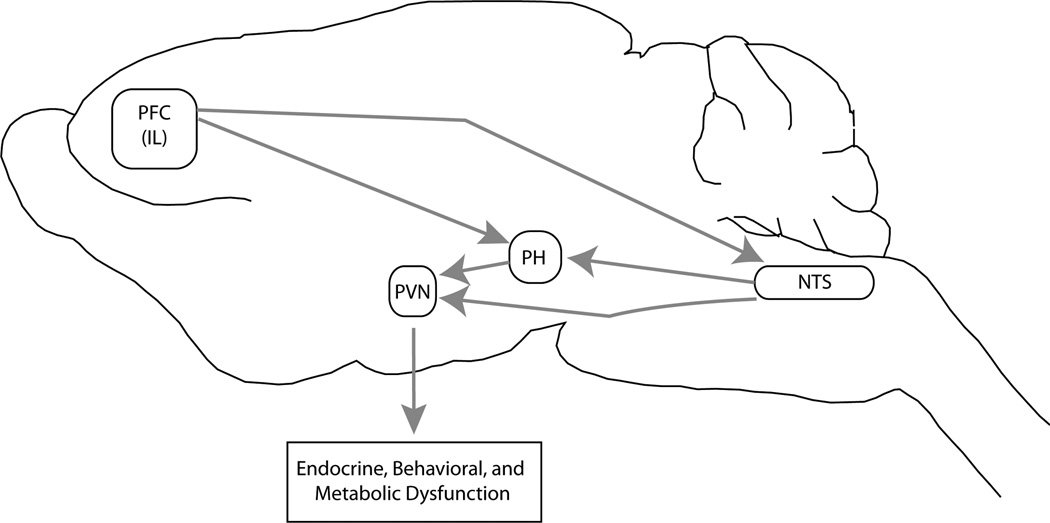
Chronic stress induces presynaptic and postsynaptic modifications in the paraventricular nucleus of the hypothalamus that are consistent with enhanced excitatory hypothalamo-pituitary-adrenocortical (HPA) axis drive. The brain regions mediating these molecular modifications are not known. We hypothesized that chronic variable stress (CVS) tonically activates stress-excitatory regions that interact with the paraventricular nucleus of the hypothalamus, culminating in stress facilitation. In order to identify chronically activated brain regions, ΔFosB, a documented marker of tonic neuronal activation, was assessed in known stress regulatory limbic and brainstem sites. Four experimental groups were included: CVS, repeated restraint (RR) (control for HPA habituation), animals weight-matched (WM) to CVS animals (control for changes in circulating metabolic factors due to reduced weight gain), and non-handled controls. CVS, (but not RR or WM) induced adrenal hypertrophy, indicating that sustained HPA axis drive only occurred in the CVS group. CVS (but not RR or WM) selectively increased the number of FosB/ΔFosB nuclei in the nucleus of the solitary tract, posterior hypothalamic nucleus, and both the infralimbic and prelimbic divisions of the medial prefrontal cortex, indicating an involvement of these regions in chronic drive of the HPA axis. Increases in FosB/ΔFosB-immunoreactive cells were observed following both RR and CVS in the other regions (e.g. the dorsomedial hypothalamus), suggesting activation by both habituating and non-habituating stress conditions. The data suggest that unpredictable stress uniquely activates interconnected cortical, hypothalamic, and brainstem nuclei, potentially revealing the existence of a recruited circuitry mediating chronic drive of brain stress effector systems.


Figures

Figure 1
Figure 1
Schematic Representation of FosB/ΔFosB immunoreactive neurons induced by chronic stress. Using modified images collected from Paxinos and Watson Atlas (Paxinos and Watson, 1997), these schematics illustrate the distribution and relative induction by CVS and RR within the medial prefrontal cotex (mPFC), dorsomedial hypothalamic nucleus (DMH), posterior hypothalamus (PH), and caudal nucleus of the solitary tract (NTS). Each dot represents one cell per unit area and corresponds to a similar density relative to the other regions despite the varying areas presented in the schematic images. Thus, the dots appear smaller in the mPFC because the displayed area is much larger than the hypothalamic regions.
Figure 2
Figure 2
Control Regions. To control for the possibility that the chronic stress regimens globally activate neural structures, we quantified FosB/ΔFosB within the interstitial nucleus of the posterior limb of the anterior commissure (IPAC) (Control n=8, WM n=9, RR n=7, and CVS n=7) and motor cortex (Control n=8, WM n=9, RR n=7, and CVS n=7). The groups did not differ in the number of FosB/ΔFosB immunoreactive nuclei within these two regions. Unit area for IPAC was in 10−3 m2 and 10−1 m2 for motor cortex.
Figure 3
Figure 3
Nucleus of the Solitary Tract. The A2 region of the nucleus of the solitary tract (NTS) supplies the majority of the medial parvocellular PVN with norepinephrine. Thus, we examined FosB/ΔFosB within the NTS (Control n=7, WM n=9, RR n=8, and CVS n=8). CVS increased the number of FosB/ΔFosB immunoreactive neurons within the NTS and especially within DBH-positive neurons (Control n=7, WM n=9, RR n=8, and CVS n=8). Since WM and RR animals did not alter FosB/ΔFosB, the data suggests that unpredictable stress recruits the NTS. * denotes group significantly different from all groups. Unit area was in 10−1 m2.
Figure 4
Figure 4
Hypothalamic Nuclei. Since the dorsomedial (DMH) and posterior hypothalamic (PH) nuclei are two regions that supply the PVN with significant amounts of glutamate, we analyzed FosB/ΔFosB within these regions. CVS increased the number of FosB/ΔFosB immunoreactive neurons within the DMH (Control n=8, WM n=9, RR n=8, and CVS n=8) and PH (Control n=4, WM n=7, RR n=6, and CVS n=7), suggesting that unpredictable recruits the DMH and PH. * denotes group significantly different from all groups.# denotes group significantly different from control group. Unit area for DMH and PH were 10−1 m2.
Figure 5
Figure 5
Medial Prefrontal Cortex. Because side and subregion of the mPFC have both been shown to differentially regulate responses to stress, we divided the mPFC into both left and right infralimbic and prelimbic mPFC to locate the specific chronic stress-activated areas of the mPFC. However, CVS elevated the FosB/ΔFosB expression within the right infralimbic (Control n=8, WM n=9, RR n=8, and CVS n=8), right prelimbic (Control n=7, WM n=9, RR n=8, and CVS n=8), left infralimbic (Control n=7, WM n=9, RR n=8, and CVS n=8), and left prelimbic (Control n=8, WM n=9, RR n=8, and CVS n=8), indicating that unpredictable stress recruits all subregions of the mPFC. * denotes group significantly different from all groups. Unit area for mPFC was in 10−3 m2.
Figure 6
Figure 6
Upstream Limbic Structures. Since chronic stress exposure classically re-wires these stress regulatory regions, we analyzed FosB/ΔFosB within the Basolateral Amygdala (Control n=7, WM n=8, RR n=8, and CVS n=8) and Dentate Gyrus (Control n=8, WM n=9, RR n=8, and CVS n=7). Chronic stress did not alter the number of FosB/ΔFosB immunoreactive neurons within the Basolateral Amygdala. However, both RR and CVS increased the number of FosB/ΔFosB immunoreactive neurons in the dentate gyrus, suggesting that both unpredictable and predictable stress recruits the dentate gyrus.# denotes group significantly different from control group. Unit area for basolateral amygdala and dentate gyrus was in 10−3 m2.
Figure 7
Figure 7
Potential Chronic stress recruited circuitry. Our data suggest the recruitment of a neural circuit underlying chronic drive of the HPA axis, but future studies will have to verify whether these recruited neurons are specifically connected. This circuit begins with the activation of the prefrontal cortex (PFC) projecting to the nucleus of the solitary tract (NTS), which drive neurons within the posterior hypothalamic nucleus (PH). The PH activates the PVN via direct glutamatergic projections to the paraventricular nucleus of the hypothalamus (PVN), known to be a player in endocrine, behavioral, and metabolic homeostasis. Via this pathway, chronic stress may produce endocrine, behavioral, and metabolic dysfunction.

Similar articles

Cited by

References

    1. Abrahamson EE, Moore RY. The posterior hypothalamic area: chemoarchitecture and afferent connections. Brain Res. 2001;889(1–2):1–22. - PubMed
    1. Akana SF, Dallman MF, Bradbury MJ, Scribner KA, Strack AM, Walker CD. Feedback and facilitation in the adrenocortical system: unmasking facilitation by partial inhibition of the glucocorticoid response to prior stress. Endocrinology. 1992;131(1):57–68. - PubMed
    1. Andrus BM, Blizinsky K, Vedell PT, Dennis K, Shukla PK, Schaffer DJ, Radulovic J, Churchill GA, Redei EE. Gene expression patterns in the hippocampus and amygdala of endogenous depression and chronic stress models. Mol Psychiatry. 2010 - PMC - PubMed
    1. Bailey TW, Dimicco JA. Chemical stimulation of the dorsomedial hypothalamus elevates plasma ACTH in conscious rats. Am J Physiol Regul Integr Comp Physiol. 2001;280(1):R8–R15. - PubMed
    1. Barsegyan A, Mackenzie SM, Kurose BD, McGaugh JL, Roozendaal B. Glucocorticoids in the prefrontal cortex enhance memory consolidation and impair working memory by a common neural mechanism. Proc Natl Acad Sci U S A. 2010;107(38):16655–16660. - PMC - PubMed

Publication types

Substances