. 2012 Oct;33(29):7028-38.
doi: 10.1016/j.biomaterials.2012.06.051. Epub 2012 Jul 11.

A hydrogel derived from decellularized dermal extracellular matrix

Affiliations

A hydrogel derived from decellularized dermal extracellular matrix

Matthew T Wolf et al. Biomaterials. 2012 Oct.

Abstract

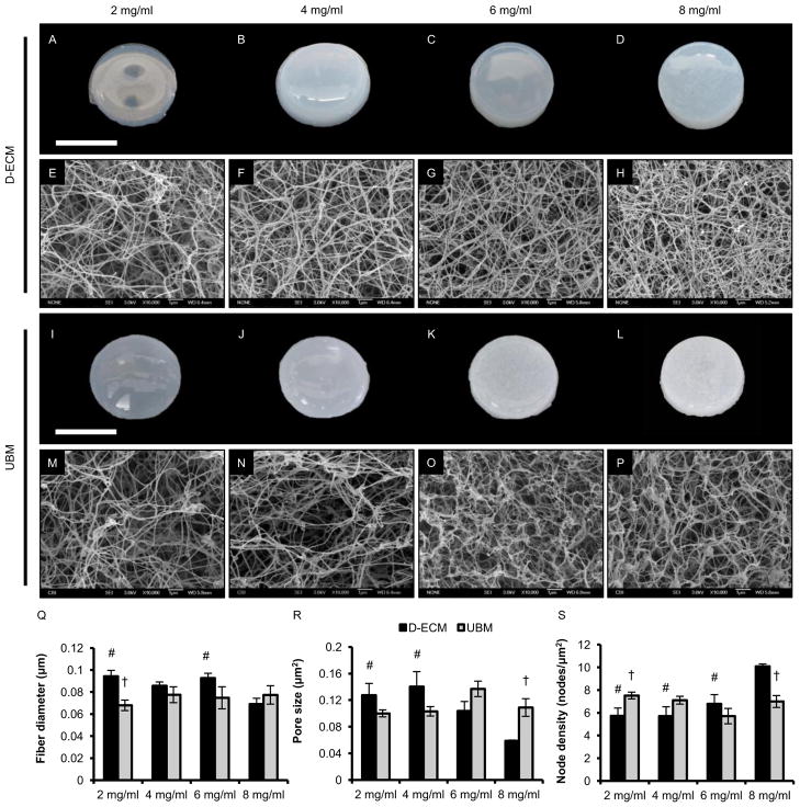
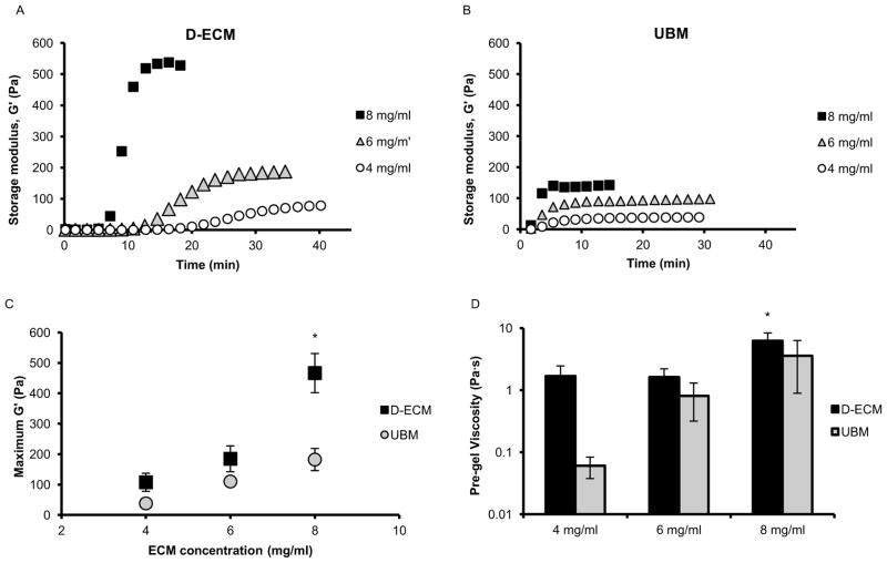
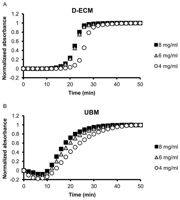
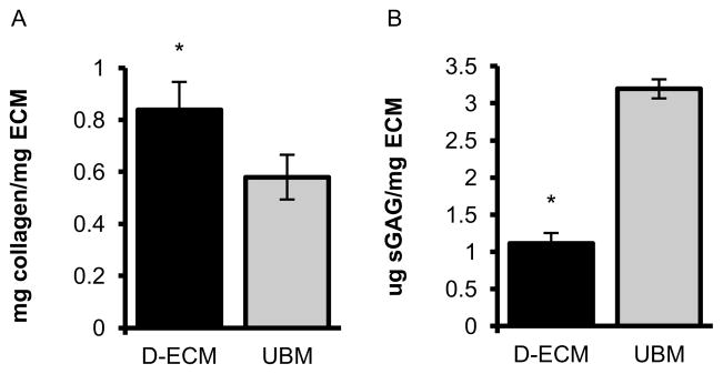
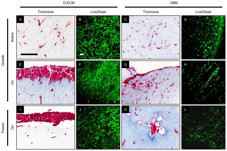
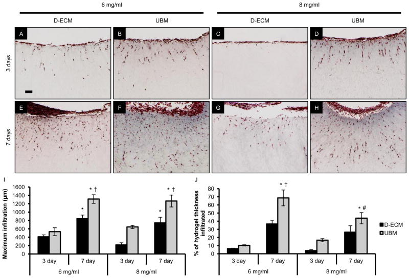
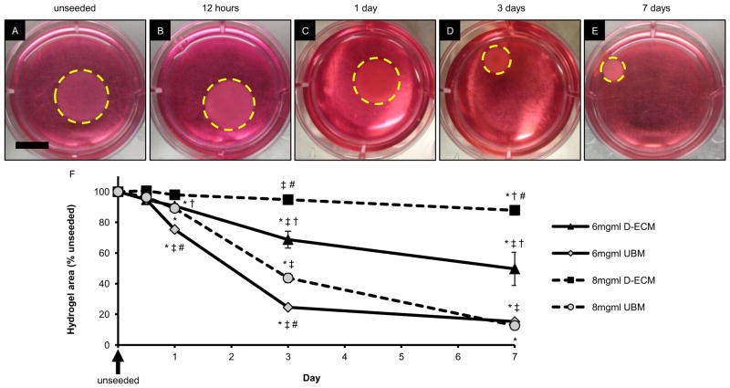
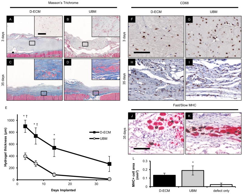
The ECM of mammalian tissues has been used as a scaffold to facilitate the repair and reconstruction of numerous tissues. Such scaffolds are prepared in many forms including sheets, powders, and hydrogels. ECM hydrogels provide advantages such as injectability, the ability to fill an irregularly shaped space, and the inherent bioactivity of native matrix. However, material properties of ECM hydrogels and the effect of these properties upon cell behavior are neither well understood nor controlled. The objective of this study was to prepare and determine the structure, mechanics, and the cell response in vitro and in vivo of ECM hydrogels prepared from decellularized porcine dermis and urinary bladder tissues. Dermal ECM hydrogels were characterized by a more dense fiber architecture and greater mechanical integrity than urinary bladder ECM hydrogels, and showed a dose dependent increase in mechanical properties with ECM concentration. In vitro, dermal ECM hydrogels supported greater C2C12 myoblast fusion, and less fibroblast infiltration and less fibroblast mediated hydrogel contraction than urinary bladder ECM hydrogels. Both hydrogels were rapidly infiltrated by host cells, primarily macrophages, when implanted in a rat abdominal wall defect. Both ECM hydrogels degraded by 35 days in vivo, but UBM hydrogels degraded more quickly, and with greater amounts of myogenesis than dermal ECM. These results show that ECM hydrogel properties can be varied and partially controlled by the scaffold tissue source, and that these properties can markedly affect cell behavior.


Figures

Figure 1
Figure 1
Macroscopic appearance, surface topology, and fiber network analysis of ECM hydrogels. ECM pepsin digests were pH neutralized and injected into 1.38 cm inner diameter rings at 37°C for one hour. Macroscopic images were obtained and hydrogels were processed for scanning electron microscopy. Scanning electron micrographs were obtained at 10,000X magnification for D-ECM and UBM hydrogels prepared at ECM concentrations of 8, 6, 4, and 2 mg/ml. SEM images were analyzed using an automated fiber tracking algorithm to determine the average fiber diameter, pore size, and node density of ECM hydrogels at each concentration. # denotes significance from the 8 mg/ml concentration of the same ECM type and † denotes significance between D-ECM and UBM at the same concentration (p < 0.05). Scale bar for macroscopic images represents 1 cm.
Figure 2
Figure 2
Rheological characterization of ECM hydrogels. Representative curves of the gelation kinetics of D-ECM (A) and UBM (B) hydrogels at ECM concentrations of 8, 6, and 4 mg/ml were determined by monitoring changes in the storage modulus (G′) after inducing gelation. The maximum storage modulus after complete gelation for each hydrogel was plotted as a function of ECM concentration (C). * denotes significance between D-ECM at 8 mg/ml from all other data points. The initial steady shear viscosity of the neutralized digest was determined under constant stress (D) prior to gelation. * denotes significance between D-ECM at 8 mg/ml and UBM at 4 mg/ml only (p < 0.05).
Figure 3
Figure 3
Turbidimetric gelation kinetics of ECM hydrogels. Representative curves of D-ECM (A) and UBM (B) hydrogels at ECM concentrations of 8, 6, and 4 mg/ml. ECM pepsin digests were pH neutralized and added to the wells of a 96-well plate at 37 °C to induce gelation. The absorbance at 405 nm was measured at 2 minute intervals and normalized between 0 (the initial absorbance) and 1 (the maximum absorbance).
Figure 4
Figure 4
Biochemical composition of ECM hydrogels. Soluble collagen (A) and sulfated GAG (B) content of D-ECM and UBM pepsin digests were determined using the Sircol and Blyscan assays, resepectively. * denotes significance between ECM types.
Figure 5
Figure 5
Myogenic potential of ECM hydrogels in vitro. C2C12 myoblasts were cultured on the surface and within D-ECM and UBM hydrogels in growth and fusion conditions. C2C12 myoblasts were evaluated via histologic analysis of Masson’s Trichrome stained cross sections and Live/Dead staining of the hydrogel surface for viable cells (green) and dead cell nuclei (red) imaged with confocal microscopy. C2C12 myoblasts were cultured for 7 days in growth media or for 7 days in growth media followed by 3 additional days in low serum fusion media to induce myotube formation. Scale bars represent 100 μm.
Figure 6
Figure 6
Cell infiltration into ECM hydrogels. NIH 3T3 fibroblasts were seeded on the surface of D-ECM (A,E,C,G) and UBM (B,D,F,H) hydrogels at ECM concentrations of 6 and 8 mg/ml after 3 and 7 days of culture. The distance infiltrated from the surface was quantified via histologic analysis of Masson’s Trichrome stained cross sections. The maximum distance infiltrated from the surface by any cell was determined (I) as well as the average infiltration across the entire hydrogel (J). * denotes significance between days 3 and 7 (for the same ECM type/concentration), † denotes significance between D-ECM and UBM (within the same timepoint/concentration), and # denotes significance between the 8 and 6 mg/ml concentrations (at the same ECM type/timepoint). Scale bar represents 100 μm.
Figure 7
Figure 7
Contraction of ECM hydrogels. NIH 3T3 fibroblasts were cultured within D-ECM and UBM hydrogels at ECM concentrations of 8 and 6 mg/ml, and imaged macroscopically after 12 hours, 1 day, 3 days, and 7 days in culture (representative images of 6 mg/ml UBM with the hydrogel border traced with a dotted yellow line, A–E). The hydrogel contraction was quantified from these images and is expressed as % area of the unseeded control for each ECM type/concentration/timepoint. * denotes significance from the unseeded control, ‡ denotes significance from the previous timepoint for an ECM type/concentration, † denotes significance between D-ECM and UBM at the same timepoint/concentration, and # denotes significance between concentration of the same ECM type/timepoint. Scale bar represents 1 cm.
Figure 8
Figure 8
In vivo response to ECM hydrogels in a rat skeletal muscle defect. D-ECM and UBM hydrogels were prepared at an ECM concentration of 8 mg/ml and then implanted in a 1×1 cm partial thickness rat abdominal wall defect for 3, 7, 14, or 35 days. The histologic appearance of Masson’s Trichrome and CD68 stained sections were determined after 3 and 35 days showing 40X and 400X (inset) magnifications. The hydrogel thickness was quantified at each timepoint from the histologic cross sections. Myogenesis was determined via immunolabeling for slow (brown) and fast (red) myosin heavy chain (MHC) after 35 days of hydrogel implantation, and the total MHC positive cell area within the defect was quantified and compared to an unrepaired control. * denotes significance from the 35 day timepoint, and † denotes significance between D-ECM and UBM within a timepoint. Scale bars represent 100 μm.

References

    1. Van Vlierberghe S, Dubruel P, Schacht E. Biopolymer-based hydrogels as scaffolds for tissue engineering applications: a review. Biomacromolecules. 2011;12(5):1387–408. - PubMed
    1. Tibbitt MW, Anseth KS. Hydrogels as extracellular matrix mimics for 3D cell culture. Biotechnol Bioeng. 2009;103(4):655–63. - PMC - PubMed
    1. Freytes DO, Martin J, Velankar SS, Lee AS, Badylak SF. Preparation and rheological characterization of a gel form of the porcine urinary bladder matrix. Biomaterials. 2008;29(11):1630–7. - PubMed
    1. DeQuach JA, Mezzano V, Miglani A, Lange S, Keller GM, Sheikh F, et al. Simple and high yielding method for preparing tissue specific extracellular matrix coatings for cell culture. PLoS One. 2010;5(9):e13039. - PMC - PubMed
    1. DeQuach JA, Yuan SH, Goldstein LS, Christman KL. Decellularized porcine brain matrix for cell culture and tissue engineering scaffolds. Tissue Eng Part A. 2011;17(21–22):2583–92. - PMC - PubMed

Publication types