Comparison of remnant size in embolized intracranial aneurysms measured at follow-up with DSA and MRA
- PMID: 22790180
- PMCID: PMC3517706
- DOI: 10.1007/s00234-012-1063-3
Comparison of remnant size in embolized intracranial aneurysms measured at follow-up with DSA and MRA
Abstract
Introduction: The possibility of recanalization and the need for retreatment are the most important limitations of intracranial aneurysm embolization. The purpose of the study was to compare the size of aneurysm remnants measured at follow-up with three-dimensional digital subtracted angiography (3D-DSA) and magnetic resonance angiography (MRA).
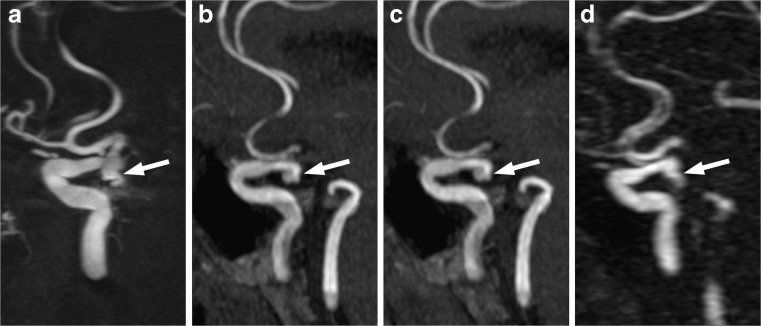
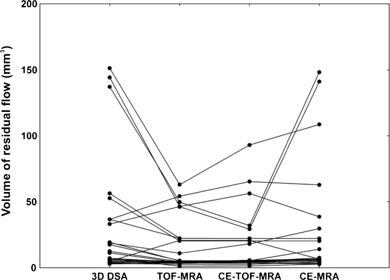
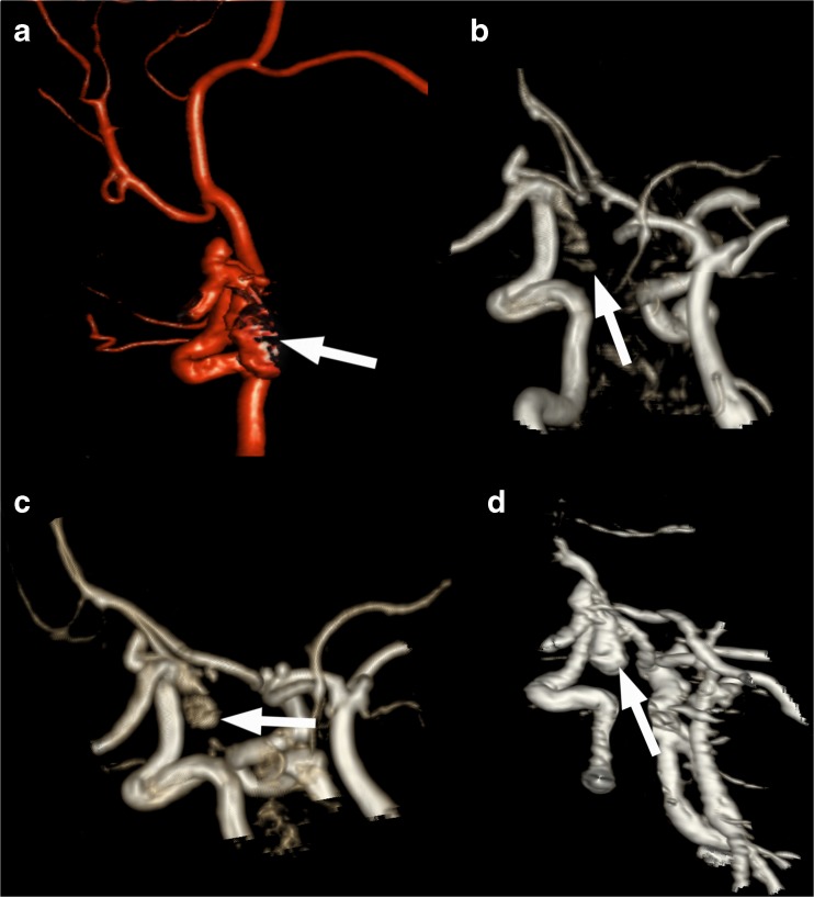
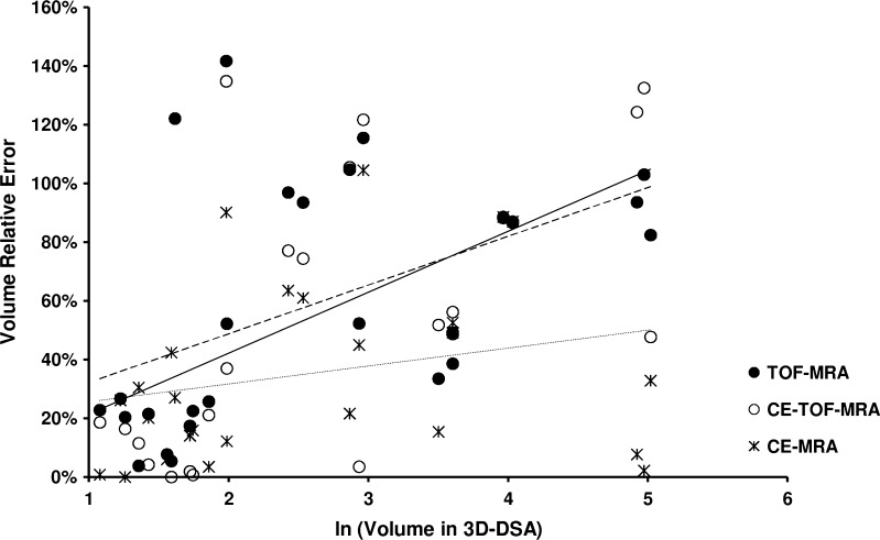
Methods: Twenty-six aneurysms were found incompletely occluded in 72 consecutively examined patients at a follow-up after 3 months. The diameters and volume of aneurysm remnants were compared between 3D-DSA, time-of-flight MRA (TOF-MRA), contrast-enhanced TOF-MRA (CE-TOF-MRA), and contrast-enhanced MRA (CE-MRA) at 1.5 T.
Results: There was a significant correlation between remnant volumes calculated based on 3D-DSA and all MRA modalities. The intraobserver variability of the measurements ranged from 3.4 to 4.1 % and the interobserver variability from 5.8 to 7.3 %. There were no significant differences in the variability between the techniques. The mean residual filling volume ranged from 16.3 ± 19.0 mm(3) in TOF-MRA to 30.5 ± 44.6 mm(3) in 3D-DSA (P < 0.04). Significant differences were found in the volumes measured with 3D-DSA and CE-MRA as compared to TOF-MRA and CE-TOF-MRA (P < 0.01). There was a moderate significant correlation between the residual filling and the relative error of measurement in the case of TOF-MRA and CE-TOF-MRA.
Conclusions: TOF-MRA seems to underestimate the size of aneurysm remnants detected at follow-up and should not be used as a sole imaging method to decide on re-embolization.
Figures


Similar articles
-
Follow-up of intracranial aneurysms treated with stent-assisted coiling: Comparison of contrast-enhanced MRA, time-of-flight MRA, and digital subtraction angiography.J Neuroradiol. 2017 Feb;44(1):44-51. doi: 10.1016/j.neurad.2016.10.004. Epub 2016 Nov 9. J Neuroradiol. 2017. PMID: 27836654
-
Follow-up after embolization of ruptured intracranial aneurysms: a prospective comparison of two-dimensional digital subtraction angiography, three-dimensional digital subtraction angiography, and time-of-flight magnetic resonance angiography.Neuroradiology. 2012 Nov;54(11):1253-60. doi: 10.1007/s00234-012-1030-z. Epub 2012 Apr 10. Neuroradiology. 2012. PMID: 22488210 Free PMC article.
-
Three-dimensional time-of-flight (3D TOF) magnetic resonance angiography (MRA) and contrast-enhanced MRA of intracranial aneurysms treated with platinum coils.Acta Radiol. 2008 Mar;49(2):190-6. doi: 10.1080/02841850701732940. Acta Radiol. 2008. PMID: 18300146 Clinical Trial.
-
Follow-up of intracranial aneurysms treated by flow diverter: comparison of three-dimensional time-of-flight MR angiography (3D-TOF-MRA) and contrast-enhanced MR angiography (CE-MRA) sequences with digital subtraction angiography as the gold standard.J Neurointerv Surg. 2016 Jan;8(1):81-6. doi: 10.1136/neurintsurg-2014-011449. Epub 2014 Oct 28. J Neurointerv Surg. 2016. PMID: 25352582
-
MRA versus DSA for the follow-up imaging of intracranial aneurysms treated using endovascular techniques: a meta-analysis.J Neurointerv Surg. 2019 Oct;11(10):1009-1014. doi: 10.1136/neurintsurg-2019-014936. Epub 2019 May 2. J Neurointerv Surg. 2019. PMID: 31048457 Review.
Cited by
-
Effectiveness of MRA on embolized intracranial aneurysms: a comparison of DSA, CE-MRA, and TOF-MRA.J Interv Med. 2019 Apr 30;1(1):32-41. doi: 10.19779/j.cnki.2096-3602.2018.01.08. eCollection 2018 Feb. J Interv Med. 2019. PMID: 34805829 Free PMC article.
-
Evaluation the Aortic Aneurysm Remodeling After a Successful Stentgraft Implantation.Pol J Radiol. 2016 Oct 13;81:486-490. doi: 10.12659/PJR.900116. eCollection 2016. Pol J Radiol. 2016. PMID: 27800038 Free PMC article.
-
Usefulness of Pointwise Encoding Time Reduction with Radial Acquisition-Magnetic Resonance Angiography after Endovascular Treatment for Intracranial Aneurysms.Interv Neuroradiol. 2023 Feb;29(1):20-29. doi: 10.1177/15910199211065906. Epub 2021 Dec 16. Interv Neuroradiol. 2023. PMID: 34913378 Free PMC article.
-
Visualization of Aneurysmal Neck and Dome after Coiling with 3D Multifusion Imaging of Silent MRA and FSE-MR Cisternography.AJNR Am J Neuroradiol. 2019 May;40(5):802-807. doi: 10.3174/ajnr.A6026. Epub 2019 Apr 4. AJNR Am J Neuroradiol. 2019. PMID: 30948372 Free PMC article.
-
Aneurysm Recurrence Volumetry Is More Sensitive than Visual Evaluation of Aneurysm Recurrences.Clin Neuroradiol. 2016 Mar;26(1):57-64. doi: 10.1007/s00062-014-0330-6. Epub 2014 Aug 27. Clin Neuroradiol. 2016. PMID: 25159038 Clinical Trial.
References
-
- Qureshi AI, Vazquez G, Tariq N, Suri MF, Lakshminarayan K, Lanzino G. Impact of International Subarachnoid Aneurysm Trial results on treatment of ruptured intracranial aneurysms in the United States. J Neurosurg. 2010;114:834–841. - PubMed
-
- Gnanalingham KK, Apostolopoulos V, Barazi S, O'Neill K. The impact of the International Subarachnoid Aneurysm Trial (ISAT) on the management of aneurysmal subarachnoid haemorrhage in a neurosurgical unit in the UK. Clin Neurol Neurosurg. 2006;108:117–123. doi: 10.1016/j.clineuro.2005.11.001. - DOI - PubMed
-
- Ringer AJ, Rodriguez-Mercado R, Veznedaroglu E, Levy EI, Hanel RA, Mericle RA, Lopes DK, Lanzino G, Boulos AS. Defining the risk of retreatment for aneurysm recurrence or residual after initial treatment by endovascular coiling: a multicenter study. Neurosurgery. 2009;65:311–315. doi: 10.1227/01.NEU.0000349922.05350.96. - DOI - PubMed
Publication types
MeSH terms
Substances
LinkOut - more resources
Full Text Sources
Medical