

Image-guided hepatopancreatobiliary surgery using near-infrared fluorescent light
- PMID: 22790312
- PMCID: PMC3501168
- DOI: 10.1007/s00534-012-0534-6
Image-guided hepatopancreatobiliary surgery using near-infrared fluorescent light
Abstract
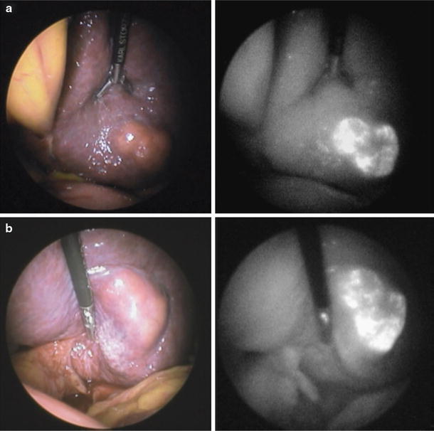
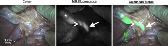
Background: Improved imaging methods and surgical techniques have created a new era in hepatopancreatobiliary (HPB) surgery. Despite these developments, visual inspection, palpation, and intraoperative ultrasound remain the most utilized tools during surgery today. This is problematic, though, especially in laparoscopic HPB surgery, where palpation is not possible. Optical imaging using near-infrared (NIR) fluorescence can be used for the real-time assessment of both anatomy (e.g., sensitive detection and demarcation of tumours and vital structures) and function (e.g., assessment of luminal flow and tissue perfusion) during both open and minimally invasive surgeries.
Methods: This article reviews the published literature related to preclinical development and clinical applications of NIR fluorescence imaging during HPB surgery.
Results: NIR fluorescence imaging combines the use of otherwise invisible NIR fluorescent contrast agents and specially designed camera systems, which are capable of detecting these contrast agents during surgery. Unlike visible light, NIR fluorescent light can penetrate several millimetres through blood and living tissue, thus providing improved detectability. Applications of this technique during HPB surgery include tumour imaging in liver and pancreas, and real-time imaging of the biliary tree.
Conclusions: NIR fluorescence imaging is a promising new technique that may someday improve surgical accuracy and lower complications.
Figures

References
-
- van der Poel HG, Buckle T, Brouwer OR. Valdes Olmos RA, van Leeuwen FW. Intraoperative laparoscopic fluorescence guidance to the sentinel lymph node in prostate cancer patients: clinical proof of concept of an integrated functional imaging approach using a multimodal tracer. Eur Urol. 2011;60(4):826–833. doi: 10.1016/j.eururo.2011.03.024. - DOI - PubMed
-
- Troyan SL, Kianzad V, Gibbs-Strauss SL, Gioux S, Matsui A, Oketokoun R, et al. The FLARE intraoperative near-infrared fluorescence imaging system: a first-in-human clinical trial in breast cancer sentinel lymph node mapping. Ann Surg Oncol. 2009;16(10):2943–2952. doi: 10.1245/s10434-009-0594-2. - DOI - PMC - PubMed
Publication types
MeSH terms
Substances
Grants and funding
LinkOut - more resources
Full Text Sources
Other Literature Sources
Medical
Miscellaneous
