. 2012 Sep 1;303(5):G635-45.
doi: 10.1152/ajpgi.00097.2012. Epub 2012 Jul 12.

HCl-induced and ATP-dependent upregulation of TRPV1 receptor expression and cytokine production by human esophageal epithelial cells

Affiliations

HCl-induced and ATP-dependent upregulation of TRPV1 receptor expression and cytokine production by human esophageal epithelial cells

Jie Ma et al. Am J Physiol Gastrointest Liver Physiol. .

Abstract

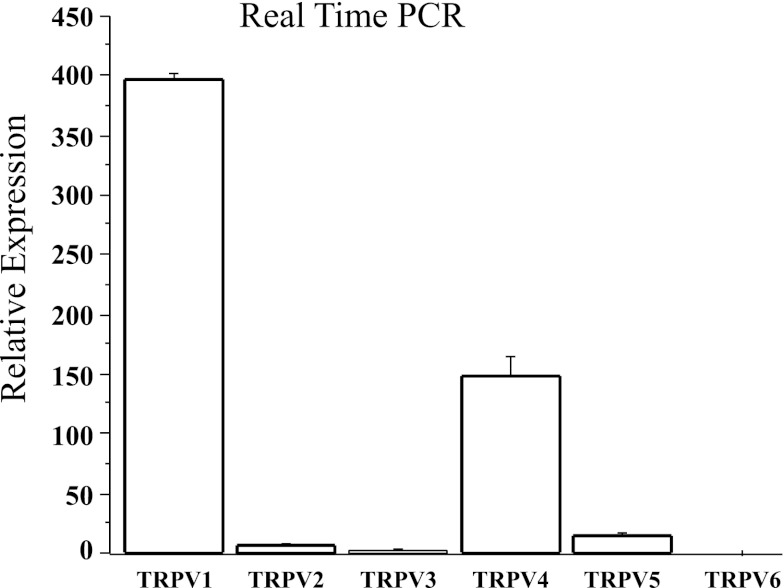
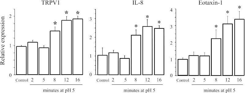
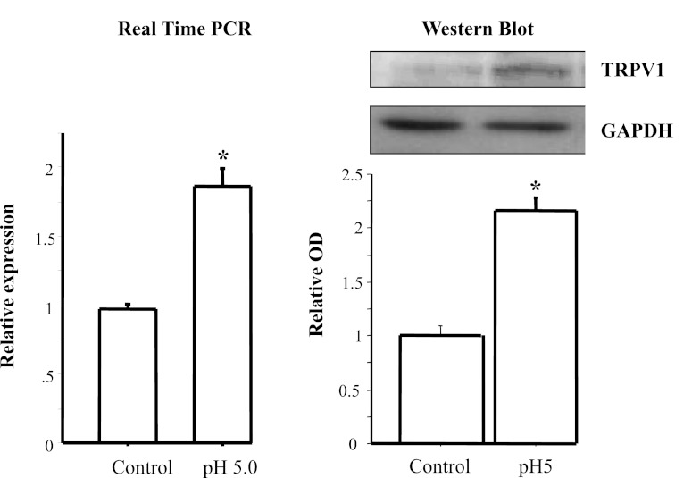
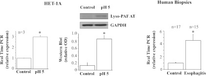
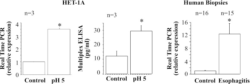
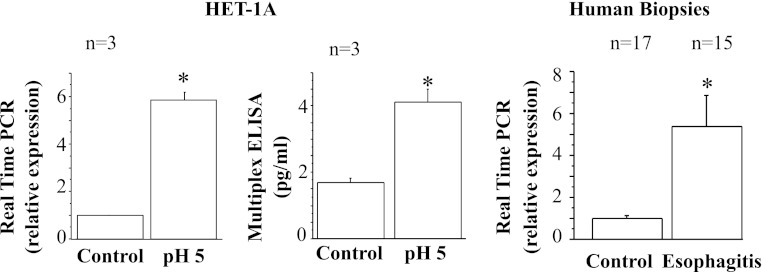
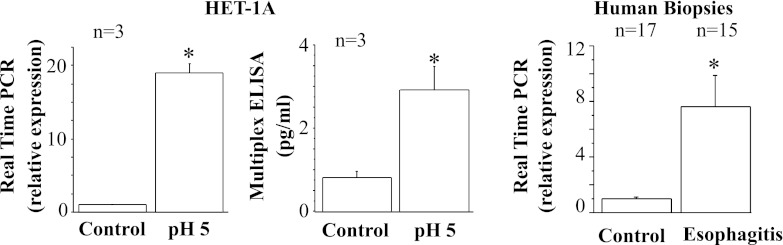
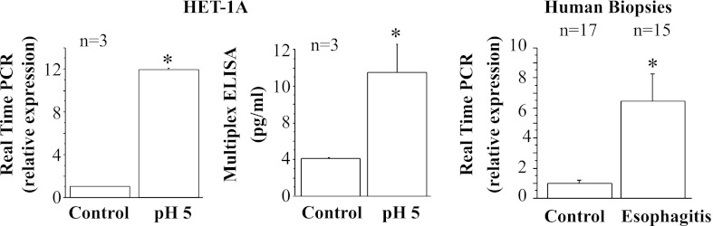
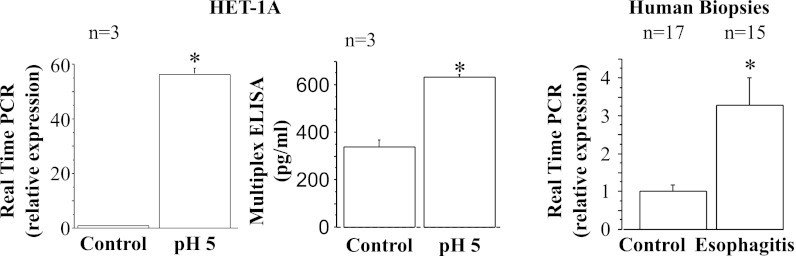
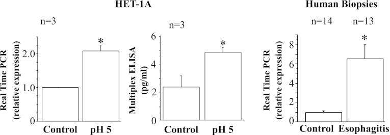
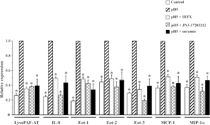
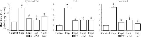
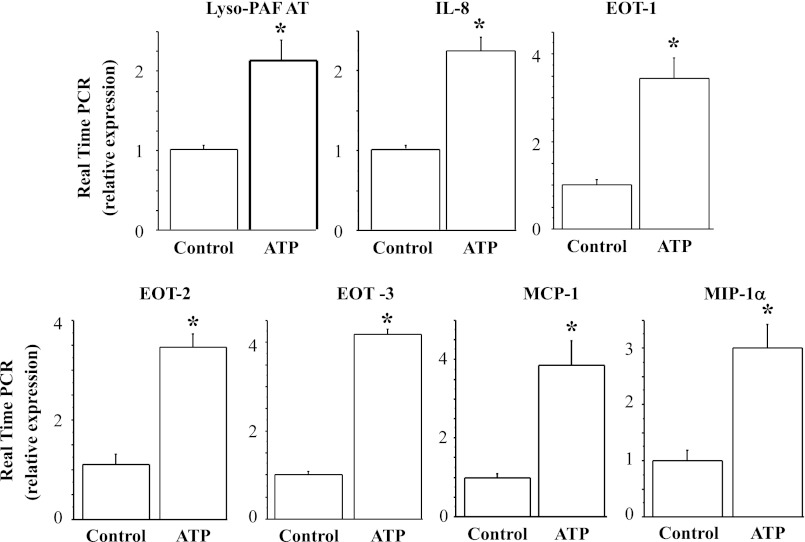
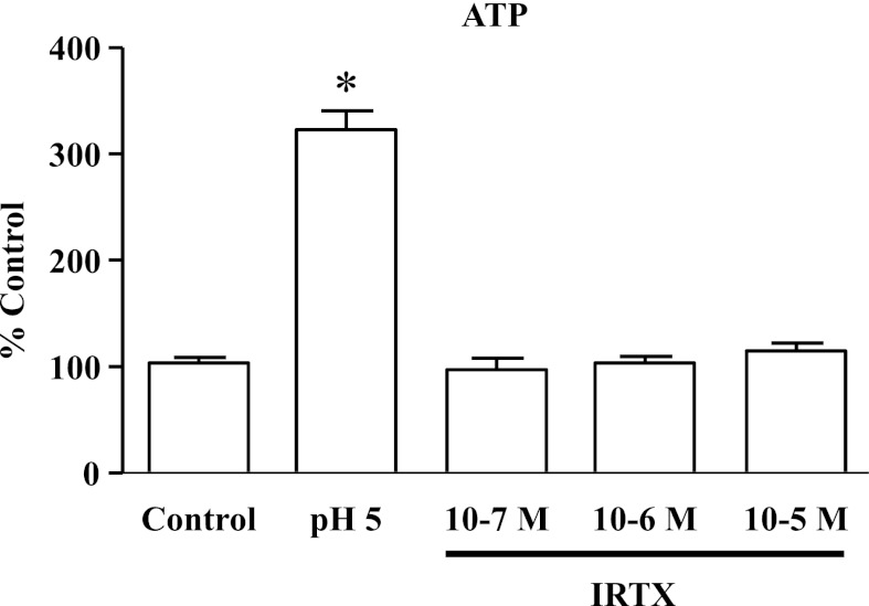
The pathogenesis of gastroesophageal reflux disease (GERD) remains elusive, but recent evidence suggests that early secretion of inflammatory cytokines and chemokines by the mucosa leads to influx of immune cells followed by tissue damage. We previously showed that exposure of esophageal mucosa to HCl causes ATP release, resulting in activation of acetyl-CoA:1-O-alkyl-sn-glycero-3-phosphocholine acetyltransferase (lyso-PAF AT), the enzyme responsible for the production of platelet-activating factor (PAF). In addition, HCl causes release of IL-8 from the esophageal mucosa. We demonstrate that esophageal epithelial cells secrete proinflammatory mediators in response to HCl and that this response is mediated by ATP. Monolayers of the human esophageal epithelial cell line HET-1A were exposed to acidified cell culture medium (pH 5) for 12 min, a total of seven times over 48 h, to simulate the recurrent acid exposure clinically occurring in GERD. HCl upregulated mRNA and protein expression for the acid-sensing transient receptor potential cation channel, subfamily vanilloid member 1 (TRPV1), lyso-PAF AT, IL-8, eotaxin-1, -2, and -3, macrophage inflammatory protein-1α, and monocyte chemoattractant protein-1. The chemokine profile secreted by HET-1A cells in response to repeated HCl exposure parallels similar findings in erosive esophagitis patients. In HET-1A cells, the TRPV1 agonist capsaicin reproduced these findings for mRNA of the inflammatory mediators lyso-PAF AT, IL-8, and eotaxin-1. These effects were blocked by the TRPV1 antagonists iodoresiniferatoxin and JNJ-17203212. These effects were imitated by direct application of ATP and blocked by the nonselective ATP antagonist suramin. We conclude that HCl/TRPV-induced ATP release upregulated secretion of various chemoattractants by esophageal epithelial cells. These chemoattractants are selective for leukocyte subsets involved in acute inflammatory responses and allergic inflammation. The data support the validity of HET-1A cells as a model of the response of the human esophageal mucosa in GERD.


Figures

Fig. 1.
Fig. 1.
Human esophageal squamous epithelial (HET-1A) cells contain mRNA for several vanilloid receptors, with greater levels of transient receptor potential cation channel subfamily vanilloid (TRPV) member 1 (TRPV1) mRNA than TRPV2–TRPV6 mRNA (P < 0.05 ANOVA). Values are means ± SE of 3 different sets of data from 3 separate experiments.
Fig. 2.
Fig. 2.
HET-1A cells were exposed 7 times to pH 5 for 0–16 min over a 48-h period. mRNA levels, obtained by real-time PCR, are shown as a function of duration of individual exposure to pH 5. mRNA increase was maximal for repeated 12-min exposures. Increasing exposure duration to 16 min did not measurably increase mRNA. For simplicity, data are shown only for TRPV1, IL-8, and eotaxin-1. Values are means ± SE; n = 3 for 3 different sets of data from 3 separate experiments. *Significantly different from control (P < 0.05).
Fig. 3.
Fig. 3.
Real-time PCR and Western blot for TRPV1. HET-1A cells were cultured in bronchial epithelial cell medium and treated with the selected HCl exposure protocol (12 min of exposure to pH 5 on 7 occasions over 48 h). Left: TRPV1 mRNA expression determined by real-time PCR. Right: TRPV1 protein expression determined by Western blotting. Relative mRNA expression was calculated with respect to mRNA from untreated cells. Western blot results are shown as relative optical density (OD) normalized to GAPDH. Exposure protocol caused a significant increase in mRNA and protein expression for TRPV1 (*P < 0.01). Values are means ± SE; n = 3 for 3 different sets of data from 3 separate experiments.
Fig. 4.
Fig. 4.
Real-time PCR and Western blot for acetyl-CoA:1-O-alkyl-sn-glycero-3-phosphocholine acetyltransferase (lyso-PAF AT), the enzyme responsible for production of platelet-activating factor. HET-1A cells were treated with the selected HCl exposure protocol and then used to determine lyso-PAF AT mRNA expression by real-time PCR (left) and protein expression (middle) by Western blotting. Relative mRNA expression was calculated with respect to mRNA from untreated cells. Repeated exposures to pH 5 over 48 h caused a significant increase in mRNA and protein expression for lyso-PAF AT (*P < 0.01). Western blot results are shown as relative optical density normalized to GAPDH. Values are means ± SE; n = 3 for 3 different sets of data from 3 separate experiments. Right: real-time PCR for human biopsies from patients with erosive esophagitis (EE) and normal controls. Increased lyso-PAF AT mRNA in EE patients parallels increase in HET-1A cells after exposure to HCl.
Fig. 5.
Fig. 5.
Real-time PCR and ELISA for IL-8. HET-1A cells were treated with the selected HCl exposure protocol and then used to determine IL-8 RNA expression by real-time PCR (left) and protein expression by ELISA (middle). Relative mRNA expression was calculated with respect to mRNA from untreated cells. Repeated exposures to pH 5 over 48 h caused a significant increase in mRNA and protein expression for IL-8 (*P < 0.02). Values are means ± SE; n = 3 for 3 different sets of data from 3 separate experiments. Right: real-time PCR for human biopsies from EE patients and normal controls. Increased IL-8 mRNA expression in EE patients parallels increase in HET-1A cells after exposure to HCl.
Fig. 6.
Fig. 6.
Real-time PCR and ELISA for eotaxin-1. HET-1A cells were treated with the selected HCl exposure protocol and then used to determine eotaxin-1 mRNA expression by real-time PCR (left) and protein expression by ELISA (middle). Repeated exposures to pH 5 over 48 h caused significant increase in mRNA and protein expression for eotaxin-1 (*P < 0.05). Relative mRNA expression was calculated with respect to mRNA from untreated cells. Values are means ± SE; n = 3 for 3 different sets of data from 3 separate experiments. Right: real-time PCR for human biopsies from EE patients and normal controls. Increased eotaxin-1 mRNA in EE parallels increase in HET-1A cells after exposure to HCl.
Fig. 7.
Fig. 7.
Real-time PCR and ELISA for eotaxin-2. HET-1A cells were treated with the selected HCl exposure protocol and then used to determine eotaxin-2 mRNA expression by real-time PCR (left) and protein expression by ELISA (middle). Repeated exposures to pH 5 over 48 h caused significant increase in mRNA and protein expression for eotaxin-2 (*P < 0.02). Relative mRNA expression was calculated with respect to mRNA from untreated cells. Values are means ± SE; n = 3 for 3 different sets of data from 3 separate experiments. Right: real-time PCR for human biopsies from EE patients and normal controls. Increased eotaxin-2 mRNA in EE patients parallels increase in HET-1A cells after exposure to HCl.
Fig. 8.
Fig. 8.
Real-time PCR and ELISA for eotaxin-3. HET-1A cells were treated with the selected HCl exposure protocol and then used to determine eotaxin-3 mRNA expression by real-time PCR (left) and protein expression by ELISA (middle). Repeated exposures to pH 5 over 48 h caused significant increase in mRNA and protein expression for eotaxin-3 (*P < 0.01). Relative mRNA expression was calculated with respect to mRNA from untreated cells. Values are means ± SE; n = 3 for 3 different sets of data from 3 separate experiments. Right: real-time PCR for human biopsies from EE patients and normal controls. Increased eotaxin-3 mRNA in EE patients parallels increase in HET-1A cells after exposure to HCl.
Fig. 9.
Fig. 9.
Real-time PCR and ELISA for monocyte chemoattractant protein 1 (MCP-1). HET-1A cells were treated with the selected HCl exposure protocol and then used to determine MCP-1 RNA expression by real-time PCR (left) and protein expression by ELISA (middle). Repeated exposures to pH 5 over 48 h caused a significant increase in mRNA and protein expression for MCP-1 (*P < 0.001). Relative mRNA expression was calculated with respect to mRNA from untreated cells. Values are means ± SE; n = 3 for 3 different sets of data from 3 separate experiments. Right: real-time PCR for human biopsies from EE patients or from normal controls. Increased MCP-1 mRNA in EE patients parallels increase in HET-1A cells after exposure to HCl.
Fig. 10.
Fig. 10.
Real-time PCR and ELISA for macrophage inflammatory protein 1α (MIP-1α). HET-1A cells were treated with the selected HCl exposure protocol and then used to determine MIP-1α RNA expression by real-time PCR (left) and protein expression by ELISA (middle). Repeated exposures to pH 5 over 48 h caused a significant increase in mRNA and protein expression for MIP-1α (*P < 0.05). Relative mRNA expression was calculated with respect to mRNA from untreated cells. Values are means ± SE; n = 3 for 3 different sets of data from 3 separate experiments. Right: real-time PCR for human biopsies from EE patients and normal controls. Increased MIP-1α mRNA in EE patients parallels increase in HET-1A cells after exposure to HCl.
Fig. 11.
Fig. 11.
Role of TRPV1 and ATP in HCl-induced upregulation of mRNA for lyso-PAF AT, IL-8, eotaxin-1 (Eot-1), eotaxin-2 (Eot-2), eotaxin-3 (Eot-3), MCP-1, and MIP-1α. HET-1A cells were treated with the selected HCl exposure protocol. Some cells were pretreated with the TRPV1 antagonists iodoresiniferatoxin (IRTX, 3 × 10⁻⁶ M) and JNJ-17203212 (10−6 M) or the nonselective purinergic receptor antagonist suramin (10−4 M) for 20 min before each exposure to acid. Real-time PCR was used for mRNA determination. Relative mRNA expression was calculated with respect to mRNA values after acid exposure (pH 5, and reported as 1). Initial mRNA values, before exposure to HCl, are shown as control. For lyso-PAF AT and all the cytokines/chemokines, initial mRNA values were significantly lower (*P < 0.05) than values after acid exposure. There was no significant difference between initial mRNA values and values after TRPV1 antagonists or after suramin pretreatment, indicating that TRPV1 antagonists and suramin inhibit the HCl-induced increase in mRNA. None of the antagonists affected cell viability as assessed by Trypan blue exclusion (>98%). Values are means ± SE; n = 3 for 3 different sets of data from 3 separate experiments. *Significantly different from pH 5 (P < 0.05).
Fig. 12.
Fig. 12.
Selective TRPV1 agonist capsaicin (Cap) was used to confirm TRPV1-mediated upregulation of mRNA for lyso-PAF AT, IL-8, and eotaxin-1. HET-1A cells were exposed for 12 min to culture medium containing capsaicin (10−5 M) on 7 occasions over 48 h. mRNA upregulation produced by capsaicin-induced TRPV1 activation was similar to that produced by HCl exposure (see Figs. 4–6). mRNA values after repeated capsaicin exposure were not significantly different from those induced by HCl exposure and significantly greater (*P < 0.001) than control values. As expected, pretreatment with IRTX (3 × 10⁻⁶ M) or JNJ-17203212 (JNJ, 10−6 M) significantly reduced capsaicin-mediated mRNA increase (#P < 0.001), confirming that the antagonists properly inhibit TRPV1 activation. Inhibition by the nonselective ATP antagonist suramin (Sur, 10−4 M) supports a role of ATP in mediating mRNA upregulation.
Fig. 13.
Fig. 13.
ATP-induced upregulation of lyso-PAF AT, IL-8, eotaxin-1, -2, and -3, MCP-1, and MIP-1α mRNA. After seven 12-min exposures to the ATP analog ATPγS (10−4 M) over 48 h, HET-1A cells were used to determine mRNA by real-time PCR. Relative mRNA expression was calculated with respect to mRNA from untreated cells. Repeated exposure to ATP over 48 h caused a significant increase (*P < 0.01) in lyso-PAF AT, IL-8, eotaxin-1, -2, and -3, MCP-1, and MIP-1α mRNA expression. Values are means ± SE; n = 3 for 3 different sets of data from 3 separate experiments.
Fig. 14.
Fig. 14.
HCl (pH 5, 5 min) induced significant release of ATP from epithelial cells (*P < 0.001) compared with untreated cells (control). IRTX (10−7–10−5 M) did not stimulate ATP release, demonstrating that IRTX concentrations used in the present study do not activate TRPV1 receptors in HET-1A cells. Values are means ± SE; n = 3 for 3 different sets of data from 3 separate experiments.

Similar articles

Cited by

References

    1. Abouheif E, Akam M, Dickinson WJ, Holland PW, Meyer A, Patel NH, Raff RA, Roth VL, Wray GA. Homology and developmental genes. Trends Genet 13: 432–433, 1997 - PubMed
    1. Azpiroz F, Baudet JS, Benages A, Canga F, Carrasco J, Ciriza C, Cucala M, Dominguez E, Faro V, Garrigues V, Giganto F, Herrerias JM, Iglesias J, Lacima G, Lopez P, Llabres M, Mearin F, Minguez M, Mones J, Mora F, Munoz C, Perez de la Serna J, Ponce J, Rodriguez-Tellez M, Romero MJ, Ruiz de Leon A, Ruiz-Cabello M, Sanchez-Gey S, Sanchiz V, Serra J, Sevilla MC, Sopena F, Soria MJ. Normal values in ambulatory oesophageal pH monitoring at two levels in Spain. Rev Esp Enferm Dig 102: 406–412, 2010 - PubMed
    1. Baggiolini M, Loetscher P, Moser B. Interleukin-8 and the chemokine family. Int J Immunopharmacol 17: 103–108, 1995 - PubMed
    1. Basseri B, Levy M, Wang HL, Shaye OA, Pimentel M, Soffer EE, Conklin JL. Redefining the role of lymphocytes in gastroesophageal reflux disease and eosinophilic esophagitis. Dis Esophagus 23: 368–376, 2010 - PubMed
    1. Bhattacharya A, Scott BP, Nasser N, Ao H, Maher MP, Dubin AE, Swanson DM, Shankley NP, Wickenden AD, Chaplan SR. Pharmacology and antitussive efficacy of 4-(3-trifluoromethyl-pyridin-2-yl)-piperazine-1-carboxylic acid (5-trifluoromethyl-pyridin-2-yl)-amide (JNJ17203212), a transient receptor potential vanilloid 1 antagonist in guinea pigs. J Pharmacol Exp Ther 323: 665–674, 2007 - PubMed

Publication types

MeSH terms