. 2012 Jul 24;107(3):469-76.
doi: 10.1038/bjc.2012.279. Epub 2012 Jul 12.

Microbubble and ultrasound radioenhancement of bladder cancer

Affiliations

Microbubble and ultrasound radioenhancement of bladder cancer

W T Tran et al. Br J Cancer. .

Abstract

Background: Tumour vasculature is an important component of tumour growth and survival. Recent evidence indicates tumour vasculature also has an important role in tumour radiation response. In this study, we investigated ultrasound and microbubbles to enhance the effects of radiation.

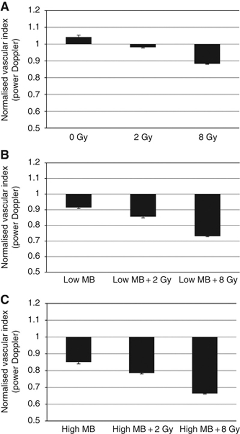
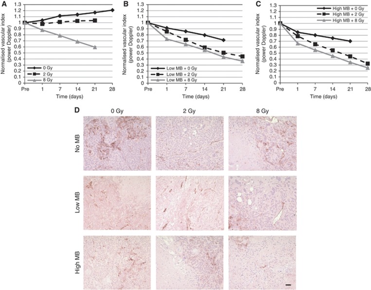
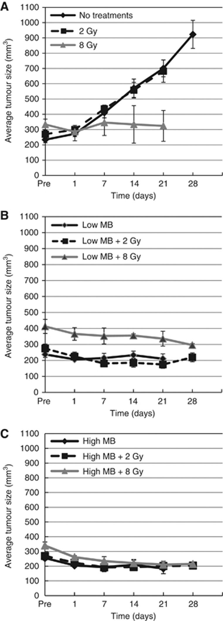
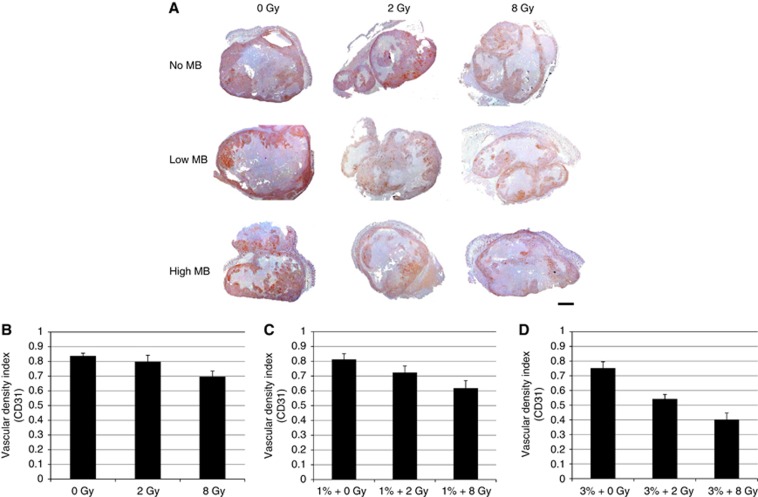
Methods: Human bladder cancer HT-1376 xenografts in severe combined immuno-deficient mice were used. Treatments consisted of no, low and high concentrations of microbubbles and radiation doses of 0, 2 and 8 Gy in short-term and longitudinal studies. Acute response was assessed 24 h after treatment and longitudinal studies monitored tumour response weekly up to 28 days using power Doppler ultrasound imaging for a total of 9 conditions (n=90 animals).

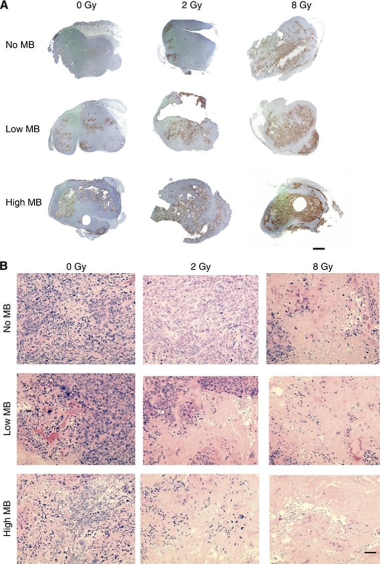
Results: Quantitative analysis of ultrasound data revealed reduced blood flow with ultrasound-microbubble treatments alone and further when combined with radiation. Tumours treated with microbubbles and radiation revealed enhanced cell death, vascular normalisation and areas of fibrosis. Longitudinal data demonstrated a reduced normalised vascular index and increased tumour cell death in both low and high microbubble concentrations with radiation.

Conclusion: Our study demonstrated that ultrasound-mediated microbubble exposure can enhance radiation effects in tumours, and can lead to enhanced tumour cell death.


Figures

Figure 1
Figure 1
Twenty four hours response monitoring of tumour vasculature using power Doppler ultrasound. (A) Tumours exposed to increasing doses of radiation demonstrated reduction in the vascularity (P<0.01, n=5). (B, C) Combination therapies with low and high concentration of ultrasound-activated microbubbles and radiation. Normalised vascular index using power Doppler ultrasound was used to measure vessel abundance (P<0.01, n=5). Error bars=s.e.m.
Figure 2
Figure 2
Long-term response monitoring of tumour vasculature. (A) Tumours treated with varying doses of radiation alone (P<0.001 for 0 and 8 Gy showing significant difference in vascularity; P>0.05 for 2 Gy showing insignificant changes in the NVI). (B, C) Low and high ultrasound-activated microbubbles in combination with radiation treatment. (P<0.001, n=5, error bars=s.e.m.). (D) Tumour tissue sections were stained with CD31 antibodies, counted and quantified after 3–4 weeks of treatment. Tumours show areas of sparse endothelial cell staining from increasing concentrations of microbubbles and radiation doses. Bar indicates 50 μm. Magnification= × 10.
Figure 3
Figure 3
Effects of ultrasound-activated microbubbles and radiation on the growth of HT-1376 human bladder cancer xenografts in SCID mice. Mice bearing HT-1376 human bladder xenografts were grouped for various combination treatments with ultrasound-activated microbubbles and radiation. Mice were also divided into control groups consisting of no treatment or non activated microbubbles or with ultrasound only. (A) No significant difference in tumour size for 2 Gy treatment, however, growth inhibition was observed in 8 Gy treatment with significant difference (P<0.001, n=4–5). (B, C) Low and high ultrasound-activated microbubbles with varying doses of radiation showed a significant increase in tumour growth inhibition (P<0.001, n=4–5). Error bars=s.e.m.
Figure 4
Figure 4
Immunohistochemical analysis in short and long-term tumours. (A) TdT-mediated dUTP-biotin nickend-labelling staining 24 h after treatment indicates tumour necrosis resulting from local treatment. Radiation treatments alone show condensed necrotic area in multiple foci around the tumour area, which is most apparent after 8 Gy. Radiation treatments in combination with low microbubble administration exhibiting viable cells at tumour periphery. High-concentration microbubble treatments with varying doses of radiation. Severe necrotic areas observed at tumour periphery and development of cystic spaces. Scale Bar indicates 2 mm. (B) Hematoxylin and eosin staining 3–4 weeks after treatment × 20 magnification at core regions of tumours. Aberrations to the tumour microenvironment as a result of local tumour ischaemia. Top row, H&E staining through the tumour specimens demonstrated expected results with single-fraction radiation treatment of 2 Gy showed no obvious regions of cell death. Tumour cells were homogenous and evenly dispersed. With 8 Gy, cells were sparse and showed regions of decreased density, suggesting cell death. Middle row, post irradiation micropetechiae as a result of microbubble treatment response. Ultrasound-activated microbubble treatment caused a heterogeneous appearance consisting of fewer intact cells at the centre and circumscribed by healthy cells at the periphery. Bottom row, post irradiation with 3% microbubbles resulting in tumour cell necrosis at the centre. Tumours treated with combined microbubble-ultrasound and radiation demonstrated the largest areas of cell death. Bar indicates 50 μm; magnification ( × 20).
Figure 5
Figure 5
Long-term CD31 staining for endothelial cell markers in tumours. Cluster of differentiation-31 antibody staining was counted and quantified in tumours with varying treatments of ultrasound-activated microbubbles and radiation. (A) Cluster of differentiation-31 staining was performed for tumours 24 h after treatment. A reduction in the total staining of endothelial cells was observed in tumours. Vascular remnants were seen with the absence of endothelial cells. Bar indicates 2 mm. (B) Mice bearing human bladder HT-1376 xenografts treated with varying doses of radiation show reduced endothelial populations 21–28 days after treatment (P>0.05, n=5). (C) Low-concentration ultrasound-activated microbubbles in combination with radiation show reduction in vasculature (P>0.05 (microbubbles alone), (P<0.01 low microbubbles+radiation), n=5). (D) High-concentration ultrasound-activated microbubbles in combination with varying radiation doses show decrease in endothelial cells at 2 Gy (P>0.05, n=5) and significant difference at 8 Gy treatments (P<0.01, n=5) after 21–28 days of treatment. Error bars=s.e.m.

Similar articles

Cited by

References

    1. Blakey DC, Westwood R, Walker M, Hughes GD, Davis PD, Ashton SE, Ryan AJ (2002) Antitumour activity of the novel vascular targeting agent ZD6126 in a panel of tumour models. Clin Cancer Res 8: 1974–1983 - PubMed
    1. Cooney MM, van Heekcheren W, Bhakta S, Ortz J, Remick SC (2006) Drug insight: vascular disrupting agents and angiogenesis-novel approaches for drug delivery. Nature Clin Prac Oncol 12: 682–692 - PubMed
    1. Dings RP, Loren M, Heun H, McNiel E, Griffioen AW, Mayo KH, Griffin RJ (2007) Scheduling of radiation with angiogenesis inhibitors anginex and avastin improves therapeutic outcome via vessel normalization. Clin Cancer Res 13(11): 3395–3402 - PMC - PubMed
    1. Duda DG, Batchelor TT, Willett CG, Jain RK (2007) VEGF-targeted cancer therapy strategies: current progress, hurdles and future prospects. Trends Mol Med 13(6): 223–230 - PMC - PubMed
    1. Feron O (2004) Targeting the tumour vascular compartment to improve conventional cancer therapy. Trends Pharmacol Sci 25(10): 536–542 - PubMed

Publication types

MeSH terms

Substances